2023 in Review: The Best of Research Using BIOPAC

As another year winds to a close, it is a holiday tradition to look back on the last 12 months and take stock. ‘Tis the season of “top 10” lists, “best of” articles, and “year in review” segments in the media. So, not to be left out, we here at BIOPAC have been assessing 2023 from our own unique vantage point. We have put together a brief list of some of our favorite research studies from the year that relied on BIOPAC products to gather and analyze data. We have nervous passengers in driverless cars, killer clowns, and individuals giving us conspicuous side-eye, all for your reading pleasure.

Backseat Drivers

While the advent of self-driving vehicles offers considerable potential for increasing the convenience of transportation and travel, this developing technology is not without controversy. Both passengers and those who would potentially share the road with autonomous vehicles worry about safety issues. A recent study funded by the French National Research Agency examined how passengers in self-driving vehicles reacted when navigating shared-space areas with large concentrations of pedestrians. The study defined shared spaces as “areas where all forms of segregation between pedestrians and vehicles (e.g., road markings, traffic signals, signs, curbs, and barriers) are minimized or eliminated.” Study participants were seated in front of a computer-based driving simulator. EDA data was gathered via a BioNomadix wireless  EDA transmitter and amplifier connected to a BIOPAC data acquisition and analysis system. Data was gathered across a variety of variables including changes in vehicle speed, concentrations of pedestrians, and proximity. Among the study’s conclusions was the suggestion that cognitive processes involved in collision risk assessments may lead to physiological adaptations that allow passengers to cope with stress related to vehicle-pedestrian interactions in shared spaces.

Send in the (Demonic) Clowns

VR HMDAnother study that caught our attention in 2023 examined how stressors created in a virtual reality (VR) environment are manifested in measurable physiological stress reactions. VR has seen a growth of applications beyond entertainment in recent years. It is being used for a wide range of applications such as training in simulated environments where it is necessary to create stressful conditions that mimic real-world experiences. A team of German researchers examined a variety of physiological signals, including electroencephalography (EEG), electrodermal activity (EDA), and peripheral skin temperature (ST) to determine which are most reliable for accurately measuring the stress levels of participants while they experienced cinematic horror scenarios in a VR environment. Study participants wore a VR headset that displayed four-minute episodic horror movies featuring demonic clowns. EEG data was gathered with an SS2L electrode lead set and general-purpose electrodes connected to the left temporal lobe. ECG recording was conducted with a second lead using an SS2L electrode lead set. The research team used an SS3LA EDA transducer to collect EDA data. All data was fed to an MP36 system running Biopac Student Lab (BSL) software. The study successfully demonstrated that stress can be triggered in VR and leads to a physiological and measurable stress reaction.

Are You Looking at Me?

The third study that sparked our interest in 2023 looked at how individuals react to the gaze of another person. A team of researchers from Finland and China used facial electromyography (EMG), skin conductance response (SCR), and heart rate (HR) to measure affective and attentional responses to faces with a direct and averted gaze. The study sought to measure both vicarious responses to watching two individuals who were looking at each other as well as individuals looking directly at the participant. SCR was recorded using a BIOPAC EDA100C amplifier connected to an MP data acquisition system. The participant’s facial muscle activity was measured and amplified with an EMG100C amplifier. The electrocardiogram (ECG) signal was measured with electrodes placed on the left and right inner forearm and amplified with an ECG100C amplifier. While the study did not find significant vicarious eye contact response, it did measure considerable affective response from direct gaze from one or more individuals. The study’s results could provide a better understanding of the role physiology plays in non-verbal communication between individuals and groups.

This is just a small sampling of the many studies that relied on BIOPAC equipment to gather and analyze data in 2023. For more ideas on how BIOPAC is leading the way in physiology, psychophysiology, neurology, and other areas of research, check out our webinars or watch one of our many online training videos. See you in 2024!


Start 2024 right by leveraging BIOPAC products in your next research study. Reach out to your regional sales representative to learn more.

Imaging is Everything: Data Acquisition in the MRI Environment

The marriage of physiological data recording and magnetic resonance imaging (MRI) has allowed scientists to unravel intricate connections between the mind and body. However, this synergy comes with its unique set of challenges that researchers must be aware of to ensure the integrity of both data and the well-being of study participants.

One of the foremost challenges in recording physiological data within the MRI lab is the strong magnetic field. Traditional monitoring equipment can be rendered useless or, worse, pose extreme safety risks due to their ferromagnetic components. Another critical consideration is the impact of radiofrequency (RF) pulses used in the imaging process. RF pulses can introduce artifacts in physiological recordings, affecting the quality of data collected. In previous posts we have discussed the basics of integrating data collection in the functional MRI (fMRI) environment as well as techniques for avoiding artifacts and signal noise while recording data in the MRI.

Researchers must also contend with challenges related to the confined space and noise levels within an MRI environment. A 2021 study conducted by the Lab for Autonomic Neuroscience, Imaging and Cognition (LANIC) at Jena University Hospital in Jena, Germany, focused on how the functional MRI (fMRI) environment itself affected outcomes in autonomic research. Specifically, the study investigated the influence of a loud and cramped environment during MRI on resting heart rate variability (HRV) measures and whether a familiarization session with MRI equipment might lower anxiety levels among participants. The research team recorded an electrocardiogram (ECG) and a photoplethysmogram (PPG) over 15 minutes from participants using a BIOPAC data acquisition and analysis system connected to finger-attached PPG sensors during non-MRI recording. For recording in the fMRI, researchers used a separate BIOPAC data acquisition system equipped with an MR-Conditional PPG module. While the study did establish a link between participant anxiety and HRV levels in the lab and fMRI environment, it did not find that the familiarization sessions had a significant impact on HRV outcomes.

gas maskDespite the challenges, the recording of physiological signals in the MRI environment provides unique opportunities for gathering critical data. A study published in the journal Military Medicine in 2020 tested the effectiveness of F MRI (not to be confused with fMRI) scans in identifying cases of War Lung Injury (WLI) in Iraq and Afghanistan war veterans. WRI refers to reductions in lung function resulting from exposure to environmental factors such as sandstorms and burn pits. F MRI employs magnetic resonance imaging with perfluorinated gases as inhaled contrast agents to evaluate various pulmonary diseases. If successful, the approach would provide an alternative to surgical biopsy for diagnosing suspected WLI. The researchers measured respiratory waveforms throughout the imaging sessions, during both normal room air breathing and PFP-oxygen gas mixture breathing using a BIOPAC MR-Conditional pneumotachometer. The study found that “F MRI provides better insight regarding lung function abnormalities” than other standard methods and that “F MRI could provide a functional tool to assist in identifying subjects with the disorder and potentially could aid in the evaluation of novel therapeutic approaches.”
BIOPAC provides a wealth of tools and information resources for gathering data in the MRI environment. This includes multiple webinars on recording data in both MRI and fMRI settings. As the field continues to evolve, BIOPAC’s innovative solutions pave the way for groundbreaking discoveries at the intersection of physiological signal recording and medical imaging.


If you are interested in recording and analyzing physiological signals in your next MRI-based research study, BIOPAC sales representatives are standing by to help you choose the right tools and resources for the job.

Put a Ring on It: Wearables Offer New Research Options

Rings, watches, bracelets, and belts—we think of them as indispensable fashion accessories. A bit of bling can put the finishing touch on an outfit for a night on the town, but what about a day in the lab? Wearable technology is transforming the landscape of physiology research by enabling real-time, noninvasive data collection in a variety of settings.

For many, their first introduction to the potential of wearable physiology technology came through smartwatches and fitness bracelets. These consumer electronics offer a convenient and affordable way for anyone from professional athletes to fitness hobbyists to monitor their health data, which often includes pulse, blood oxygen levels, steps, and other basic physiological signals.

As the underlying technology has become more sophisticated, researchers have embraced the convenience offered by such devices. Portable, unobtrusive, and wireless wearables have entered the research device market and now allow a range of signals to be monitored under a variety of circumstances and environments.

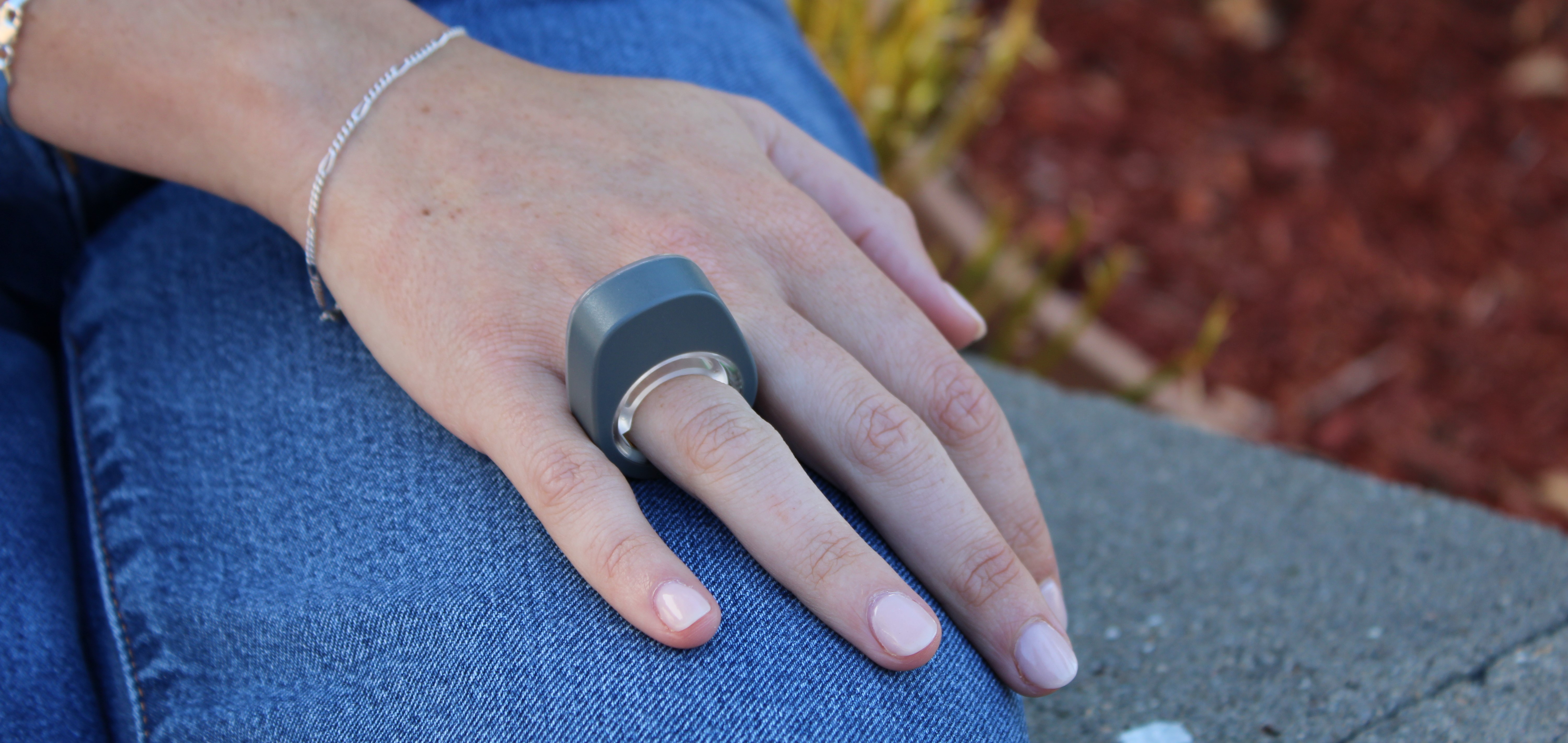
smartphoneBIOPAC recently expanded its established line of wireless devices by introducing the Research Ring, a wearable device with a specific focus on physiological signals such as Photoplethysmography (PPG), Electrodermal Activity (EDA), Electrocardiography (ECG), and body temperature. Products like the Research Ring allow the capture of high-quality data via an unintrusive and accessible wearable device, making them ideal for a wide range of potential applications. These include psychophysiology research, sports and exercise studies, sleep and stress research, and aging and wellness studies, to name just a few.

BIOPAC is no newcomer to providing the research and education communities with wireless and portable solutions designed for gathering physiological data. The Bionomadix line of wearable remote monitoring devices set the standard for such products and have been used in thousands of cited studies. For example, researchers conducting a study of on-the-job stress among healthcare professionals collected EDA and PPG signals using a BIOPAC wireless PPG and EDA amplifier and a wireless respiration with ECG amplifier. Wireless technology proved to be a key component of the study’s success as it allowed the data to be gathered without interfering with the immersive quality of the simulated work environment.

A team of researchers from the University of Porto in Portugal utilized a BIOPAC wearable physiological signal logger to collect EDA and ECG data while studying the correlation between physiological signals and expressive writing. In the study, published in the journal Experimental Psychology, participants were able to engage in writing exercises without the distraction of more cumbersome and intrusive wired solutions.

BIOPAC’s latest entry into its line of wearable wireless data collection devices promises to add greater convenience and versatility to this burgeoning sector. For more information on the new Research Ring, check out the recent webinar BIOPAC hosted with ring developer and partner, Senstream.


If you are interested in adding wearable devices to your next physiology study, reach out to your regional sales representative today to learn which BIOPAC wireless solutions are right for you.

ENS: Signals for Studying the Body’s “Second Brain”

We have all heard the saying “thinking with one’s stomach,” a cliché that suggests basing one’s critical thought processes on the whims of their digestive system. However, there may be more than a little truth to this saying. Often referred to as the body’s “second brain,” the enteric nervous system (ENS) comprises a complex network of neurons and ganglia embedded within the walls of the gastrointestinal tract. In recent years, researchers have increasingly recognized the significance of the ENS and its application in physiological research.

The ENS is a division of the autonomic nervous system (ANS) responsible for regulating the digestive process. It consists of millions of neurons spread throughout the esophagus, stomach, small intestine, and large intestine. These neurons form an intricate network that can function independently of the central nervous system (CNS) but also communicates with it. The ENS controls essential gastrointestinal functions, such as peristalsis, secretion of digestive enzymes, and blood flow regulation within the gut.

Several effective techniques are available to researchers to evaluate and measure ENS activity for their studies. Electrogastrography (EGG) provides a noninvasive approach to measuring the electrical activity of the stomach muscles. EEG can help researchers understand the rhythm and coordination of stomach contractions, aiding in the diagnosis of motility disorders.

In addition to EGG, a range of other physiological signals may be applied to ENS research. A recent study published in the June 2023 edition of Nature Communication investigated the gut-brain relationship by measuring physiological response to stimulation of the digestive system via different noninvasive techniques: EGG, electroencephalogram (EEG), skin conductance response (SCR), heart rate (HR), and heart rate variability (HRV). Research participants ingested a small vibrating probe that provided varying levels of stimulation as it passed through the lower digestive tract. EGG, EEG, and SCR signals were recorded along with hemodynamic signals via a BIOPAC data acquisition and analysis system. Researchers concluded that these techniques provided a viable alternative to more invasive approaches to measuring the body’s response to sensory stimulation of the digestive tract with, according to the published report, “implications for understanding gut feelings and gut-brain interactions in healthy and clinical populations.”

ENS ResearchMore invasive techniques for studying the ENS can still provide researchers with valuable data. A 2023 study by a team of South Korean researchers investigated how radial stretching affects intestinal contractions. The study also examined how drugs targeting the ENS affect such contractions to develop treatments for gastrointestinal disorders. Samples of intestinal tissue were taken from participants undergoing aggressive treatment for colorectal cancer. Samples were preserved in a tissue chamber and connected to a BIOPAC isometric force transducer to measure contractions, with data recorded and analyzed with a BIOPAC MP160 system running AcqKnowledge software. The study was able to detect regional differences in the stretch reflex of colon tissue via electrophysiological recordings of intestinal smooth muscles.

These are some of the ways in which researchers are currently using BIOPAC technologies to better understand the relationship between the ENS and other neurophysiological processes. For additional information on this topic, see our page on AcqKnowledge’s Gastric Myoelectric Activity feature and our EGG citations.


If you would like to know more about how BIOPAC can help you plan and equip your ENS study, reach out to your regional sales representative today.

Noise Proof: Attack Artifacts Before They Ruin Data

After investing months of time, funds, and personal effort into your electrodermal activity (EDA) study, you begin combing through your data recordings when something catches your eye. Several of the recorded signals don’t look quite right. Rather than a narrow baseline in the signal graph, the baseline is thick and fuzzy, a sure sign of signal noise. To make matters worse, the recordings are broken by random peaks, artifacts unrelated to the EDA signal you sought to record. Noise and artifacts can corrupt research data, often rendering it useless. While it may be possible in some cases to clean up and salvage recorded data, such results often mean going back to square one and starting over.

The best option is to avoid bad data from the beginning. Several best practices can save researchers untold effort and expense by avoiding the pitfalls that can result in noise and artifacts. Noise and artifacts can be defined as unwanted changes to a signal caused by sources other than the target signal during recording. Noise and artifacts can originate from a range of sources—environmental (interference from other electromagnetic sources) or biological (signal changes from muscle activity in a participant), to name a few. The key is not to wait until all your data is recorded to tackle signal problems. Careful planning and testing procedures when designing and experimenting can avoid later headaches.

Most artifacts are caused by some form of electrical impedance interfering with a clean connection between the participant and the electrodes linking the data-gathering equipment. Sources could include dead skin or hair at the connection sight, dirty or improperly prepared electrodes, or electrodes that have come loose due to movement.

Using an impedance detector can help identify problematic signals before they corrupt data. An example of this can be found in a 2020 study at Ghent University in Belgium that examined how expert and novice map readers managed their cognitive loads using electroencephalogram (EEG) and eye tracking data. Researchers used a BIOPAC EL-CHECK impedance detector to ensure a good skin-electrode interface before recording data with AcqKnowledge software. This allowed the research team to keep the whole EEG circuit (ground, active EEG, and reference electrodes) at less than the recommended 10 K ohms (Herman, 2015; Teplan, 2002) to prevent unwanted signal noise.

noise checkStrategies for avoiding artifacts can vary depending on the type of signal being recorded. Some signals, such as EEG, may require light abrading of the skin at the point of electrode attachment for optimal conductance, while other signals, like EDA, do not require skin abrasion. Proper site preparation is recommended for most signals, skin should be clean and free of obstructions, such as excess hair. When gel is to be applied, make sure it is the correct type for the electrodes being used. Finally, the preparation of study participants can be critical to successfully recording clean data. In general, participants should be in an environment that is free of distractions and stimuli originating outside the study (sudden noises, distracting conversations, etc.).

For detailed information on how to proactively prevent signal noise and artifacts, check our AcqKnowledge Bootcamp webinar in which we discuss how to set up experiments to ensure quality data gathering. You can also consult our forthcoming guide with detailed instructions for the collection of optimal data.  Don’t wait until it’s too late; eliminating signal noise should be the first step in setting up a successful study.


Do you need help eliminating artifacts and signal noise in your recorded research data? BIOPAC’s regional sales representatives are ready to assist with tools and strategies to ensure you get only the best data possible. Contact us today!

4T|Phys 2023: BIOPAC Shares Tools, Trends, Techniques & Technology at UCSB

They hailed from across the globe, from Arkansas to Zanzibar and all points between. Students, teachers, researchers, and innovators gathered on the campus of UC Santa Barbara, overlooking the Pacific Ocean. They came to learn, share, and explore breakthroughs and best practices in physiology and related fields. 4T|Phys kicked off July 17 under clear California summer skies, an idyllic setting for discovering the Tools, Trends, Techniques, and Technology driving physiology education and exploration.

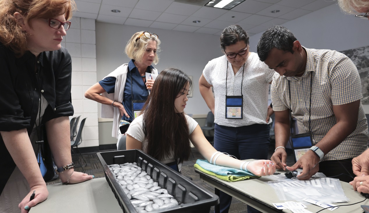
4T|Phys 2023 was the third installment in BIOPAC’s highly successful series of biennial industry conferences that began in 2017. After a two-year hiatus during the global Covid-19 pandemic, 4T came roaring back this summer with more workshops, more tracks, more sponsors, more presenters, and more countries represented than ever before. Over the course of three days, nearly 100 attendees participated in hands-on sessions with some of the top minds in physiology research, experiencing the latest tools for gathering and analyzing data.

BIOPAC’s in-house experts were joined by an array of authorities from the fields of physiology, psychology, imaging (fNIRS and fMRI), neuroscience, cardiovascular research, eye tracking, virtual reality (VR), biology, and engineering. Participants took part in workshop schedules drawn from five separate tracks that included fundamentals, which focused on the basics of data acquisition and analysis. The research track took attendees on a deeper dive into techniques and technologies, including recording evoked potentials, screen-based eye tracking, impedance cardiography using trans-radial bioimpedance velocimetry (TREV), stimulus presentation, exercise physiology, and many others. Participants were also able to tour UC Santa Barbara’s state-of-the-art Brain Imaging Lab to see up close how BIOPAC products are being used in both functional MRI (fMRI) and TREV studies.

“It was an incredible experience that left a lasting impact on me by helping me to understand more about physiological data collection and analysis,” said Dr. Parth Trivedi, a 4T|Phys attendee. “The choice of presenters and the topics covered were truly inspiring, offering valuable knowledge and fresh perspectives that enriched my understanding.”

4T|Phys 2023 Attendee MapThis year’s conference featured several new tracks such as a dedicated VR track led by partner company WorldViz. The WorldViz team showcased virtual reality solutions in a series of workshops that introduced how VR is being integrated in physiology and psychology research, explained how to create effective and immersive VR experiments, and demonstrated their latest PRISM technology for creating fully immersive projected 3D virtual environments.

Several other partner companies introduced their technologies and products both in the 4T exhibition hall and in workshop sessions. Argus Science presented their ETVision eye-tracking glasses that synch seamlessly with AcqKnowledge. Cognixion demonstrated the Cognixion One Axon-R—Research Edition wearable BCI-AR computing platform, which is being used to help individuals with neurodegenerative conditions and severe motor impairments communicate and interact with their environments assisted by augmented reality. Seenel Imaging showed off their MedelOpt Systems for whole-head EEG with fNIRS, including new VR integration. Participants also got a chance to try on the Research Ring by Senstream, a new user-friendly wearable research ring, which allows easy recording of EDA, PPG, Temperature, and ECG signals. CNSystems showed off the company’s NIBP continuous noninvasive blood pressure (NIBP) and hemodynamics devices.

If you missed the educational and networking opportunities at 4T|Phys 2023, don’t despair. We will be back again in a few years, bigger and better than ever. Watch this space for updates on our next installment. In the meantime, see our Video Tutorials and Webinars On Demand for trainings and seminars on our products and partners.


Make sure you never miss a BIOPAC event and keep up with the latest Tools, Trends, Techniques, and Technologies by contacting your local BIOPAC sales representative.

A Heartbeat Away: HRV Unlocks Autonomic Insights

The beating of the heart holds the key to a myriad of physiological questions that are of interest to researchers. The measured variation in time intervals between consecutive heartbeats, otherwise known as Heart Rate Variability (HRV), provides valuable insights into the activity of the autonomic nervous system, reflecting the balance between sympathetic and parasympathetic influences on the heart.

HRV allows researchers to assess cardiac health, stress levels, and overall autonomic function, making it an invaluable component of physiological studies to investigate numerous areas such as cardiovascular health, mental health, stress, and exercise physiology to name just a few.

A recent study applied HRV to examine the effectiveness of yoga and Ayurveda, a form of alternative medicine traditionally practiced in India and Nepal, to regulate and treat lymphedema. Lymphedema is tissue swelling caused by an accumulation of protein-rich fluid that’s usually drained through the body’s lymphatic system. Because lymph drainage has been connected to autonomic function, researchers postulated that HRV would provide meaningful insight into the effectiveness of yoga and Ayurveda programs in treating lymphedema. A BIOPAC two-channel BSL data acquisition system was employed to gather HRV and respiration data of participants who underwent an intensive regimen of yoga exercise, limb soaking, herbal baths, and manual lymph drainage. Initial comparisons of HRV data between both the test and control groups showed significant improvement in the lymphedema patients following treatment.

beating heartIn many cases, researchers choose to combine HRV with other signals to provide a more detailed picture of a participant’s autonomic response to stimuli. A 2020 study sought to identify an effective noninvasive measure of how the sympathetic nervous system (SNS) responds to stimuli. Typically, such measurements involve the invasive recording of muscle sympathetic neural activity at the peroneal nerve. This study examined HRV and electrodermal activity (EDA), both separately and in combination, to see which provided the most effective measurement of SNS activity. Researchers measured participants’ SNS response to both a cold-pressor test (submerging hand in ice water) and while viewing emotionally challenging video images. Throughout both tests, continuous ECG and EDA signals were acquired and analyzed using a BIOPAC four-channel data acquisition system. An ECG100C and EDA100C were used to acquire the ECG and EDA signals, respectively. From their results, researchers concluded that the combination of HRV and EDA provided a better metric for SNS activity than HRV alone.

Whether alone or in combination with other signals, HRV offers researchers a powerful tool for measuring how the autonomic nervous system responds to a wide range of stimuli and conditions with applications across a broad spectrum of fields. Visit our Webinars page for multiple examples of how HRV is being used in research and check our HRV citations as well.


Are you designing a study that features HRV? BIOPAC’s regional sales reps can help you access the right tools for the job to ensure the best possible results. Contact us today!

Faster, Stronger, Further—Unlocking the Body’s Potential with Exercise Physiology

Every athlete seeks the winning formula that will make them more competitive. Add coaches, trainers, weekend warriors, and everyday health enthusiasts to the mix, all looking for an edge to improve fitness, shorten recovery times, and avoid injuries. Exercise physiology plays a key role in gaining such advantages and insights.

Exercise physiology encompasses the scientific study of how the body responds and adapts to exercise and physical activity. It covers a wide spectrum of systems including the cardiovascular, respiratory, and musculoskeletal as well as metabolic function and overall performance. Today’s exercise physiologists benefit from a range of tools and techniques to measure, collect, and analyze such responses and acquire a better understanding of the ways in which exercise affects both humans and other animals, including solutions that integrate the latest virtual reality and eye tracking technology.

This type of research is not without its challenges. Study participants are often required to be physically active during data acquisition. The tools employed in such instances must allow participants to move freely without introducing signal artifacts or noise into the data. A 2021 study into the physiological progression of fatigue shows how researchers can resolve such problems by employing wireless devices such as BIOPAC mobile EMG amplifiers, used in this case to measure strain to the biceps and triceps during exercise. Researchers successfully gathered a range of EMG data with no negative effects from the physical exertion of the study participants.heavy lifting

Exercise physiology research has also been applied to the treatment and management of exercise- and sports-related injuries, both acute and chronic. Additionally, studies have investigated the connection between physical activity and pathology, revealing new ways to prevent or reduce the progress of certain diseases.

A 2020 study by researchers at the University of Udine examined the effects of both exercise alone and exercise combined with a high-protein diet on patients with late-onset Pompe disease, a neuromuscular disorder that degrades a person’s tolerance to physical activity. The study explored which routine had the greatest impact, if any, on a patient’s exercise tolerance, muscle and pulmonary functions, and quality of life. A BIOPAC isometric dynamometer measured muscle force generated by the right leg and arm during flex and extension tests. Data gathered with a BIOPAC data acquisition and analysis system running AcqKnowledge software demonstrated that the combination of exercise and a high-protein diet did improve exercise tolerance, muscle enzymes, pulmonary function, and quality of life for the patients.

Exercise physiology research also sheds light on the ways in which exercise impacts mental and physical wellness. A study published at the Ho Chi Minh University of Technology used heart rate variability (HRV) to evaluate the influence of stress, both physical and mental, on the performance of stationary bike trainees. During the experiment, electrocardiogram (ECG) signal data was gathered from participants using an MP36 data acquisition unit running Biopac Student Lab (BSL) software over a two-month exercise regimen. Based on the results of the study, researchers concluded that HRV provided a useful window into how stress impacts performance and that HRV data could be used as a management tool to monitor stress levels during training.

The techniques and tools available to today’s exercise physiology researchers are as diverse and varied as the areas in which they are being employed, from athletic performance monitoring and enhancement to wellness studies. For more information on how such tools can benefit your exercise physiology research, watch BIOPAC’s webinar on Recording Great Data from Exercising and Mobile Subjects.


Our regional BIOPAC sales reps are ready to help you find the right tools to design your next exercise physiology study. Contact them today to learn more.

Body Electric: Bioimpedance in Physiology Research

With nearly 70 percent of our bodies made up of water as well as a variety of dissolved salts, humans are natural conductors of electrical current. However, various elements of the human body restrict or impede the flow of electrical current, a phenomenon termed “bioimpedance.” Variations in restrictiveness to the flow of electrical current through an organism can provide researchers with a wealth of information on a range of physiological conditions.

Bioimpedance can be defined as the response of an organism to an externally applied electrical current. For more than a century, researchers have used bioimpedance as a safe and noninvasive metric for a range of physiological data, from measuring body composition through bioimpedance analysis (BIA) to investigating various biological systems such as cardiovascular function via impedance cardiography.

While not new, breakthroughs in technology and techniques for recording and analyzing bioimpedance have made it increasingly accessible to researchers. Through impedance cardiography and cardiac output, researchers gain access to a wide range of cardiovascular and hemodynamic data, including heart rate (HR), cardiac output (CO), acceleration index (AI), mean blood pressure (MBP), stroke index (IA), stroke volume (SV), and systemic vascular resistance, to name just a few.

The recent development of trans-radial electrical bioimpedance velocimetry (TREV) has provided researchers with a heightened level of accessibility when attempting to record high-quality data. Unlike traditional transthoracic methods of recording impedance cardiography signals, TREV measures blood movement through the radial and ulnar arteries of the forearm. The approach streamlines electrode placement, making it less prone to complications associated with other impedance cardiography methods.

bioimpedanceOne example of the practical application of impedance cardiography is in research on stress as it relates to coronary health. A 2019 study published by researchers at the University of Illinois at Urbana-Champaign compared the stress response of a group of young participants to that of participants over the age of 55 who had been diagnosed with coronary artery disease (CAD). Researchers recorded physiological signals, including electrocardiogram (ECG), impedance cardiogram (ICG), and continuous BP during rest, stress, and recovery. ECG and ICG signals were collected using BIOPAC BN-NICO wireless amplifiers and BN-EL50-LEAD4 leads. Researchers found ICG provided a reliable metric for stress response.

Recently, a team from the University of California, Santa Barbara (UCSB) employed TREV to demonstrate how this newer bioimpedance metric can be used to track event-related contractility in a maximal grip force experiment. The experiment employed TREV electrodes attached to the participant’s forearm and amplified by a NICO100D smart amplifier. Additionally, ECG electrodes were amplified by an ECG100D while the respiration cycle was recorded via a TSD221-MRI respiration belt. All data signals were acquired and analyzed using an MP160 system running AcqKnowledge software. The experiment confirmed the effectiveness of TREV as a reliable metric for connecting the cardio-sympathetic drive to other physiological conditions, such as force production.

While not new to physiological research, the application of bioimpedance continues to flourish and evolve. To learn more about how bioimpedance is being applied in the research setting as well as new techniques, check out BIOPAC’s webinar on bioimpedance and cardiac output focusing on TREV.


To learn more about incorporating impedance cardiography and TREV in your study, contact your regional BIOPAC sales representative today.

Effort Rewarded: Tools for Measuring Motivation

A job well done may be its own reward, but the mechanisms that drive humans to produce better results are far more complex. Much research has been devoted to the study of motivation and the psychophysiological processes that drive our behavior in pursuit of a goal, whether in relationships, on the job, or in education.

The nature of motivation is controversial, and a variety of competing theories have been put forward regarding what a motivational state comprises. Some researchers have argued that motivation is driven by specific human needs, like food, safety, or respect from one’s peers. Motivation may be categorized as intrinsic, meaning an individual pursues an activity because the activity itself is enjoyable or interesting, or extrinsic, which is when the pursuit offers some external reward.

Research into motivation has drawn interest across a wide range of fields, particularly in the areas of business and education. In the realm of business, corporations are especially focused on the motivation to work, which impacts productivity. In an era where buzzwords such as “quiet quitting” and “bare minimum Mondays” have entered the lexicon, companies are keen to know what motivates workers. Similarly, educators are looking to what types of intrinsic and extrinsic motivational factors are most effective in helping students achieve their learning goals. A key metric for measuring motivation is effort, which can be measured with a range of tools readily available to researchers.

work harderA 2020 Harvard University study published by the American Psychological Association investigated the differences in how adolescents and adults translated motivation into action based on the physical effort exerted for financial reward. Participants age 12 to 23 exerted force by squeezing a BIOPAC hand dynamometer in response to visual cues indicating monetary rewards from $.05 to $.75. The dynamometer, connected to a DA100C amplifier, measured grip force, which the participant saw displayed on a screen as a vertical bar. These tools provided the research team with a means of accurately demonstrating quantitative variations in motivational behavior across various age groups in pursuit of effort-based rewards.

A similar approach was used in a 2019 study from the Netherlands that examined eating behavior by “quantifying the dynamic aspects of appetitive motivation.” As with the above example, researchers relied on a BIOPAC device to measure grip effort, in this case, a bulb transducer connected to an MP36 data acquisition system running Biopac Student Lab software. By squeezing the bulb, participants were able to self-administer sips of chocolate milk via a tube in response to stimuli while experiencing various states of hunger and satiation. The process developed for this study, termed the “Grab-to-Eat Task”, or GET, successfully showed that handgrip effort provided an accurate metric for “evaluating motivational aspects of food consumption” with implications for further research into eating behavior and weight control.

Techniques for measuring motivation are not limited to grip effort. A 2019 study published by the National Institute of Health relied on cardiovascular signals to measure the willingness of older adults to engage in cognitively demanding tasks. Blood pressure and heart rate data were collected using a CNAP monitor connected to a BIOPAC MP150 system, which provided a detailed measure of effort exerted through the participants’ physiological response to each task.

These examples just begin to touch on the ways various psychophysiological measures are being applied to the study of motivation and effort. For additional insight, view the BIOPAC webinar on tools for measuring effort and motivation in the lab.


For more information on how to gather and integrate effort-related data into your motivation study, contact your regional BIOPAC sales representative.

error: Content is protected !!