Under the Skin: Digging Deeper into the Physiology of Fear

From the thrill of cinematic jump scares to debilitating phobias that can turn daily life for some into a waking nightmare, our sense of fear can feel like both a blessing and a curse. Fear is one of the most fundamental human emotions, serving as a necessary safeguard for recognizing and responding to danger. Despite its primal origins, the mechanisms of fear remain complex. For decades, psychophysiologists have studied fear using measures such as the startle reflex and skin conductance, building a deep understanding of how the body reacts to threats. Today, however, researchers are beginning to look beyond these traditional markers. Technologies such as virtual reality (VR) expand how researchers can stimulate the fear response. At the same time, a broad range of signals and techniques can measure not only the body’s autonomic responses, but also how people allocate attention to threats and how the brain processes fear-related information.

One reason fear remains such a compelling topic is that it sits at the intersection of cognition, emotion, memory, and physiology. Fear responses can be observed through changes in sweating, heart activity, breathing patterns, pupil size, neural activity, and even social behavior. As researchers seek to gain a better understanding of anxiety disorders, phobias, post-traumatic stress disorder (PTSD), and related conditions, they increasingly rely on multiple synchronized signals to capture a more complete picture of how fear is learned, remembered, and generalized.

Among the most widely used measures is electrodermal activity (EDA), often recorded as skin conductance response (SCR), which reflects changes in sympathetic nervous system arousal. More recently, eye tracking and pupillometry have emerged as valuable tools for measuring vigilance and attention, while functional near-infrared spectroscopy (fNIRS) has enabled monitoring of changes in cortical activity during fear-related tasks in more naturalistic settings. Together, these approaches help researchers move beyond viewing fear as a single physiological reaction and toward understanding it as a coordinated brain-body process.

A team of researchers at South China Normal University in China explored how the duration of memory retrieval influences the reconsolidation and extinction of fear memories. Using a four-day fear-conditioning paradigm, participants were conditioned to associate visual cues with mild electric shocks. Simultaneously, skin conductance responses were recorded using an MP36 running Biopac Student Lab System. The experiment was designed and implemented with E-Prime stimulus presentation software. The team found that briefly reactivating a fear memory before training participants to view the stimulus as safe was most effective at preventing the fear from returning. Longer periods of memory reactivation produced weaker effects. The study highlights how electrodermal activity can reveal subtle changes in fear-memory processing and may ultimately contribute to improved behavioral interventions for anxiety-related disorders.

fingers and faceA study conducted at the University of Würzburg in Germany examined how anxiety sensitivity influences attention and memory during threat and safety learning. Participants learned associations between neutral faces and aversive outcomes while researchers recorded both electroencephalography (EEG) and pupil diameter. Pupillometry data were acquired using a Tobii eye tracker, allowing researchers to measure moment-to-moment changes in attention. Individuals with higher anxiety sensitivity showed larger pupil responses and enhanced neural markers of attention when viewing both threatening and safe cues, reflecting a generalized pattern of hypervigilance. Interestingly, this heightened attentional engagement did not translate into better memory performance. The findings suggest that pupillometry provides a valuable window into how anxious individuals process potential threats and may help researchers better understand mechanisms underlying pathological anxiety.

Another perspective on fear comes from research at Sichuan Normal University in China, where investigators look at how trait anxiety influences observational fear learning. Unlike traditional conditioning studies, participants learned about danger by watching another person experience threat-related events. Researchers measured skin conductance using a BioNomadix wireless EDA plus data acquisition and analysis system and monitored prefrontal brain activity using fNIRS. The study found that individuals with high trait anxiety exhibited stronger physiological fear responses and increased medial prefrontal cortex activation, even when observing threats indirectly. The work demonstrates that fear can be acquired socially and that neural measures such as fNIRS can reveal aspects of fear processing that are not readily captured by autonomic measures alone. These findings may have important implications for understanding how fear and anxiety spread through social environments and how vulnerability to anxiety disorders develops.

These studies illustrate how fear research leverages multimodal approaches to incorporate multiple physiological perspectives. Skin conductance continues to provide valuable information about autonomic arousal, while pupillometry reveals how attention is divided between threat and safety cues, and fNIRS offers insight into the neural processes involved in fear learning and regulation. As multimodal research platforms become increasingly accessible, researchers are gaining unprecedented insight into how fear is acquired, maintained, and modified. By combining signals from the body, the eyes, and the brain, modern psychophysiology is shining a light on the shadowy labyrinth that is one of humanity’s most powerful and important emotions. To learn more about how to construct a fear stimulation lab for your next fear-conditioning research project, check out our webinar on the subject.


If you are planning a multimodal fear conditioning study, our staff can help identify the right equipment and software for stimulus presentation and data recording and analysis.


 

The Nose Knows: Brain-Body Research and the Science of Olfactory Cognition

“Smell is a potent wizard that transports you across thousands of miles and all the years you have lived.” — Helen Keller

There is something uniquely intimate about the sense of smell. Unlike vision or hearing, olfactory signals travel a remarkably direct neural route from receptors in the nasal epithelium through the olfactory bulb, and straight into the brain’s limbic system, the seat of emotion and memory. A single scent can transport us instantly to a childhood kitchen, sharpen our alertness, or dissolve stress within seconds. Yet for all its power, olfaction has historically been one of the most understudied human senses. That landscape is changing. A new generation of multimodal brain-body research platforms, capable of simultaneously recording signals from the brain and the body, is giving scientists insight into how smell shapes thought, emotion, and cognition.

The olfactory system is the only sensory system that bypasses the thalamus, the brain’s central relay station, and connects directly to the amygdala and hippocampus, two structures critical to emotional processing (affect) and memory. This anatomical shortcut explains why odors are so potent at triggering emotional memories, often more vividly than visual or auditory cues. Beyond memory, researchers are increasingly interested in how odor exposure alters measurable physiological and neurological states in real time, ranging from modulating cognitive performance and stress to the early detection of neurodegenerative disease, in which olfactory dysfunction often appears years before other symptoms emerge.

What makes this research technically challenging is also what makes it scientifically rich: the olfactory experience simultaneously engages the central nervous system and the autonomic nervous system, meaning that studying it well requires capturing signals from both brain and body at once. Electroencephalography (EEG) provides direct data on the brain’s electrical activity with millisecond-level temporal resolution, making it well-suited to detecting rapid shifts in cognition and arousal following odor exposure. Functional near-infrared spectroscopy (fNIRS) offers a complementary picture, using near-infrared light to detect changes in cortical oxygenation, a measure of the brain’s metabolic response to stimulation. Wearable and tolerant of movement, fNIRS is particularly well-suited to naturalistic experimental designs. Electrodermal activity (EDA), or skin conductance, captures the autonomic nervous system’s arousal response through sweat gland activity, while electrocardiography (ECG) and heart rate variability (HRV) add a cardiovascular layer that can distinguish sympathetic from parasympathetic activity in fine detail. Today’s lab setups use a range of sophisticated scent delivery systems to integrate precise, controllable olfactory stimuli into experiments. The real power of modern multimodal platforms lies in their ability to synchronize all these channels within a single setup, time-locking each physiological response to a specific odor event and revealing the full brain-body picture that no single signal could show alone.

A research team at the All India Institute of Medical Sciences in Jodhpur investigated whether citrus odor could buffer the cognitive and physiological impact of working memory load. Thirty participants completed a demanding working memory test under both control and citrus-odor conditions, with the odor delivered via an aroma diffuser. ECG, photoplethysmography (PPG), and EDA were recorded using a BIOPAC data acquisition system with AcqKnowledge software for analysis, and dual-signal BioNomadix wireless EDA and PPG module. For EDA recording, disposable silver/silver chloride disc electrodes were applied to the hypothenar and thenar eminences, and the PPG sensor was placed on the index finger. Citrus odor significantly improved task accuracy while suppressing the rise in skin conductance typically associated with cognitive load. HRV measures, including pNN50, RMSSD, and HF power, shifted toward parasympathetic dominance, indicating a calmer, more regulated physiological state. The findings, published in the Annals of Neurosciences, suggest that citrus odor acts as a cognitive stress buffer and point to practical applications in educational, occupational, and clinical settings with high sustained cognitive demands.

A counterintuitive question motivated a study at Nagaoka University of Technology in Japan: could black pepper, widely known as a stimulant, actually calm the body’s stress response? A group of university students performed a 30-minute arithmetic calculation task under three aroma conditions: black pepper, ginger, and a scentless control. Scents were delivered at precise, reproducible intervals through an olfactometer. A BIOPAC data acquisition system recorded continuous ECG and skin conductance level (SCL) at 200 Hz with 16-bit resolution, with high-frequency HRV (HF-HRV) derived as a measure of cardiac parasympathetic activity. Electrodes were placed beneath the right clavicle and on the lower left abdomen following the Lead II placement protocol for ECG measurements using multi-lead ECG cables. SCL was recorded using skin conductance sensors placed on the palmar side of the middle phalanx of the index and ring fingers of the participant’s non-dominant hand. The results, published in the Journal of Physiological Anthropology, found that black pepper aroma reduced the stress-induced rise in heart rate by 38.9% compared to the scentless control, attenuated the decline in HRV by 32.9%, and blunted the increase in SCL compared to ginger. Crucially, subjective stress ratings did not differ across conditions, demonstrating that the body can respond to olfactory stimuli at a physiological level even when conscious perception does not register a difference. These findings have meaningful implications for workplace wellness and aromatherapy research.

lavenderA French consortium of researchers from the Université Bourgogne Franche-Comté and the Université Grenoble Alpes tested whether music, lavender odor, or their combination would accelerate autonomic recovery following acute cognitive stress. Ninety-nine participants completed a battery of cognitively demanding tasks. They were then randomly assigned to one of four 30-minute recovery conditions: silence, relaxing classical music, lavender essential oil, or music and lavender together. A data acquisition system running AcqKnowledge continuously recorded ECG and EDA using Ag-AgCl finger electrodes, with HF-HRV as an index of parasympathetic activity and EDA as a measure of sympathetic arousal. Published in Psychophysiology, the study found that both music and lavender independently facilitated autonomic recovery. The surprising finding, however, was the absence of any multisensory advantage: combining the two stimuli produced no greater benefit than either alone, suggesting that when multiple sensory channels converge on the same relaxation goal, they may share rather than combine their neural resources. The study underscores both the value of multimodal physiological recording in parsing the mechanisms of stress recovery and the importance of rigorously testing assumptions about how the senses work together.

These studies illustrate how much remains to be discovered at the intersection of smell, brain, and body, and how essential it is to measure all three at once. As synchronized multimodal platforms continue to evolve, the science of olfactory cognition continues to shape our fundamental understanding of how the brain works and the development of practical tools to support cognitive health and wellbeing. To learn more about the convergence of olfaction with brain-body research, be sure to check out our webinar on how scents influence cognition.


If you are planning a brain-body study that integrates multimodal data collection with stimulus presentation like scent delivery, our expert staff is ready to help find everything you need to get started.

Healing Hands: EDA and Affective Touch

“Nothing eases suffering like human touch.” — Bobby Fischer

The gentle caress of a mother to her child. A reassuring pat on the shoulder from a friend. The touch of another human is one of the first forms of communication we experience in life, preceding the development of spoken language. Touch can have a remarkable effect on our emotional, psychological, and physiological states. It shapes our relationships with others and helps us build social connections.

The positive effects of touch are primarily associated with affective touch, characterized by comforting, caress-like stroking that activates C-tactile afferent (CT-afferent) neurons in the skin of mammals (including humans). Affective touch has attracted the interest of researchers seeking to decode how the sympathetic nervous system confronts and manages a variety of challenges, including pain regulation, stress management, and social interactions. The subject has drawn attention in the study of neurodevelopmental disorders, such as Autism Spectrum Disorders (ASD), where it is playing an essential role in understanding the sensory and social challenges facing individuals with atypical neurocognitive function.

While studies on touch, and affective touch specifically, utilize a range of physiological signals to gather data, electrodermal activity (EDA) has proven particularly useful in this area. Also referred to as Galvanic Skin Response (GSR), EDA provides a direct window into the sympathetic nervous system, which is responsible for emotional or affect-related arousal. When someone experiences pleasant or meaningful touch (e.g., a gentle stroke or supportive hand), the brain triggers subtle changes in sweat gland activity manifested as shifts in skin conductance. These changes occur when people aren’t fully aware of their emotional response. This makes EDA a sensitive, objective marker of how the body reacts to social and tactile experiences. In studies of affective touch, researchers track EDA to see whether a touch feels calming, engaging, or stressful, and how these responses differ across individuals or clinical groups such as autistic youth. Because EDA reflects genuine physiological arousal tied to emotional processing, it helps scientists understand the underlying autonomic mechanisms that shape how we experience social touch.

Researchers at the University of Tours in France examined how children with ASD respond to gentle, affective touch to determine whether they exhibit physiological patterns that differ from those of their neurotypical peers under the same conditions. The team measured multiple aspects of the autonomic nervous system, including EDA and electrocardiogram (ECG). These signals were collected using a BIOPAC MP36 data acquisition and analysis system to capture subtle changes in sympathetic arousal. Researchers collected EDA data using two 8 mm Ag/AgCl cup electrodes placed on the second phalanges of the index and middle fingers of the right hand. ECG was collected via two disposable vinyl electrodes placed on the sternum and on the right shoulder.  The study found that autistic children exhibited atypical EDA responses, showing either blunted or inconsistent autonomic reactions relative to controls, even when their outward behavior appeared typical. These results suggest that affective touch may not regulate internal arousal in autistic children the same way it does in others, providing an essential insight for therapies and sensory-based interventions that rely on touch.

In a similar study, a team from the University of Turin in Italy explored how autistic adults process affective touch by comparing their subjective ratings of pleasantness with their physiological responses. The researchers used a BIOPAC data acquisition research system with an EDA amplifier module to record EDA. At the same time, participants received gentle, slow stroking intended to activate the brain’s affective touch pathways. The goal was to determine whether autistic adults show a mismatch between what they say they feel and how their bodies respond. The findings revealed an apparent dissociation. While autistic participants often rated the touch similarly to neurotypical participants, their EDA responses differed significantly, indicating altered sympathetic arousal. This physiological-subjective mismatch provides compelling evidence that affective touch is processed differently in ASD, which may help explain challenges with social touch and sensory integration in everyday interactions.hands

Researchers from the University of Liverpool and Manchester Metropolitan University examined how brief, naturalistic social touch alters brain activity, autonomic physiology, and emotion recognition performance. Although this multimodal study did not focus on an autism-only sample, it is relevant to social-touch research more broadly and provides methodological examples that apply to ASD studies. To capture changes in prefrontal cortex function, the team employed a BIOPAC fNIRS imager system, with signals sent to a data acquisition unit. Further analysis of fNIRS signals was handled via fNIRS visualization software. Both EDA and photoplethysmography (PPG) were recorded at 2 kHz, using shielded Ag/AgCl electrodes on the index and fourth fingers and a PPG sensor on the middle finger, and were acquired and analyzed in AcqKnowledge. The results showed that social touch increased prefrontal activation, modulated cardiac and vascular dynamics captured via PPG, and produced measurable changes in sympathetic arousal, as indexed by EDA. Participants also demonstrated improved emotion-recognition accuracy following touch. Together, these findings suggest that social touch engages coordinated central and peripheral systems in ways that may support social cognition and emotional understanding.

These are just a few examples of how EDA is being used in research into affective touch. For additional information on integrating EDA in your study, see our multiple webinars on the subject.


Are you designing a study into affective touch or just planning a research project that will focus on EDA? Our customer service staff can help you streamline the process to produce best possible data.

Game On: Applying Physiological Data in Video Game Design and Research

Ever since players first started feeding quarters into arcade games and began plugging in home systems in the late twentieth century, researchers have looked for practical tools for both shaping video game design and measuring their effects on players. Physiological signals filled this niche area of research. Researchers began investigating the connection between physiological science and gaming in the 1980s and 1990s, when early studies explored the effects of video games on heart rate and arousal. These first studies focused on issues such as aggression and overstimulation, but research has since expanded to encompass the cognitive and emotional aspects of gameplay. An early research project by NASA, for example, examined how biofeedback could be applied to game design to enhance traditional game control methods while also improving players’ cognitive abilities.

Today, physiological monitoring is not only a method for psychological inquiry but also a vital asset in improving user experience (UX) research, game accessibility, and even driving therapeutic game development. As wearable sensors and game engines grow more sophisticated, the integration of physiological signals into game design is opening new frontiers for immersive, responsive, and health-conscious gaming. By analyzing real-time bodily signals, such as heart rate (HR), electroencephalography (EEG), electromyography (EMG), electrodermal activity (EDA), respiration, photoplethysmography (PPG), and electrooculography (EOG), researchers and developers can gain a deeper understanding of how players emotionally and physically respond to game elements.

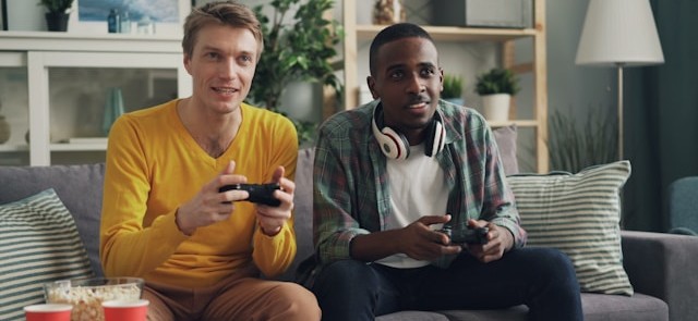
A study published in the International Journal of Human-Computer Studies investigated whether players’ self-reported preferences for first-person shooter (FPS) game dynamics match their emotional reactions during both gameplay and viewing. Researchers recruited 24 active gamers and divided them into two groups: those who enjoyed the fast-paced action of FPS games and those who didn’t. While participants played an FPS game and later watched a recording of it, the team measured electrodermal activity (EDA), heart rate, and facial muscle responses, specifically expressions associated with frowning (corrugator supercilii) and smiling (zygomaticus major), to capture emotional arousal and valence. Physiological signal data were recorded using BIOPAC EDA, PPG, and EMG amplifiers, which were connected to a data acquisition and analysis system running AcqKnowledge software. Although both groups reported more positive feelings and higher excitement after actively playing rather than watching, the data revealed a deeper story: players who disliked the game showed more substantial rises in arousal during play. Those who favored the dynamics, however, showed steadier emotional responses across both conditions. Muscle activity patterns also differed. Those who enjoyed FPS dynamics exhibited sharper increases in frowning-related muscle activity during play than watchers, indicating nuanced emotional engagement. Overall, the study found that what people say they prefer does reflect in their bodies’ emotional reactions. Still, those preferences also shape whether playing or spectating feels more intense, highlighting how combining self-reports with physiological signals can deepen an understanding of game design and player experience.

controllerA team of researchers from Bournemouth University’s Centre for Digital Entertainment investigated how players’ bodies react during intense engagement, or flow, in virtual reality (VR) games. The goal was to capture objective physiological signals, such as heart rate and eye-blink patterns, alongside subjective self-reports to see how well a game’s design induced flow versus boredom or anxiety. The researchers had participants play a custom VR “tower defense” game while monitored with BIOPAC equipment, including a two-channel Biopac Student Lab (BSL) data acquisition system and physiological modules to record HRV via ECG, as well as EOG and EEG signals. The team then utilized clustering and machine-learning classifiers to decode flow states with high accuracy. The research team also successfully identified that high flow correlated with fewer blinks and longer blink intervals, indicating a state of deep concentration. The study not only demonstrates the value of combining physiological and self-report data but also shows how wearable sensors can inform design decisions in VR games by providing real-time feedback on player experience.
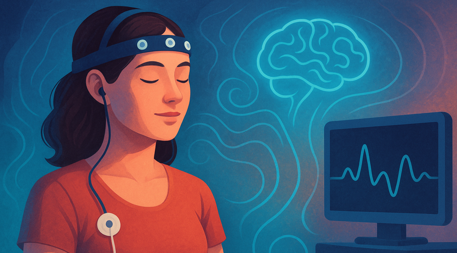
Beyond game design, a primary area of research focus has been on the long-term psychological, neurological, and physiological impact that games have on players. A study published in the Journal of Media Psychology explored how experiences with violent video games might reduce prosocial behavior, such as the motivation to help others, through physiological changes. Researchers recruited 155 Chinese college students who reported their exposure to violent games (in terms of violence, bloodiness, and duration) and completed simple self-reports measuring their willingness to help and the time they took to help others in controlled scenarios. Researchers used a BIOPAC data acquisition unit running AcqKnowledge software to record relevant physiological data. A three-lead ECG amplifier, respiration module, and respiration belt were used to record ECG and breathing signals across baseline, violent-film task, and during recovery periods. Researchers calculated the reduction in respiratory sinus arrhythmia (RSA), a marker of parasympathetic nervous system engagement. They found that higher levels of violent-game experience predicted less RSA reduction during stress, which in turn was linked to reduced prosocial behavior. The study’s findings suggest that playing violent video games may impair physiological readiness to respond in an empathetic manner to others, showing RSA reduction to be a key influence. The study’s findings demonstrate how physiological and psychophysiological signals provide a deeper understanding of the impacts of video games, while also guiding design decisions.

Physiological signals help researchers identify moments of excitement, stress, immersion, or frustration. Such insights are increasingly being used to refine gameplay mechanics, balance difficulty, and enhance player engagement through adaptive or personalized game experiences. They also reveal the short- and long-term effects gameplay has on both individuals and society. For more ideas on how to apply these signals in your research, visit our Webinars page for information on wearable devices, mobile data acquisition, and other helpful topics.


Are you planning a research project related to video games? Reach out to our customer service specialists to get all the help you need in mapping out and equipping your study team.

Mind-Body Connection: Biofeedback in Research

The connection between the mind and body has long intrigued scientists, driving research into how our thoughts, emotions, and mental states can influence physical health and well-being. Biofeedback has become a recognized approach for helping people manage stress, reduce pain, and even enhance cognitive and physical performance. Biofeedback involves using sensors and real-time data to monitor physiological functions that are typically automatic, such as heart rate, breathing, muscle tension, and skin conductance. With this information, individuals can learn to gain greater awareness and, in some cases, voluntarily control these bodily processes.

Common physiological signals used in biofeedback include electrodermal activity (EDA), which reflects sweat gland activity and emotional arousal; electroencephalogram (EEG), which measures electrical activity in the brain; electrocardiogram (ECG), which measures heart electrical activity; heart rate variability (HRV), an index of autonomic nervous system flexibility; and respiration rate, among others. These signals offer researchers a window into the body’s stress responses, regulatory mechanisms, and even aspects of cognition and emotional control.

To complement physiological monitoring, researchers are increasingly integrating tools like stimulus presentation systems, virtual reality (VR) environments, and eye tracking technology into biofeedback studies. These tools allow scientists to study how people react to controlled stimuli, immerse participants in realistic scenarios, and track where and how long someone focuses visually, all while collecting detailed data on their physiological responses. This combination of immersive experiences and real-time monitoring helps researchers investigate how the body responds to stress or cognitive challenges and whether individuals can be trained to influence those responses.

Swiss researchers at ETH Zurich investigated whether individuals could voluntarily control their arousal states using real-time feedback on their pupil size. Pupil size is a physiological marker closely linked to the brain’s arousal systems, particularly the locus coeruleus. Study participants employed mental strategies to adjust their pupil diameter, receiving visual feedback through a dynamically changing circle that reflected their pupil size. The participants’ cardiovascular data, including ECG and respiration, were recorded to provide insights into the physiological effects of pupil self-regulation. The research team employed a BIOPAC data acquisition and analysis system running AcqKnowledge software to record ECG data continuously at a sampling rate of 1,000 Hz. Additionally, a BIOPAC respiratory effort transducer was used to measure respiration, although these respiratory data were not reported in the publication. The study’s findings demonstrated that pupil-based biofeedback enabled participants to modulate not only their pupil size but also associated neural activity and cardiovascular responses, such as heart rate. The results suggest a non-invasive method for individuals to control their autonomic functions, with potential applications in managing stress and anxiety disorders.

mind-bodyVagally mediated heart rate variability (vmHRV) refers to the natural changes in the time between heartbeats that are controlled by the vagus nerve, which is closely tied to how well a person can adapt to stress and regulate their body. Researchers from Université Paris Cité explored whether a brief session of vmHRV biofeedback could help people better control their memory, specifically whether it could help them tell the difference between real and false memories. In the study, 71 healthy young adults completed a VR memory task after receiving either vmHRV biofeedback or a simple breathing exercise. To track their physiological responses, the team used a BIOPAC data acquisition and analysis system and recorded heart activity via a three-lead ECG configured according to Einthoven’s triangle using Lead II. The results showed that this short biofeedback session had no immediate effect on memory accuracy or on vmHRV levels during the task. This suggests that a single session of HRV biofeedback may not be enough to influence memory-related brain functions, underlining the complex relationship between the body’s stress responses and cognitive control.

​A study published in the Journal of Human Kinetics looked at whether just one 10-minute session of HRV biofeedback, using a special breathing technique called resonance frequency breathing, could affect the level of activity of certain nerve cells. Researchers measured this by looking at a reflex in the lower leg muscle (the soleus) called the Hoffman reflex (H-reflex). Twelve healthy men took part in both the HRV biofeedback session and a regular breathing session in a random order. To measure muscle activity, researchers used a BIOPAC surface electrode on the leg and a reference electrode on the hand. The signals were recorded with a BIOPAC data acquisition system and analyzed with AcqKnowledge software, which helped line up the timing of muscle activity with electrical stimulation. The results showed that HRV biofeedback lowered the strength of the reflex and a related ratio (Hmax/Mmax), suggesting the spinal nerve cells were less excitable. These findings point to the potential of short HRV biofeedback sessions to affect the nervous system in ways that could be useful for physical therapy or improving movement control.

To learn more about how to integrate biofeedback techniques into your next research project, check out BIOPAC’s webinar, Biofeedback: Theory, Techniques, and Tools for Research.


Are you planning a research project that will utilize biofeedback data? Our expert customer service representatives can help you find the right tools for all your biofeedback needs.


Aesthetically Pleasing: Exploring How the Brain Perceives the Arts

Beauty is in the eye of the beholder—a mostly subjective experience, so the saying implies. While we may share many common ideals about beauty and aesthetics, our reactions to art, music, and literature can diverge, converge, and overlap to a myriad of degrees. Neuroaesthetics explores how the brain perceives and responds to art, music, and other aesthetically evocative experiences. Combining neuroscience with psychology and the arts, researchers seek to understand why humans find certain images, sounds, or designs appealing and how these perceptions influence our emotions, cognition, and behavior. This interdisciplinary science has practical applications across a wide range of fields, including design, marketing, education, and mental health therapy, where understanding aesthetic experience can inform and enhance user engagement, learning, and emotional well-being.

Biometric signals provide insight into emotional and cognitive processing, allowing researchers to gain valuable data on how the brain creates and responds to aesthetically engaging stimuli. Commonly used techniques include electrodermal activity (EDA), which measures changes in skin conductance linked to emotional arousal; electroencephalography (EEG), which tracks brainwave patterns associated with attention and engagement; functional near-infrared spectroscopy (fNIRS), which monitors blood flow changes in the brain; and functional magnetic resonance imaging (fMRI), which provides high-resolution images of neural activity.

Additionally, the use of virtual reality (VR) and augmented reality (AR) is expanding in neuroaesthetics research, allowing scientists to create controlled, immersive environments that mimic real-world experiences. These technologies offer new ways to investigate how individuals engage with art and design in dynamic, interactive settings, pushing the boundaries of how we understand and harness aesthetic experiences.

Researchers in Shanghai, China, for example, investigated how different visual modes—2D and 3D VR—affect emotional arousal, demonstrating how immersive environments influence emotional experiences. The research team utilized EEG to monitor brain activity and EDA to measure skin conductance responses, both indicators of emotional arousal. EDA and EEG data were recorded while study participants wore VR headsets to view 2D and 3D environments. Data was collected and analyzed using a BIOPAC MP system with AcqKnowledge software. The findings revealed that 3D VR environments elicited more intense emotional stimulation than 2D settings, as evidenced by higher beta (21–30 Hz) EEG power and increased EDA responses. According to the published findings, results suggest that enhanced perception in 3D environments leads to greater emotional engagement, highlighting the potential of 3D VR in applications such as immersive therapy, virtual training, and interactive entertainment.

MusicA team of German researchers recently examined the intersection of neurology, psychology, music, and literature. Their findings, published in the journal NeuroImage, explored how melodic features influence the neural processing of spoken poems and songs, examining the interplay between language and music. Researchers employed fMRI to investigate the neural mechanisms underlying the processing of musical and nonmusical verse. The fMRI data revealed that both spoken poems and songs activated bilateral temporal auditory areas of the brain. One aspect of the study was the measurement of participants’ emotional responses to the stimulus, i.e., whether they liked the song or poem to which they were listening while the fMRI scan was conducted. To gather this data, participants interacted with a BIOPAC differential pressure transducer connected to the participants’ right and left index fingers to record the degree to which they enjoyed the music. The researchers stated the findings suggest that melodic properties are central to the aesthetic experience of both speech and music.

A study published by Britain’s Royal Society looked at the connection between aesthetic appreciation and emotional states, specifically how exposure to art or music can shift feelings of anxiety to curiosity. To understand how the brain perceives beauty, the researchers used EDA to assess participants’ physiological arousal during exposure to aesthetically pleasing stimuli, such as music. EDA data was gathered using a BIOPAC EDA amplifier connected to a data acquisition and analysis system while participants were administered anxiety-inducing electrical tactile stimulation during music listening sessions. The study demonstrated that aesthetic appreciation can reduce anxiety and promote curiosity by analyzing the EDA data alongside self-reported measures. The research team concluded that their results “might have potential applications across different human activities such as learning and therapy. The idea that aesthetic appreciation might affect anxious states and that expecting aesthetic rewards can transform anxiety into arousal and curiosity plays a central role in some psychotherapeutic models of human change.”

These examples provide a rough outline of a bigger picture, illustrating the many ways researchers use physiological signals to better understand the human mind’s response to art, music, and literature. For more information on the tools BIOPAC provides to assist in neurological and physiological research, see our online training and webinars.


Are you planning a research project to investigate the intersection of aesthetics and neuroscience? Contact a BIOPAC representative in your area to identify the right resources and equipment for a successful study.

Only the Lonely: The Human Impact of Social Isolation

Humans by their nature are social creatures. A sense of community, daily interactions, and shared experiences are tied to our overall mental and physical well-being. Few events in modern history have shut down such interactions more thoroughly than the outbreak of COVID-19, which forced millions into prolonged isolation. As lockdowns and social distancing became the norm, an increasing number of people reported heightened feelings of loneliness, anxiety, and depression. Beyond the pandemic, other modern social factors contribute to isolation, including the growing reliance on social media and virtual interactions in place of face-to-face connections. While technology offers new ways to stay in touch, excessive screen time and digital communication can sometimes deepen feelings of loneliness by reducing meaningful, in-person social engagement. In the wake of these societal shifts, researchers are working to understand the long-term consequences of social isolation on both mental and physical health.

Studies suggest that loneliness isn’t just an emotional experience—it has measurable physiological effects on the body. Using advanced biometric signals, researchers are trying to uncover how isolation influences our nervous system, stress responses, and overall wellness. Heart rate variability (HRV) provides insights into autonomic nervous system balance, while electrodermal activity (EDA) reflects changes in stress and emotional arousal. Respiration patterns, electromyography (EMG) readings, and other physiological markers help paint a complete picture of how isolation affects both mind and body. By combining psychological assessments with physiological data, scientists aim to better understand how chronic loneliness can contribute to conditions such as cardiovascular disease, weakened immune function, and mental health disorders.

In one such study published in Britain’s Royal Society Open Science journal in 2024, researchers looked at the effect of social isolation on human adolescents, specifically their ability to learn to perceive threats. Study participants between the ages of 16 and 19 underwent sessions of complete isolation and isolation combined with virtual social interaction, as well as baseline sessions. Following each session, study participants reported their psychological state and completed a threat learning task (participants learned to associate neutral shapes with an aversive sound) to measure physiological response to learned threats and safety cues. During these sessions, researchers recorded participants’ EDA signals using BIOPAC EDA finger transducers connected to an MP36R data acquisition and analysis system running AcqKnowledge software. Results showed heightened EDA levels during threat extinction following isolation compared to the baseline. Researchers reported that their findings suggest that isolation and loneliness among adolescents might increase vulnerability and increase the instances of emotional disorders such as phobias, anxiety, and obsessive-compulsive disorder (OCD).

A team of researchers in Brazil compared how giving and receiving of social touch as well as heart rate variability serve as accurate predictors of loneliness in college-age adults. Participant loneliness levels were measured using the UCLA Loneliness Scale. Participants completed a psychiatric screening questionnaire that reported loneliness, social support, and social touch levels. Participant HRV measurements were taken via electrocardiographic (ECG) signal processing using a BIOPAC ECG amplifier connected to a 16-channel MP data acquisition and analysis system running AcqKnowledge. According to the researchers’ published findings, “social support and social touch, specifically receiving touch, accounted for a significant proportion of the variance in loneliness. However, neither giving touch nor resting HRV was a significant predictor of loneliness.”alone

A study by researchers from the University of Bonn examined the link between loneliness and how males and females process trauma and react to fear-inducing stimuli. Over three days, participants were exposed to experimental trauma and evoked intrusive thoughts. Researchers employed functional magnetic resonance imaging (fMRI) scans to assess neural habituation to fearful faces and fear learning (conditioning and extinction) before trauma exposure. Fear conditioning and extinction tasks used a BIOPAC MP system to deliver stimulus in the form of electric impulses. EDA and respiration data were recorded using the BIOPAC data acquisition and analysis system. Study results revealed “a significant interaction between loneliness and sex such that loneliness is associated with more intrusions in men, but not in women.” The implication is, according to the study’s authors, “that loneliness may confer vulnerability to intrusive memories after trauma exposure in healthy men and that this phenotype relates to altered limbic processing of fear signals.”

As the physiological and psychological effects of the COVID-19 pandemic continue to resonate and we grapple with the isolating influence of technology, increasing numbers of research studies are being launched to measure these impacts. Studies into human isolation and loneliness represent just one small corner of a broad spectrum of research on the topic. For information on how BIOPAC equipment is being used in such research areas, check out our on-demand webinars for additional ideas on how to deploy BIOPAC resources in your next study.


Are you planning a research project investigating the effects of isolation, loneliness, or other related subjects? Reach out to our team of experts to help you get started with the right resources and equipment.


Pour One Out: The Physiology of Alcohol Consumption

“Dry January,” the cutting out of alcohol consumption during the first month of the calendar year, entered the public vernacular a little over a decade ago. The idea was first promoted in 2013 by a British charity to encourage alcohol abstinence, or at the very least, healthy moderation. The idea took off as a counterbalance to the excesses of indulgence that often characterize the end-of-year holiday season. As if on cue, the U.S. surgeon general released a report earlier this month linking increased cancer risk to even moderate alcohol use and called for alcohol products to carry a warning label like those found on tobacco products.

Since the early 1990s, the prevailing belief among some in the medical community held that moderate amounts of alcohol may not necessarily be detrimental to overall health and may even have beneficial effects. Despite the surgeon general’s report, the consensus in the benefits versus risks debate remains elusive, a fact summarized well in a recent article in The Atlantic and an accompanying podcast by journalist Derek Thompson. Without wading further into the controversy, it is worth pointing out that there is a rich and growing body of research into the effects of alcohol on human physiology, cognitive and psychological well-being, and longevity, all of which rely on some form of life science metrics to measure these effects.

Physiological signals offer a powerful window into the biological and neurological effects of alcohol consumption, providing objective, real-time insights into the body’s and brain’s response. By measuring electrical, chemical, and physical signals generated by the body, researchers can examine changes in heart rate, brain activity, muscle tone, and more. Commonly used signals in alcohol studies include electroencephalography (EEG) to monitor brain activity, electromyography (EMG) for muscle function, electrocardiography (ECG) for heart rhythms, and electrodermal activity (EDA) and galvanic skin response (GSR) for stress and arousal levels. Each tool captures aspects of how alcohol influences the human system, from cognitive processing impairments to autonomic regulation shifts. In research settings, these signals are applied to explore both short- and long-term effects of alcohol. For example, EEG studies can track alterations in brainwave patterns, shedding light on reduced attention, memory disruption, or impaired decision-making during intoxication. ECG and GSR data, meanwhile, reveal how alcohol modulates cardiovascular activity and stress responses, highlighting its impact on the autonomic nervous system (ANS).

A 2021 study by researchers at Wake Forest University and John Hopkins University School of Medicine used heart rate variability (HRV) to examine the relationship between alcohol abstinence, ANS function, and whole brain functional brain networks among everyday drinkers. Whole brain functional brain networks demonstrate how different parts of the brain work together as a connected system. This study combined functional brain wave mapping generated with resting-state MRI scans during three-day periods of alcohol consumption and abstinence. The effects of abstinence were measured using HRV. Alcohol craving in patients with an alcohol use disorder (AUD) has been associated with high-frequency heart rate variability (HF-HRV) with acute consumption being related to decreasing HF-HRV. During the study, ECG data was collected via a BIOPAC data collection and analysis system. The study concluded that “while brain networks do differ across drinking states in risky drinkers, the change is primarily driven by HRV.”woman drinking

A current study by a team of German researchers explores the effectiveness of Virtual Reality (VR) in Cue Exposure Therapy (CET), a strategy in Cognitive Behavioral Therapy (CBT) used for treating AUD. Research participants are exposed to VR scenarios with alcohol-related cues. VR with integrated eye tracking offers advantages over real-life scenarios when developing new treatment options, according to the study authors. Participants wore head-mounted displays (HMD) to immerse them in three different scenarios while heart rate, HRV, pupillometry, and EDA were measured. Respiration and ECG data are gathered via a wireless BioNomadix BN-RSPEC while PPG and EDA are gathered with a BN-PPGED. Physiological signal data is then fed to a BIOPAC data acquisition system running AcqKnowledge software for analysis. While the study is ongoing, researchers believe it will lead to a better understanding of the induction of craving in VR cue exposure…” and that it could offer “new diagnostic and therapeutic perspectives…providing a basis for future biofeedback training in VR exposure therapy.”

These are a few of the ways physiological signals are being applied in alcohol-related research. For more information on integrating HRV analysis into your next research study, see our webinar on the topic. Better yet, get hands-on training on a full range of research equipment and techniques at our biannual conference on Tools, Trends, Techniques, and Technologies in Human Physiology, 4T|Phys.


Are you planning an alcohol or addiction-related study? BIOPAC has the tools and our regional sales staff is ready to help you with everything you need.

Everybody Hurts: Studying the Physiological and Neurological Components of Pain

A Buddhist saying states, “Pain is inevitable, but suffering is optional.” Pain’s dual nature has presented a conundrum for philosophy and the sciences. As a warning system, it protects us from harm, but it can also be the source of profound psychological and emotional damage. Pain is a complex and deeply subjective experience, yet it is one of the most critical areas of study in neuroscience.

Understanding pain involves not only exploring its physical origins but also decoding the neural circuits and physiological signals that contribute to its perception and regulation. Neuroscientific research into pain aims to answer fundamental questions about how pain is processed in the brain, how it varies from person to person, and how we can develop better treatments for chronic conditions.

This research uses various physiological signals to provide insights into pain mechanisms. Electroencephalography (EEG) captures brainwave activity, revealing how different regions of the brain respond to painful stimuli. Photoplethysmography (PPG) tracks blood flow changes, while electrocardiography (ECG) and skin conductance response (SCR) measure autonomic nervous system activity, offering clues about how the body reacts to stress and discomfort. Respiration patterns, too, are key indicators, as breathing can change in response to pain. Together, these signals help scientists map the body’s response to pain, shedding light on the underlying neural pathways and guiding the development of more effective pain management strategies. Understanding these physiological markers is a critical step toward personalized pain care and improving the quality of life for millions worldwide.

Pain research commonly focuses on the neurological mechanisms of pain to better understand its causes and how it can be treated. Researchers publishing their findings in the Journal of Biomedical and Health Informatics developed a device for studying pain perception biomarkers using thermal stimulation. Their device generates minutely controlled thermal stimuli using a computer-driven Peltier cell while simultaneously capturing EEG and PPG signals. Researchers identified the heat pain threshold (HPT) for each subject, defined as the maximum the subject can withstand when the Peltier cell gradually increases the temperature. The team used two-dimensional spectral entropy (SE) obtained from both the EEG and PPG signals to differentiate the condition of pain. EEG and PPG signals were fed to a BIOPAC MP36 data acquisition and analysis system running Biopac Student Lab (BSL) software. The study concluded that hemodynamics, brain dynamics, and their interactions can be used to discriminate thermal pain perception, which could have practical applications in monitoring pain perception. Possible applications include the study and treatment of “pathologies that affect the peripheral nervous system, such as small fiber neuropathies, fibromyalgia, or painful diabetic neuropathy.”

The pain mechanism associated specifically with fibromyalgia, one of the most common chronic pain disorders, was the focus of a study at the University of Jae´n in Spain. Despite its prevalence, much is still unknown about the underlying cause of this condition. Clinical and experimental studies of individuals diagnosed with fibromyalgia syndrome (FMS) have revealed autonomic nervous system (ANS) deficiencies. While both the parasympathetic and sympathetic components of the ANS have been studied, less is known about the sympathetic component as it relates to FMS. In this study, researchers used SCR as an indirect measure of the sweat response, which is controlled by the sympathetic mechanisms of the ANS. Researchers used a BIOPAC MP36 running AcqKnowledge to collect SCR data via an EDA lead set connected to participants’ fingers. The study found reduced skin conductance levels (SCL) and SCR in FMS patients compared to the non-FMS control group, indicating lower levels of sweating and reduced sympathetic activity in the skin. These factors provided the research team with a better understanding of the sympathetic nervous system’s role in FMS, with the potential for revealing new routes to better pain management and treatment.

For several decades, researchers and clinicians have studied the use of hypnosis to treat chronic pain. More recently, new technologies such as virtualpills reality have been incorporated into these techniques. A team of researchers from the University of Strasbourg in France studied the application of virtual reality hypnosis (VRH) to regulate HPT in healthy adult participants. Researchers used several physiological signals to measure autonomic function during the experiment, including ECG, respiration, SCR, and EEG. ECG signals were recorded using the BIOPAC ECG amplifier, while respiration rate was measured by a thermistor transducer that determined the difference in temperature between inhaled and exhaled air. SCRs were measured from the index and middle fingers of the dominant hand with a BIOPAC skin conductance transducer. Physiological signals were fed into a BIOPAC data acquisition system for analysis. A computer-generated immersive environment was created in VR and combined with a hypnotic auditory script. As with classical hypnosis treatments, the VRH scheme in this study began with an “induction period” followed by a “dissociation” period (separation between the mental state and the environment), followed by suggestions of pain reduction as analgesia. The study results supported a positive correlation between VRH and an increase in the participants’ pain threshold with promising implications for new non-pharmacological approaches to pain management and treatment.

These are just some of the ways that BIOPAC equipment is being used to explore the frontier of pain research and help ease the suffering of millions who deal with chronic pain and its physical and psychological effects. See our webinars page for additional information on how physiological signals can be applied to your next study.


Are you planning a study on pain or any related neurological or physiological area of research? Our team of experts is ready to help you find the tools to ensure success.

Facing Fear: Measuring What Scares Us

In many countries, October ushers in the season of ghosts, goblins, and all manner of frightening things that inhabit our deepest fears. While the popularity of Halloween reflects the thrills many derive from a good scare, facing one’s fears is not always fun and games. Our fears can protect us from danger, but they can also hamper individuals from performing the simplest tasks and negatively impact one’s quality of life and mental health.

The study of fear conditioning and fear learning sheds light on how humans and animals develop responses to threatening stimuli. Research often relies on psychophysiological measurements—techniques that capture the body’s physical responses to fear. Key among these measurements are skin conductance response (SCR), electrodermal activity (EDA), heart rate variability (HRV), respiration, and electromyography (EMG).

SCR and EDA track changes in sweat gland activity, providing a direct measure of emotional arousal when a person experiences fear. HRV and respiration rate reveal the autonomic nervous system’s reaction to stress, indicating shifts between sympathetic (fight-or-flight) and parasympathetic (rest-and-digest) states. EMG measures muscle activity, especially facial muscles, to detect subtle expressions or physical tension associated with fear. Understanding these mechanisms has practical applications in understanding and treating anxiety disorders, phobias, and post-traumatic stress disorder (PTSD). By analyzing the body’s automatic fear responses, scientists can develop targeted therapies to help individuals recondition or dampen these reactions.

A study published in the Journal of Behavior Therapy and Experimental Psychiatry used a range of indices to assess learned fear (unconditioned stimulus expectancy or US-expectancy) in individuals. These included both self-reporting and measurements of autonomic response such as skin conductance. Researchers using E-Prime Stimulus Presentation Systems, paired various neutral visual stimuli with unconditioned stimuli in the form of electrical shocks delivered via a constant voltage stimulator coupled with a BIOPAC STM100C amplifier. After a brief break, both neutral stimuli were administered without shock. Skin conductance response data was gathered and analyzed during each phase with a BIOPAC data acquisition and analysis system and software. Results showed a positive correlation between US-expectancy ratings and skin conductance response.

facing fearResearchers at the University of Bologna in Italy took a different approach to measuring the psychophysiological mechanisms of fear learning by focusing on HRV. In their study, published in the journal Psychophysiology, they explained that “understanding transient dynamics of the autonomic nervous system during fear learning remains a critical step to translate basic research into treatment of fear-related disorders.” Researchers gathered SCR and electrocardiogram (ECG) data. As with the previous study, mild electrical shocks were applied as unconditioned stimuli and paired with neutral or conditioned stimuli (CS). SCR signals were acquired using a skin conductance transducer attached to the participants’ fingertips connected to a BIOPAC EDA amplifier. ECG data was collected using disposable surface electrodes connected to an ECG100C amplifier. EDA and ECG signals were processed with a BIOPAC data acquisition and analysis system running AcqKnowledge software, which calculated HRV from the ECG data. Researchers concluded that “learned fear elicited a transient heart rate deceleration in anticipation of noxious stimuli.” They added that the study’s findings “provide a proximal measure of the involvement of cardiac vagal dynamics into the psychophysiology of fear learning and extinction, thus offering new insights for the characterization of fear in mental health and illness.”

Another study published in the journal Psychophysiology examines fear learning by applying a broad range of physiological signals and responses to determine which provides the “optimal measurement of human trace fear conditioning.” For this study, researchers compared SCR, pupil size response, heart period response (via ECG), respiration amplitude response, and electromyography (EMG) to measure participants’ responses to electrical current, audio, and visual stimulation. EDA data was gathered via a BIOPAC EDA amplifier, while ECG, EMG, and respiration data were fed to BIOPAC ECG100C, EMG100C, and RSP100C amplifiers, respectively. All data was processed using BIOPAC data acquisition and analysis systems and software. Results of the experiment suggested that startle eye-blink response through EMG provided the most sensitive metric for measuring fear conditioning.

Fear is just one of the emotions that can be studied through the body’s affective response to stimuli. Read more about the relationship between affect and emotion or watch our webinar on Emotion and Objective Physiological Measures.


Are you facing the reality of putting together your own fear-based study? Don’t be afraid to contact your local BIOPAC customer service representative for help finding the right tools for the job.

error: Content is protected !!