The Nose Knows: Brain-Body Research and the Science of Olfactory Cognition

“Smell is a potent wizard that transports you across thousands of miles and all the years you have lived.” — Helen Keller

There is something uniquely intimate about the sense of smell. Unlike vision or hearing, olfactory signals travel a remarkably direct neural route from receptors in the nasal epithelium through the olfactory bulb, and straight into the brain’s limbic system, the seat of emotion and memory. A single scent can transport us instantly to a childhood kitchen, sharpen our alertness, or dissolve stress within seconds. Yet for all its power, olfaction has historically been one of the most understudied human senses. That landscape is changing. A new generation of multimodal brain-body research platforms, capable of simultaneously recording signals from the brain and the body, is giving scientists insight into how smell shapes thought, emotion, and cognition.

The olfactory system is the only sensory system that bypasses the thalamus, the brain’s central relay station, and connects directly to the amygdala and hippocampus, two structures critical to emotional processing (affect) and memory. This anatomical shortcut explains why odors are so potent at triggering emotional memories, often more vividly than visual or auditory cues. Beyond memory, researchers are increasingly interested in how odor exposure alters measurable physiological and neurological states in real time, ranging from modulating cognitive performance and stress to the early detection of neurodegenerative disease, in which olfactory dysfunction often appears years before other symptoms emerge.

What makes this research technically challenging is also what makes it scientifically rich: the olfactory experience simultaneously engages the central nervous system and the autonomic nervous system, meaning that studying it well requires capturing signals from both brain and body at once. Electroencephalography (EEG) provides direct data on the brain’s electrical activity with millisecond-level temporal resolution, making it well-suited to detecting rapid shifts in cognition and arousal following odor exposure. Functional near-infrared spectroscopy (fNIRS) offers a complementary picture, using near-infrared light to detect changes in cortical oxygenation, a measure of the brain’s metabolic response to stimulation. Wearable and tolerant of movement, fNIRS is particularly well-suited to naturalistic experimental designs. Electrodermal activity (EDA), or skin conductance, captures the autonomic nervous system’s arousal response through sweat gland activity, while electrocardiography (ECG) and heart rate variability (HRV) add a cardiovascular layer that can distinguish sympathetic from parasympathetic activity in fine detail. Today’s lab setups use a range of sophisticated scent delivery systems to integrate precise, controllable olfactory stimuli into experiments. The real power of modern multimodal platforms lies in their ability to synchronize all these channels within a single setup, time-locking each physiological response to a specific odor event and revealing the full brain-body picture that no single signal could show alone.

A research team at the All India Institute of Medical Sciences in Jodhpur investigated whether citrus odor could buffer the cognitive and physiological impact of working memory load. Thirty participants completed a demanding working memory test under both control and citrus-odor conditions, with the odor delivered via an aroma diffuser. ECG, photoplethysmography (PPG), and EDA were recorded using a BIOPAC data acquisition system with AcqKnowledge software for analysis, and dual-signal BioNomadix wireless EDA and PPG module. For EDA recording, disposable silver/silver chloride disc electrodes were applied to the hypothenar and thenar eminences, and the PPG sensor was placed on the index finger. Citrus odor significantly improved task accuracy while suppressing the rise in skin conductance typically associated with cognitive load. HRV measures, including pNN50, RMSSD, and HF power, shifted toward parasympathetic dominance, indicating a calmer, more regulated physiological state. The findings, published in the Annals of Neurosciences, suggest that citrus odor acts as a cognitive stress buffer and point to practical applications in educational, occupational, and clinical settings with high sustained cognitive demands.

A counterintuitive question motivated a study at Nagaoka University of Technology in Japan: could black pepper, widely known as a stimulant, actually calm the body’s stress response? A group of university students performed a 30-minute arithmetic calculation task under three aroma conditions: black pepper, ginger, and a scentless control. Scents were delivered at precise, reproducible intervals through an olfactometer. A BIOPAC data acquisition system recorded continuous ECG and skin conductance level (SCL) at 200 Hz with 16-bit resolution, with high-frequency HRV (HF-HRV) derived as a measure of cardiac parasympathetic activity. Electrodes were placed beneath the right clavicle and on the lower left abdomen following the Lead II placement protocol for ECG measurements using multi-lead ECG cables. SCL was recorded using skin conductance sensors placed on the palmar side of the middle phalanx of the index and ring fingers of the participant’s non-dominant hand. The results, published in the Journal of Physiological Anthropology, found that black pepper aroma reduced the stress-induced rise in heart rate by 38.9% compared to the scentless control, attenuated the decline in HRV by 32.9%, and blunted the increase in SCL compared to ginger. Crucially, subjective stress ratings did not differ across conditions, demonstrating that the body can respond to olfactory stimuli at a physiological level even when conscious perception does not register a difference. These findings have meaningful implications for workplace wellness and aromatherapy research.

lavenderA French consortium of researchers from the Université Bourgogne Franche-Comté and the Université Grenoble Alpes tested whether music, lavender odor, or their combination would accelerate autonomic recovery following acute cognitive stress. Ninety-nine participants completed a battery of cognitively demanding tasks. They were then randomly assigned to one of four 30-minute recovery conditions: silence, relaxing classical music, lavender essential oil, or music and lavender together. A data acquisition system running AcqKnowledge continuously recorded ECG and EDA using Ag-AgCl finger electrodes, with HF-HRV as an index of parasympathetic activity and EDA as a measure of sympathetic arousal. Published in Psychophysiology, the study found that both music and lavender independently facilitated autonomic recovery. The surprising finding, however, was the absence of any multisensory advantage: combining the two stimuli produced no greater benefit than either alone, suggesting that when multiple sensory channels converge on the same relaxation goal, they may share rather than combine their neural resources. The study underscores both the value of multimodal physiological recording in parsing the mechanisms of stress recovery and the importance of rigorously testing assumptions about how the senses work together.

These studies illustrate how much remains to be discovered at the intersection of smell, brain, and body, and how essential it is to measure all three at once. As synchronized multimodal platforms continue to evolve, the science of olfactory cognition continues to shape our fundamental understanding of how the brain works and the development of practical tools to support cognitive health and wellbeing. To learn more about the convergence of olfaction with brain-body research, be sure to check out our webinar on how scents influence cognition.


If you are planning a brain-body study that integrates multimodal data collection with stimulus presentation like scent delivery, our expert staff is ready to help find everything you need to get started.
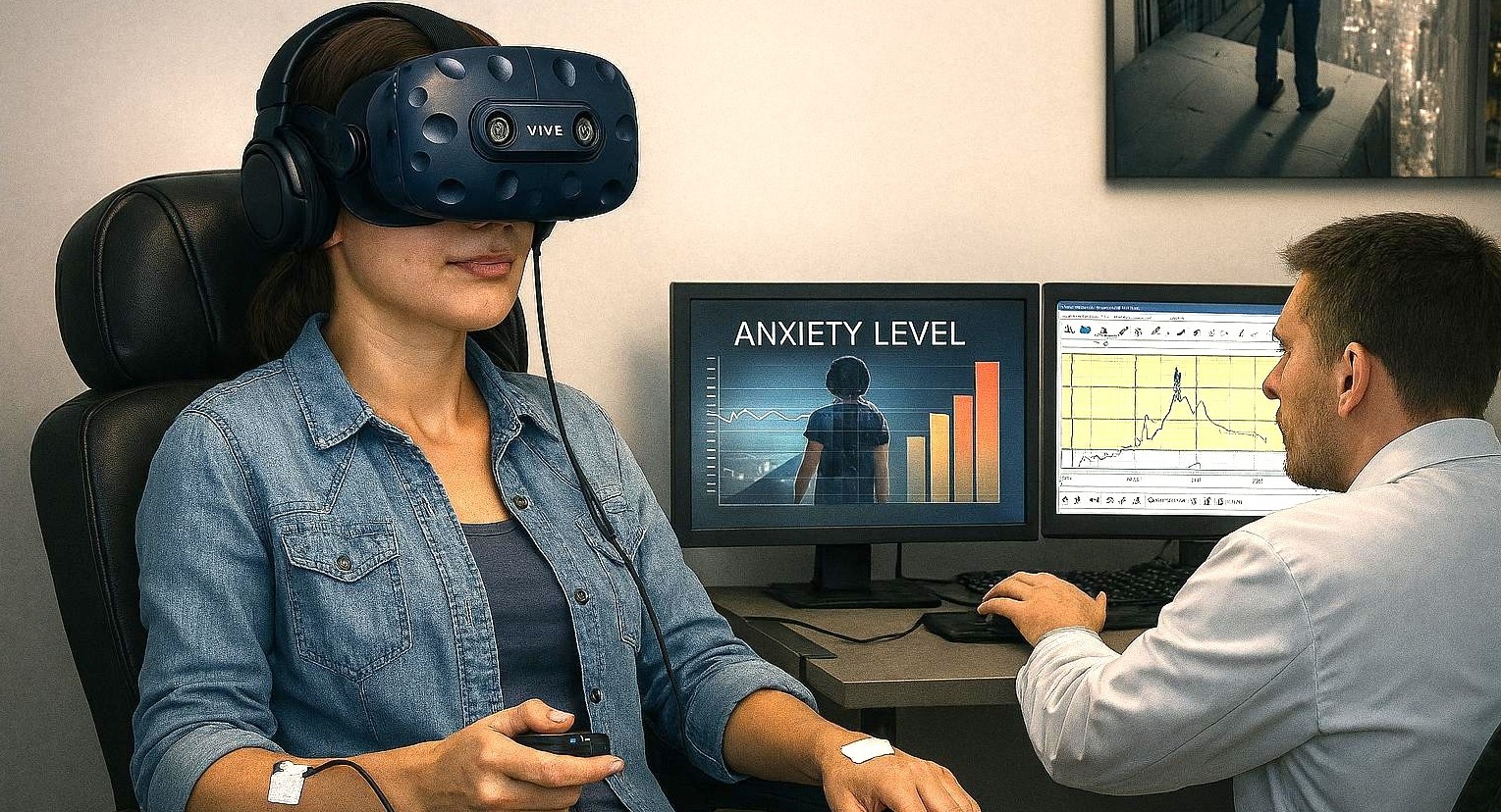

Virtual Phobia: How VR is Transforming Anxiety and Fear Research

“Do the thing you fear and the death of fear is certain.”

These words by American poet and philosopher Ralph Waldo Emerson assert a head-on approach to overcoming life’s anxieties and phobias. It’s a strategy backed by the latest research methodology, which, thanks to technology, can leverage the relative safety of virtual environments to study how participants react to and manage anxiety.

Anxiety disorders are among the most prevalent mental health conditions and are often maintained through avoidance of fear‑provoking situations. A cornerstone of evidence‑based treatment is exposure therapy, in which individuals gradually and safely confront feared experiences to reduce anxiety over time. In recent decades, virtual reality (VR) has emerged as an effective way to deliver exposure therapy and to study anxiety under controlled conditions. Virtual reality exposure therapy (VRET) uses immersive, computer‑generated environments to recreate anxiety‑triggering situations, such as public speaking, social interaction, or medical procedures, while allowing researchers and clinicians to control intensity, pacing, and repetition.

Research shows that VRET can be as effective as traditional, real‑world exposure for many anxiety‑related conditions, including specific phobias, social anxiety, panic disorder, and symptoms of post‑traumatic stress disorder (PTSD). One of VR’s key advantages is its balance between realism and experimental control. Environments can be standardized across participants, exposure levels can be adjusted in small steps, and scenarios can be repeated across sessions without the logistical challenges of invivo exposure. At the same time, well‑designed VR experiences reliably evoke emotional and stress responses, making them valuable for both treatment and research.

The scientific impact of VR is amplified when immersive environments are combined with multimodal psychophysiological measurements—such as electrocardiography (ECG), electroencephalography (EEG), functional near‑infrared spectroscopy (fNIRS), respiration, electrodermal activity (EDA), and eye tracking. In multimodal studies, researchers gain a deeper, objective understanding of how the brain and body respond to emotional challenges. By synchronizing VR events with physiological data, researchers can observe how the body responds moment by moment as fear is triggered, maintained, and eventually reduced.

Researchers at the University of Cyprus investigated whether a single session of VR exposure could meaningfully reduce public speaking anxiety in university students. Participants were placed in a virtual auditorium and asked to give a speech in front of realistic virtual audiences. The researchers could adjust audience size and behavior, allowing them to gradually increase the social challenge while maintaining full experimental control. To capture objective stress responses, heart activity was recorded using a Lead I electrocardiogram configuration, with two disposable silver/silver‑chloride electrodes placed on the forearms. Signals were amplified with a BIOPAC ECG100C bio‑amplifier, acquired using a MP data acquisition system with AcqKnowledge software for recording and analysis. This setup enabled precise measurement of heart rate and heart rate variability (HRV) throughout the task. The virtual environment was created using the Vizard Virtual Reality Toolkit, which was integrated with the BIOPAC system so that physiological data could be synchronized with key moments in the VR experience, such as the start of the speech. Results showed significant reductions in both self‑reported anxiety and physiological arousal following a single VR exposure session, demonstrating how VR and physiological monitoring can be combined to assess rapid emotional change.

Researchers at the University of Macau examined how social rewards and emotional cues influence anxiety during interpersonal interaction. Participants engaged in a virtual reality social interaction game designed to mimic real social exchanges while allowing researchers to control feedback and outcomes. During the task, heart activity was recorded using ECG with an MP data acquisition system. By synchronizing physiological data with events in the virtual environment, the research team could examine how anxiety‑related responses evolved as social interactions unfolded. The findings suggested that anxiety responses were more strongly related to sensitivity to others’ emotional cues than to reward outcomes themselves. This study highlights how immersive VR combined with synchronized physiological recording can provide insight into the subtle emotional dynamics of social anxiety, processes that are difficult to capture using self‑report measures alone.

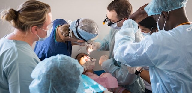
dentalanxietyIn a multi‑institutional effort led by researchers at the University of Toledo, researchers developed a study protocol to better understand how dental fear develops and how early exposure might help prevent it. The study uses a virtual reality dental clinic to expose participants to dental‑related sights and sounds across multiple sessions and contexts. The virtual environment was programmed using Vizard software, and participants interacted with it using an HTC Vive Pro headset and handheld controllers, allowing natural movement and exploration. Skin conductance responses (SCR) were recorded using a BIOPAC MP system paired with a wireless BioNomadix EDA transmitter, enabling participants to move freely while skin conductance and other signals are collected. This wireless, immersive setup is especially well suited for fear‑learning research, where comfort and natural movement are critical. Although the publication focuses on study design rather than completed outcomes, it demonstrates how VR combined with physiological monitoring can support translational research aimed at understanding and preventing the development of fears and anxiety.
These studies show how VR and synchronized physiological measurement are expanding the study of anxiety, stress, and fear. VR provides immersive, repeatable experiences that reflect real‑world challenges, while physiological signals reveal how the body responds beneath conscious awareness. For additional information on how to integrate VR into multimodal research, visit our Webinar page for instructive trainings on this topic and others.


If you are planning to integrate VR into your next multimodal study, reach out to our skilled sales staff who can assist you through each step of the process.

Hands-on Learning: How Real‑Time Physiological Data Deepens Clinical Education

Physicist Albert Einstein noted, “The only source of knowledge is experience.” This principle is particularly relevant in healthcare education, where hands-on learning is vital for cultivating the skills needed to care for patients effectively. The practice of recording and analyzing physiological signals in the learning lab serves as a powerful teaching tool, enabling students to immerse themselves in real-world scenarios and reveal autonomic responses, cardiovascular patterns, and emotional states that are often hidden from view.

For students in nursing, dentistry, and pharmacology, engaging with these signals transforms abstract concepts into concrete, measurable experiences. By analyzing heart activity, visual attention, stress markers, and pharmacodynamic effects, students not only enhance their critical thinking skills but also prepare themselves for clinical challenges with greater confidence.

Physiological signals across the health sciences teach learners to interpret subtle features embedded in physiological waveforms. Modern teaching laboratories increasingly incorporate multimodal systems to help students better understand the complexity of human physiology.

Nursing programs emphasize accurate patient monitoring and interpretation of vital signs, including electrocardiograms (ECG) and heartrate variability (HRV). A 2025 study published in Sensors evaluated the validity of Apple Watch Series 6 HRV data against laboratory‑grade ECG recordings using a BIOPAC 3‑lead ECG system to provide a comparative baseline for measurement. By synchronizing event markers between the Apple Watch and BIOPAC timeline, researchers ensured precise temporal alignment for comparing R‑R intervals and HRV indices. The results showed near-perfect agreement in R-R interval timing under resting conditions, indicating that such wearable devices can serve as a reasonable tool for certain wellness-oriented assessments. At the same time, the BIOPAC system remains the gold standard for precise physiological analysis.

For nursing students, this study demonstrated that consumer wearables must be validated against clinical-grade systems before being relied upon for assessment or decision-making. By examining the comparative data, students can explore how artifacts, sampling frequencies, and sensor placement influence the fidelity of HRV measurements. Moreover, because HRV relates directly to autonomic balance and stress physiology, the study provides a meaningful touchpoint for nursing courses in pathophysiology and patient assessment. Through hands‑on lab sessions using BIOPAC ECG amplifiers, students can visualize waveform morphology, measure beat‑to‑beat variability, and learn how cardiovascular signals reflect underlying homeostatic regulation.

Dental students should understand not only the mechanics of clinical procedures but also the emotional and perceptual experiences of their patients, especially children. A 2025 cross-sectional pilot study in the European Archives of Paediatric Dentistry used an eye tracking and  data acquisition and analysis system from BIOPAC to measure children’s gaze behavior and physiological stress when viewing images of dentists, dental chairs, and operatory designs.

Using eye-tracking metrics such as mean dwell time, fixation count, time to first fixation, and total fixation time, researchers found that study participants stared longer at dentists wearing white coats, indicating a perception of compitance and safety, but also indimidation. In addition, gender matching mattered, as children showed longer fixation on dentists of the same gender. The study also found that warm‑colored dental chairs (especially yellow) attracted greater visual attention, particularly among younger children. Simultaneously, HRV and ECG signals recorded with the BIOPAC system revealed that younger children exhibited greater autonomic arousal than older peers when exposed to dental images.

These findings offer dental students actionable insights into clinic design, caregiver attire, and emotional perception, all of which influence pediatric patient comfort. The study demonstrates how combining eye tracking with physiological monitoring can reveal otherwise hidden psychological responses. For dental educators, integrating such physiological signal analysis into coursework helps students develop empathetic, patient‑centered care strategies.

colored pillsIn pharmacology courses, students learn how drugs influence physiological systems. A study published in Brain Sciences investigated the lingering cardiovascular effects of caffeine, challenging the assumption that its effects dissipate within six hours. Using BIOPAC respiratory transducers, noninvasive blood pressure modules, bioimpedance amplifiers, and ECG amplifiers connected to a data acquisition and analysis system researchers monitored participants before and after consumption of caffeinated and decaffeinated coffee. Results showed measurable changes in heart rate, respiratory patterns, and thoracic impedance well beyond the expected metabolic window, suggesting caffeine’s physiological effects outlast its pharmacokinetic half‑life.

For pharmacy students, these findings reinforce critical distinctions between pharmacokinetics (drug metabolism) and pharmacodynamics (physiological response). By analyzing real‑world signal data, learners gain a more nuanced understanding of how stimulants affect autonomic regulation. Such insights prepare future pharmacists to counsel patients on medication timing, drug interactions, and individual responses to common agents such as caffeine.

Across nursing, dentistry, and pharmacy, physiological signals provide potent educational value. They bridge the gap between theory and clinical application by giving students concrete, measurable access to physiological processes such as autonomic reactivity, cardiovascular regulation, and perceptual attention. Using both research and education-based lab systems, learners can visualize real-time physiological data, develop analytical skills, and cultivate a deeper understanding of the human body under stress, stimulation, and clinical observation.

As healthcare grows more data‑driven, integrating hands-on experience with signal acquisition into professional training helps future clinicians interpret complex physiological information with confidence. Whether validating wearable devices, improving pediatric patient experiences, or exploring drug‑induced autonomic effects, physiological signals empower students to connect scientific principles with evidence‑based, care. Check out our webinar, Advancing Practical Learning: Biosignal Essentials, to learn more.


If you’re exploring ways to incorporate physiological signals into your curriculum or simulation lab, our team is ready to help identify the best tools and workflows for your program.

Tick, Tock: Pulse Transit Time Offers Noninvasive Physiological Insights

A split second can reveal much about the cardiovascular system and the other physiological systems it influences. Pulse Transit Time (PTT) captures one of those critical moments, the brief interval it takes for the pressure wave generated by contraction of the left ventricle to travel from the heart to a peripheral site in the body. That tiny stretch of time tells us something important about the arteries—namely, how stiff or flexible they are in that moment. When blood pressure rises and the vessel walls tighten, the pulse wave travels more quickly, so PTT gets shorter. When the vessels are relaxed and more elastic, the wave slows down, and PTT becomes longer. Because of this back-and-forth relationship, PTT gives researchers a simple, noninvasive way to track changes in vascular tone, blood flow dynamics, and autonomic nervous system activity.

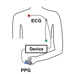
PTT is determined by measuring the volume or pressure pulse at a proximal artery (such as the carotid or brachial) and at a distal artery (such as the finger or toe). To prevent confusion in PTT nomenclature across different methods, some researchers use labels formed from the first one or two letters of the starting and arrival signals. PTT-PP refers to PTT recorded using PPG between a proximal and a distal artery, PTT-EP refers to PTT measured between electrocardiogram (ECG) proximally and photoplethysmogram (PPG) distally and PTT-IP refers to PTT recorded between Impedance Cardiogram proximally and PPG distally.

PTT-EP is recorded by simultaneously recording ECG and PPG. PPG is usually recorded at the finger using a small infrared light to detect changes in blood flow with each heartbeat. As blood pulses into the area, the amount of light absorbed changes, creating a waveform with a clear rise and peak. To calculate PTT-EP, researchers measure the time between the ECG spike and a specific point on the PPG wave, typically the point at which the wave first rises or reaches its peak. Strictly speaking PTT-EP is not a true PTT as it includes pre-ejection period, a short delay between electrical activation of the ventricles and actual ejection of blood. For that reason, it is sometimes referred to as Pulse Arrival Time (PAT). Even so, the ECG-to-PPG approach remains widely used because it is noninvasive, reliable, relatively inexpensive, and straightforward to implement across a range of study designs.

Typical lab configurations focus on integrated, multimodal data acquisition systems that allow researchers to record ECG and PPG signals simultaneously using dedicated amplifier modules, with analysis performed via software. These systems enable precise channel synchronization, automated waveform feature detection, beat-by-beat PTT computation, and data export for statistical analysis. In some studies, researchers also integrate noninvasive blood pressure (NIBP) modules to directly examine the relationship between PTT and systolic or mean arterial pressure, strengthening the interpretation of vascular changes.

Obtaining high-quality PTT data requires attention to detail and careful planning. Participants should rest in a quiet, temperature-controlled room, since vascular tone can be affected by stress, ambient temperature, and sympathetic activation. Avoid testing immediately after meals or the consumption of caffeine or nicotine, which can alter cardiovascular dynamics. Proper sensor placement and stable contact pressure are critical for minimizing motion artifacts and waveform distortion. Consistent measurement timing, accounting for diurnal variation, helps ensure consistent data quality across sessions. Signal processing choices also matter. Careful filtering (see example study below), accurate event marking, and careful peak or foot detection are essential to ensure that the measured interval truly reflects physiological timing rather than processing artifacts.

When these best practices are followed, PTT becomes a versatile research tool. It has been used to study stress reactivity, autonomic regulation, arterial stiffness, posture-related hemodynamics, exercise physiology, sleep physiology, and the development of cuffless blood pressure estimation technologies. Recent studies illustrate both the promise and the practical considerations of PTT measurement.PTT Diagram

At the Georgia Institute of Technology, researchers explored whether noninvasive nerve stimulation applied at the ear, neck, or wrist could reduce acute stress responses in healthy young adults. During laboratory sessions that included mental arithmetic and cold exposure tasks, cardiovascular signals were recorded using a BIOPAC MP data acquisition unit and a PPG100C to capture ECG and fingertip PPG. PTT served as a key marker of sympathetic (“fight-or-flight”) activation. The research team found that stimulation at the ear and wrist significantly reduced stress-related cardiovascular changes compared to a sham condition, supporting the potential for wearable systems that integrate neuromodulation and real-time cardiovascular monitoring.

In another study published in Nature, researchers at Columbia University tackled a common problem in cuffless blood pressure monitoring: when wearable sensors sit above or below heart level, changes in arm position create hydrostatic pressure errors that skew estimates based on PTT. Cuffless systems frequently use PTT to estimate blood pressure, but arm movements can distort that timing and reduce accuracy. To investigate this, the team collected synchronized physiological data using a BIOPAC data acquisition system to record high-resolution ECG signals via a BIOPAC ECG amplifier and fingertip PPG signals via BIOPAC PPG100C, while a BIOPAC NIBP module measured reference blood pressure using upper-arm and finger cuffs. Participants also wore wrist and upper-arm inertial sensors (IMUs) to track how the arm was positioned relative to the heart. The research team then developed a method called IMU-Track, which uses motion data and deep learning to correct PTT for arm height effects, improving the accuracy of blood pressure predictions across different postures. Their findings showed that accounting for sensor position dramatically reduced errors in cuffless blood pressure estimation, suggesting that combining motion tracking with PTT measurements could make wearable BP monitoring more reliable in everyday use.

When applied correctly, PTT provides a potent, noninvasive tool for assessing cardiovascular function—one measured in milliseconds but capable of revealing meaningful physiological change. For more information on integrating PTT into research, see our PTT webinar.


If you are planning to use PTT in your next study, don’t hesitate to reach out for our highly capable team of experts for assistance in identifying the equipment and procedures to ensure your success.

TREV Q&A: Unpacking Trans-radial Electrical Bioimpedance Velocimetry

As researchers look for cleaner, more direct ways to capture sympathetic nervous system (SNS) activity, Trans-radial Electrical Bioimpedance Velocimetry (TREV) has emerged as a powerful new approach for measuring beat-by-beat changes in cardiac contractility to index sympathetic drive.headshot To explain how the technique works and where it fits alongside more established measures, we sat down with Viktoriya Babenko, PhD, BIOPAC’s in-house TREV expert and research specialist. Babenko helps researchers implement advanced cardiovascular and autonomic measurements, combining her extensive hands-on experience with bioimpedance technologies and a strong grounding in the interpretation of physiological signals. In the Q&A below, she describes the physiological principles behind TREV, how it differs from traditional electrodermal activity (EDA), electrocardiogram (ECG), and impedance cardiography (ICG) approaches, and why it offers a uniquely direct and practical window into sympathetic drive for both laboratory and neuroimaging research.
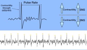
At a high level, what is Trans-radial Electrical Bioimpedance Velocimetry (TREV), and what does it measure?

TREV is a noninvasive method for estimating cardiac contractility as a continuous, beat-by-beat index of SNS activity. Rather than measuring bioimpedance across the chest like traditional ICG, TREV measures impedance changes across the radial and ulnar arteries of the forearm.

As the heart contracts, it generates a pressure wave that travels through the arterial system and causes red blood cells to momentarily align. This alignment reduces electrical resistance in the blood, which TREV continuously tracks. The impedance signal measured by TREV provides velocity, acceleration, and ultimately a contractility index, where stronger contractions reflect greater sympathetic drive.

Why is cardiac contractility such a useful index of SNS activity?

Cardiac contractility reflects the vigor with which the heart contracts during each heartbeat and is strongly modulated by sympathetic tone, either through direct neural innervation or circulating catecholamines like epinephrine. Increased sympathetic activation leads to stronger, faster contractions, while reduced sympathetic drive results in weaker contractile force.

Because of this close physiological relationship, contractility is considered one of the most direct and objective noninvasive measures of SNS activity. Unlike heart rate alone, which reflects both sympathetic and parasympathetic influences, contractility provides a more targeted window into sympathetic drive specifically.

How does TREV differ from traditional ICG- or ECG-based approaches?

Traditional approaches for indexing sympathetic drive, such as combining ECG and thoracic ICG to extract the pre-ejection period (PEP), are powerful but complex. They typically require 10 electrodes placed across the neck, chest, and torso, along with careful preprocessing and time-based feature extraction between both ICG and ECG to get PEP.waveform

TREV dramatically simplifies this process by using just four strip electrodes placed on the forearm. The resulting contractility waveform produces a single sharp peak per heartbeat, where the amplitude directly reflects contractile strength. Analysis is as simple as extracting peak amplitudes, rather than identifying precise timing relationships across ECG and ICG signals, making TREV faster to set up and easier to analyze.

What is the physiological rationale for using the forearm and radial and ulnar arteries as the measurement site?

The radial and ulnar arteries provide a relatively simple and accessible vascular model that is well-suited for bioimpedance measurements. When the heart contracts, a pressure wave propagates rapidly through the arterial tree and reaches the forearm, aligning red blood cells even before bulk blood flow arrives.

This pressure-wave-driven alignment is sufficient to cause measurable drops in impedance, allowing TREV to estimate cardiovascular dynamics without the anatomical and physiological complexity of the thorax. Measuring at the forearm also reduces sensitivity to respiratory and pulmonary effects that complicate thoracic impedance measurements.

What cardiovascular or hemodynamic parameters can be derived from TREV?

TREV provides estimates of blood velocity, acceleration, and the integral of velocity (blood position) derived from the impedance waveform. Most importantly, the second derivative of the impedance signal yields a contractility index (blood flow “jerk”) that reflects sympathetic modulation of the heart on a beat-by-beat basis.

While multiple parameters are available, BIOPAC primarily recommends TREV as a tool for tracking cardiac contractility over time, particularly in studies interested in stress, arousal, motivation, and cognitive or emotional processes linked to sympathetic activation.

What does a typical TREV measurement session look like, from setup to data output?equipment

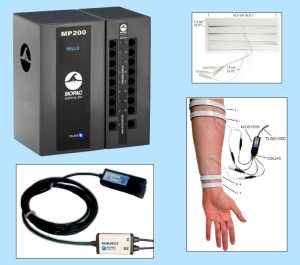
Once the arm is clean and dry, four hydrogel strip electrodes (EL526) are placed on the forearm and connected via the CBL246 adapter to the NICO100D system. I recommend watching the TREV Webinar titled “A Simple Next-Gen Method for Stress Research: TREV” for step-by-step guidance on electrode placement.

During data collection, the participant’s arm must remain fully supported and relaxed, ideally with the elbow and forearm stabilized through the wrist to minimize muscle activity. When collected under proper conditions, TREV produces a clean, continuous contractility waveform with one peak per heartbeat, allowing for straightforward beat-by-beat analysis of sympathetic drive.

What advantages does TREV offer compared to ECG, EDA, or other stress-related measures?

Many commonly used physiological signals, such as heart rate, blood pressure, or heart rate variability, are influenced by multiple factors, including hydration, temperature, respiration, and both branches of the autonomic nervous system (parasympathetic and sympathetic nervous systems). EDA provides a sympathetic index, but it is relatively slow, delayed, and indirect, reflecting eccrine sweat gland activity modulated by the SNS rather than cardiovascular dynamics.

TREV fills an important gap by offering a fast, dynamic, and more direct measure of sympathetic drive through cardiac contractility. It combines the physiological specificity of ICG-derived measures with a much simpler setup and analysis pipeline, making it especially attractive for experimental and applied research settings

What are the main challenges in acquiring clean, interpretable TREV data?

The primary challenge with TREV is its sensitivity to motion artifact. Muscle activity in the forearm generates electromyography (EMG) signals that are much larger than the impedance changes of interest and can easily overwhelm the TREV signal.

For this reason, careful experimental control is essential. The participant’s arm should be fully supported, relaxed, and free from movement or strain, while other body movements are generally permissible. When these conditions are met, TREV data quality is excellent, but the method does demand attention to posture and comfort.

In what research or applied settings is TREV showing the most promise?

TREV is particularly well-suited for research at the intersection of stress, cognition, emotion, and autonomic physiology. It has been successfully used in laboratory studies examining sympathetic responses to cognitive and emotional stressors, as well as in MRI environments to enable the simultaneous study of neural activity and SNS dynamics.

Across both standard laboratory settings and neuroimaging environments, TREV provides a continuous, beat-by-beat index of sympathetic activity, making it especially valuable for event-related experimental designs where precise timing and dynamic changes in autonomic responses are of interest. In the MRI specifically, this temporal resolution allows researchers to directly link moment-to-moment fluctuations in sympathetic drive with neural processes measured using functional MRI (fMRI), offering a powerful framework for studying how brain activity and cardiovascular sympathetic responses interact during task performance, affective processing, or stress-related appraisals. As with most MRI-based physiological measurements, specific hardware configurations and safety considerations apply, and researchers should contact the BIOPAC support team before data collection to ensure proper setup and compliance.

How does TREV perform in dynamic or real-world conditions, such as movement or exercise?

At present, TREV performs best in controlled conditions where the forearm can remain fully supported and at rest. Simple movements while seated may be permissible, provided that the arm containing the TREV electrodes remains immobile and relaxed. Due to its sensitivity to motion, TREV is not currently recommended for continuous data collection during active movement or exercise.

That said, TREV can still be extremely valuable in movement- or exercise-related research by comparing pre- and post-task states. This approach allows researchers to quantify sympathetic changes associated with physical or psychological challenges without collecting data during movement itself.

Looking ahead, how do you see the TREV method evolving over the next five to ten years?

As validation studies continue and more researchers adopt the method, TREV is likely to become a standard tool for indexing sympathetic drive in both basic and applied research. Ongoing work, including peer-reviewed publications, will further refine interpretation, best practices, and experimental design considerations.

In the longer term, improvements in signal processing, hardware integration, and multimodal data collection may expand TREV’s use into more naturalistic settings, helping bridge the gap between controlled laboratory studies and real-world physiological monitoring.


If you are planning a study and would like to learn more about integrating TREV, our team of experts can help guide you to the right equipment and strategies.

Related posts: Body Electrictric: Bioimpedance in Physiology Research and ‘Tis the Season for Stress: Research Tools for Anxious Times

Healing Hands: EDA and Affective Touch

“Nothing eases suffering like human touch.” — Bobby Fischer

The gentle caress of a mother to her child. A reassuring pat on the shoulder from a friend. The touch of another human is one of the first forms of communication we experience in life, preceding the development of spoken language. Touch can have a remarkable effect on our emotional, psychological, and physiological states. It shapes our relationships with others and helps us build social connections.

The positive effects of touch are primarily associated with affective touch, characterized by comforting, caress-like stroking that activates C-tactile afferent (CT-afferent) neurons in the skin of mammals (including humans). Affective touch has attracted the interest of researchers seeking to decode how the sympathetic nervous system confronts and manages a variety of challenges, including pain regulation, stress management, and social interactions. The subject has drawn attention in the study of neurodevelopmental disorders, such as Autism Spectrum Disorders (ASD), where it is playing an essential role in understanding the sensory and social challenges facing individuals with atypical neurocognitive function.

While studies on touch, and affective touch specifically, utilize a range of physiological signals to gather data, electrodermal activity (EDA) has proven particularly useful in this area. Also referred to as Galvanic Skin Response (GSR), EDA provides a direct window into the sympathetic nervous system, which is responsible for emotional or affect-related arousal. When someone experiences pleasant or meaningful touch (e.g., a gentle stroke or supportive hand), the brain triggers subtle changes in sweat gland activity manifested as shifts in skin conductance. These changes occur when people aren’t fully aware of their emotional response. This makes EDA a sensitive, objective marker of how the body reacts to social and tactile experiences. In studies of affective touch, researchers track EDA to see whether a touch feels calming, engaging, or stressful, and how these responses differ across individuals or clinical groups such as autistic youth. Because EDA reflects genuine physiological arousal tied to emotional processing, it helps scientists understand the underlying autonomic mechanisms that shape how we experience social touch.

Researchers at the University of Tours in France examined how children with ASD respond to gentle, affective touch to determine whether they exhibit physiological patterns that differ from those of their neurotypical peers under the same conditions. The team measured multiple aspects of the autonomic nervous system, including EDA and electrocardiogram (ECG). These signals were collected using a BIOPAC MP36 data acquisition and analysis system to capture subtle changes in sympathetic arousal. Researchers collected EDA data using two 8 mm Ag/AgCl cup electrodes placed on the second phalanges of the index and middle fingers of the right hand. ECG was collected via two disposable vinyl electrodes placed on the sternum and on the right shoulder.  The study found that autistic children exhibited atypical EDA responses, showing either blunted or inconsistent autonomic reactions relative to controls, even when their outward behavior appeared typical. These results suggest that affective touch may not regulate internal arousal in autistic children the same way it does in others, providing an essential insight for therapies and sensory-based interventions that rely on touch.

In a similar study, a team from the University of Turin in Italy explored how autistic adults process affective touch by comparing their subjective ratings of pleasantness with their physiological responses. The researchers used a BIOPAC data acquisition research system with an EDA amplifier module to record EDA. At the same time, participants received gentle, slow stroking intended to activate the brain’s affective touch pathways. The goal was to determine whether autistic adults show a mismatch between what they say they feel and how their bodies respond. The findings revealed an apparent dissociation. While autistic participants often rated the touch similarly to neurotypical participants, their EDA responses differed significantly, indicating altered sympathetic arousal. This physiological-subjective mismatch provides compelling evidence that affective touch is processed differently in ASD, which may help explain challenges with social touch and sensory integration in everyday interactions.hands

Researchers from the University of Liverpool and Manchester Metropolitan University examined how brief, naturalistic social touch alters brain activity, autonomic physiology, and emotion recognition performance. Although this multimodal study did not focus on an autism-only sample, it is relevant to social-touch research more broadly and provides methodological examples that apply to ASD studies. To capture changes in prefrontal cortex function, the team employed a BIOPAC fNIRS imager system, with signals sent to a data acquisition unit. Further analysis of fNIRS signals was handled via fNIRS visualization software. Both EDA and photoplethysmography (PPG) were recorded at 2 kHz, using shielded Ag/AgCl electrodes on the index and fourth fingers and a PPG sensor on the middle finger, and were acquired and analyzed in AcqKnowledge. The results showed that social touch increased prefrontal activation, modulated cardiac and vascular dynamics captured via PPG, and produced measurable changes in sympathetic arousal, as indexed by EDA. Participants also demonstrated improved emotion-recognition accuracy following touch. Together, these findings suggest that social touch engages coordinated central and peripheral systems in ways that may support social cognition and emotional understanding.

These are just a few examples of how EDA is being used in research into affective touch. For additional information on integrating EDA in your study, see our multiple webinars on the subject.


Are you designing a study into affective touch or just planning a research project that will focus on EDA? Our customer service staff can help you streamline the process to produce best possible data.

Brain and Body: SPA-fNIRS Solves Physiological Signal Noise

Functional near-infrared spectroscopy (fNIRS) offers researchers a powerful noninvasive tool for studying brain activity by measuring changes in blood oxygen levels. These changes reflect how various brain regions respond to stimuli such as tasks that involve thinking, emotion, movement, and social interaction. These are often in natural, real-world settings that are not possible with other brain imaging methods. However, fNIRS is not without its own limitations. Systemic fluctuations in the body, such as breathing patterns, cardiovascular changes, and autonomic responses, can result in signal noise that is misinterpreted as brain activity by traditional fNIRS. The solution: a visit to the SPA.

Systemic Physiology Augmented fNIRS (SPA-fNIRS) is an advanced approach to fNIRS that goes beyond measuring local changes in cerebral oxygenation. While standard fNIRS measures cortical hemodynamics by tracking oxygenated (HbO) and deoxygenated (HbR) hemoglobin, noise originating from systemic physiological changes can corrupt those signals. Combining data from both neural and systemic physiological signals, SPA-fNIRS provides researchers with a more diverse and accurate view of how the brain and body interact in real time.

The method is being applied across various fields, including psychophysiology, cognitive neuroscience, exercise physiology, mental health, and human performance research. Studies have utilized SPA-fNIRS to investigate a range of topics, including meditation and emotional regulation, movement control, and social interaction. The broader goal is to move beyond studying the brain in isolation and to understand it as part of an integrated mind-body system. This holistic view is transforming how scientists interpret brain-function data, opening new paths for applied research in education, rehabilitation, and human-machine interface design.

soundResearchers in India investigated the impact of a traditional sound-based meditation technique, known as the Mind-Sound Resonance Technique (MSRT), on both brain and heart activity. Using fNIRS to track changes in oxygen levels in the prefrontal cortex, and a BIOPAC data acquisition and analysis system with AcqKnowledge software to record and analyze heart rate variability (HRV), the study employed a SPA-fNIRS design, integrating neural and physiological data in a single, synchronized dataset. HRV analysis allowed the researchers to quantify shifts in autonomic balance during and after meditation. The results showed increased oxygenation in the right prefrontal cortex and enhanced low-frequency HRV components, reflecting greater parasympathetic regulation. Together, these findings suggest that sound-based meditation not only improves brain oxygenation but also promotes calmer, more balanced heart-brain dynamics, illustrating how SPA-fNIRS methods can reveal the comprehensive physiological impact of mindfulness practices.

In her doctoral research at the Université de Lille in France, neuroscientist Aurore Guérin examined how the brain controls movement speed using a multimodal combination of fNIRS and physiological data recording systems. Participants performed repetitive arm movements at fast, natural, and slow tempos. At the same time, the study captured oxygenation in the prefrontal and motor cortices using fNIRS, alongside heart rate and respiration signals collected with a BIOPAC data acquisition system. A respiratory effort transducer measured breathing patterns, while a BioNomadix ECG module recorded cardiac activity for HRV analysis. All physiological signals were synchronized and analyzed in AcqKnowledge software, allowing the team to identify and remove systemic influences (e.g., heartbeat and breathing rhythms) from the fNIRS signals, following a SPA-fNIRS approach. The results revealed that faster movements primarily engaged motor regions, while slower, more deliberate pacing resulted in higher prefrontal activation, reflecting greater cognitive control. By integrating physiological monitoring with fNIRS, the study provided a clearer picture of how the brain and body coordinate timing and control during movement.

These studies illustrate how SPA-fNIRS is reshaping the way scientists investigate the interaction between brain and body. By combining neural and physiological data in a single synchronized system, researchers can separate brain activity from systemic noise and uncover the interplay between cognition, emotion, and autonomic regulation. For more information on this topic, watch our webinar on Multimodal SPA-fNIRS and check our our latest multimoldal fNIRS research products.


Are you planning a study that blends fNIRS with other physiological signals? Contact your local BIOPAC customer service representative to learn more about how SPA-fNIRS can benefit your research.

Back to the Wild: Measuring the Effects of Human Interaction with Nature

“I think that I cannot preserve my health and spirits, unless I spend four hours a day at least—and it is commonly more than that—sauntering through the woods and over the hills and fields, absolutely free from all worldly engagements.” —Henry David Thoreau

In his 1861 essay “Walking,” transcendentalist writer and philosopher Henry David Thoreau extolled the virtues, both physical and mental, of communing with nature. While the psychological and physiological benefits of human connection with the natural environment are well established, modern science has taken a closer look at these interactions. Today’s researchers often rely on interdisciplinary approaches that blend psychology, physiology, neuroscience, and environmental science to investigate the mechanisms behind nature’s restorative effects on mind and body. With continued urban growth and shrinking access to wild rural spaces, as well as the digitization of our daily lives, this field has become more relevant than ever.

Researchers study human interactions with nature to better understand the tangible health and psychological benefits associated with exposure to natural environments, such as reduced stress, improved mood, and enhanced cognitive function. Theories such as the Biophilia Hypothesis and Attention Restoration Theory (ART) suggest that humans have a built-in affinity for nature, which can support mental and emotional well-being. Psychophysiological research seeks to uncover the processes underlying these effects and to compare how different environments—whether urban, natural, or virtual—impact our bodies and minds.

The study of human-nature interaction encompasses a wide range of practical applications, including healthcare and mental wellness. Beyond health applications, this field informs decisions and policies regarding urban planning and architecture, such as the design of biophilic buildings and green spaces in cities. Research also aims to help individuals focus their attention, enhance creativity, and promote emotional well-being in both work and educational settings.

A study published in the Journal of Environmental Psychology examined how immersion in nature and exposure to nature-related images affect attention and could be used in attention restoration therapy and related applications. Researchers measured participants’ neural activity before, during, and after a four-day group camping trip in Utah using electroencephalography (EEG) to capture reward positivity (RewP), a brain signal associated with reward processing. During these periods, researchers measured how participants responded to extrinsic rewards, including monetary incentives. To ensure data integrity and minimize motion artifacts in the outdoor setting, the team utilized BIOPAC’s wireless BioNomadix EEG system and verified electrode contact using the EL-CHECK impedance checker. The study’s findings showed a significant decrease in neural responsiveness to extrinsic (monetary) rewards following immersion in nature. This suggests that increased time spent immersed in natural environments may recalibrate the brain’s reward sensitivity and promote intrinsic motivation. These findings have implications for a variety of behaviors related to extrinsic stimuli, such as the treatment of addiction.mountaintop

Not all research into human-nature interactions focuses on immersion in real-world environments. With the evolution of virtual reality (VR) technology, new research leverages artificial environments to elicit biophilic responses. A study by researchers at the Southwest Jiaotong University School of Architecture in China, published in Frontiers in Public Health, investigated how exposure to biophilic design elements affects stress recovery in older adults. A group of 25 participants over the age of 60 were exposed to stressors and then underwent sessions with randomized VR environments that included green walls and outdoor views. The researchers measured two key physiological signals: heart rate variability (HRV), which assesses parasympathetic activity, and skin conductance level (SCL), a marker of sympathetic arousal. Electrocardiogram (ECG) data were collected using a BIOPAC ECG amplifier, while electrodermal activity was gathered via an EDA amplifier. Both signals were recorded using a BIOPAC data acquisition and analysis system. Results found that participants exposed to environments with green features demonstrated significantly higher HRV and lower SCL, indicating enhanced autonomic recovery and reduced stress. These findings suggest that the biophilic benefits of nature exposure can be made available to individuals virtually when real-world immersion is not possible or available.

As these studies demonstrate, there exists a wide and varied frontier to explore how human interaction with nature shapes our physiological, neurological, and psychological well-being. For more information on the tools to assist with such research, visit our Webinars library for detailed video trainings on how to use these technologies in your nature-related study.


Whether your study participants are hitting the trail, climbing a mountain, or just relaxing in the garden, BIOPAC customer service reps are ready with the right solutions to take your next research project back to nature.

Pour One Out: The Physiology of Alcohol Consumption

“Dry January,” the cutting out of alcohol consumption during the first month of the calendar year, entered the public vernacular a little over a decade ago. The idea was first promoted in 2013 by a British charity to encourage alcohol abstinence, or at the very least, healthy moderation. The idea took off as a counterbalance to the excesses of indulgence that often characterize the end-of-year holiday season. As if on cue, the U.S. surgeon general released a report earlier this month linking increased cancer risk to even moderate alcohol use and called for alcohol products to carry a warning label like those found on tobacco products.

Since the early 1990s, the prevailing belief among some in the medical community held that moderate amounts of alcohol may not necessarily be detrimental to overall health and may even have beneficial effects. Despite the surgeon general’s report, the consensus in the benefits versus risks debate remains elusive, a fact summarized well in a recent article in The Atlantic and an accompanying podcast by journalist Derek Thompson. Without wading further into the controversy, it is worth pointing out that there is a rich and growing body of research into the effects of alcohol on human physiology, cognitive and psychological well-being, and longevity, all of which rely on some form of life science metrics to measure these effects.

Physiological signals offer a powerful window into the biological and neurological effects of alcohol consumption, providing objective, real-time insights into the body’s and brain’s response. By measuring electrical, chemical, and physical signals generated by the body, researchers can examine changes in heart rate, brain activity, muscle tone, and more. Commonly used signals in alcohol studies include electroencephalography (EEG) to monitor brain activity, electromyography (EMG) for muscle function, electrocardiography (ECG) for heart rhythms, and electrodermal activity (EDA) and galvanic skin response (GSR) for stress and arousal levels. Each tool captures aspects of how alcohol influences the human system, from cognitive processing impairments to autonomic regulation shifts. In research settings, these signals are applied to explore both short- and long-term effects of alcohol. For example, EEG studies can track alterations in brainwave patterns, shedding light on reduced attention, memory disruption, or impaired decision-making during intoxication. ECG and GSR data, meanwhile, reveal how alcohol modulates cardiovascular activity and stress responses, highlighting its impact on the autonomic nervous system (ANS).

A 2021 study by researchers at Wake Forest University and John Hopkins University School of Medicine used heart rate variability (HRV) to examine the relationship between alcohol abstinence, ANS function, and whole brain functional brain networks among everyday drinkers. Whole brain functional brain networks demonstrate how different parts of the brain work together as a connected system. This study combined functional brain wave mapping generated with resting-state MRI scans during three-day periods of alcohol consumption and abstinence. The effects of abstinence were measured using HRV. Alcohol craving in patients with an alcohol use disorder (AUD) has been associated with high-frequency heart rate variability (HF-HRV) with acute consumption being related to decreasing HF-HRV. During the study, ECG data was collected via a BIOPAC data collection and analysis system. The study concluded that “while brain networks do differ across drinking states in risky drinkers, the change is primarily driven by HRV.”woman drinking

A current study by a team of German researchers explores the effectiveness of Virtual Reality (VR) in Cue Exposure Therapy (CET), a strategy in Cognitive Behavioral Therapy (CBT) used for treating AUD. Research participants are exposed to VR scenarios with alcohol-related cues. VR with integrated eye tracking offers advantages over real-life scenarios when developing new treatment options, according to the study authors. Participants wore head-mounted displays (HMD) to immerse them in three different scenarios while heart rate, HRV, pupillometry, and EDA were measured. Respiration and ECG data are gathered via a wireless BioNomadix BN-RSPEC while PPG and EDA are gathered with a BN-PPGED. Physiological signal data is then fed to a BIOPAC data acquisition system running AcqKnowledge software for analysis. While the study is ongoing, researchers believe it will lead to a better understanding of the induction of craving in VR cue exposure…” and that it could offer “new diagnostic and therapeutic perspectives…providing a basis for future biofeedback training in VR exposure therapy.”

These are a few of the ways physiological signals are being applied in alcohol-related research. For more information on integrating HRV analysis into your next research study, see our webinar on the topic. Better yet, get hands-on training on a full range of research equipment and techniques at our biannual conference on Tools, Trends, Techniques, and Technologies in Human Physiology, 4T|Phys.


Are you planning an alcohol or addiction-related study? BIOPAC has the tools and our regional sales staff is ready to help you with everything you need.

Going for Gold: Physiology Research Powers Olympic Athletes

As the world’s top athletes gather for the 2024 Olympic Games in Paris, they hope that years of rigorous preparation and training to ensure peak performance will pay off with a trip to the winner’s podium. Physiology research plays a critical role in these preparations, providing athletes and their coaches with valuable insights into how to best optimize training regimens and enhance athletic performance. With the introduction of new sports in the competition, understanding the specific physiological demands of each sport is critical. Advanced tools and techniques, like those developed by BIOPAC, allow researchers to study the body’s responses to different types of movement, providing tailored training programs that cater to the unique needs of each athlete.

A range of physiological signals is pivotal in sports and exercise research. Electromyography (EMG) is used to analyze muscle activation patterns, helping athletes refine their movements for efficiency and power. Electrodermal activity (EDA) provides insights into stress and arousal levels, which are essential for mental preparation and focus. Monitoring respiration rates and patterns can optimize breathing techniques, while electrocardiography (ECG) and heart rate variability (HRV) assess heart health and endurance capacity. By integrating these physiological signals, researchers and coaches can develop comprehensive training strategies that improve performance and minimize injury risk, giving athletes the best chance at Olympic gold.

One of the newest sports introduced at the Paris Olympics is breaking or competitive breakdancing. Olympic breaking competitions will feature teams of male and female competitors facing off with opposing teams in one-on-one dance battles. Dance, whether competitive or recreational, has been the subject of extensive study as researchers have examined this intersection of music and rhythmic bodily movement. A study published in 2021 in the journal Nature examined how younger and older adults timed bodily movement to both metronomic rhythms and music. The study compared participants’ responses to auditory cues from a metronome and music with finger tapping, toe tapping, and dance steps. The pace of whole-body stepping movements was recorded using BIOPAC heal-toe strike sensors attached to participants’ shoes with data fed to a data acquisition and analysis system running AcqKnowledge software. The study found that whole-body movement with musical accompaniment positively influenced participants’ ability to time movements accurately with encouraging implications for the treatment of rhythmically impaired individuals, such as those with developmental disorders.

Paris 2024Surfing debuted at the 2020 Olympics in Tokyo and is returning for the Paris games where it will be joined by two new aquatic additions, kiteboarding and kayak cross. The surfing competition for 2024 will be held in the French territory of Tahiti. Physiology studies have played a key role in preparing competitive surfers for the ultimate test of their skills. A study published in a 2021 issue of Nature investigated how surfers’ paddling cycles affect fatigue. Paddling is a primary component of a surfer’s ability to catch the best waves during competition. Researchers measured participant velocity as they paddled in a pool while tethered to a cable that measured propulsive force. Velocity data was recorded using a BIOPAC data acquisition and analysis system running AcqKnowledge. Untethered velocity was also measured using a cable-based speedometer device with data also transferred to the data acquisition system. According to the researchers, the data gathered should help in the development of “a powerful and endurable metabolic base while reducing energy cost during surfing paddling,” which could play a key role in future training programs.

Boxing’s inclusion in the Olympics dates back to the original games in Ancient Greece. Modern trainers look to research conducted in the physiology lab to enhance their fighters’ competitive edge. A 2023 study by a group of researchers in Pakistan used EMG to measure muscle activity when comparing the effectiveness of two types of punches: the jab and the cross. Researchers used a force plate to measure the impact force of each type of punch, while electrodes attached to the participants’ biceps and triceps recorded EMG data via a BIOPAC data acquisition system.  While both punches delivered similar force, the triceps carried most of the workload during the jab. The research may provide insight into how to best manage fatigue during competitive bouts.

These are just a few examples of how sports physiology research powered by BIOPAC tools and techniques is preparing today’s athletes to compete at the highest levels. For additional information on how BIOPAC can help your sports-related study, see our webinars on exercise physiology using our BioNomadix and Research Ring wireless recording tools and product lines.


Are you planning sports or exercise study? BIOPAC’s regional sales reps are standing by to assist you in finding the right tools for the best results possible.

error: Content is protected !!