Muscles in Motion: Electromyography in Physiological Research

When we think about the human muscular system, movement often comes to mind. Activities such as running, lifting, and maintaining posture all rely on coordinated skeletal muscle activity, while cardiac muscle drives circulation and smooth muscle supports essential functions like digestion. Yet muscles do more than move us from point A to B; they also reflect what is happening inside the body. Patterns of muscle activation can reveal information about physical effort, fatigue, coordination, and recovery, as well as emotional expression, stress, and cognitive engagement. Electromyography (EMG) is a widely used technique for measuring the electrical signals generated by muscles during contraction, providing researchers with a direct and objective means to study both physiological function and the links among the nervous system, behavior, and mental state. By capturing muscle activity that may be too subtle to observe visually, EMG enables precise investigation of processes ranging from motor control and performance to emotional and stress-related responses.

There are several types of EMG, each suited to different research needs. Surface EMG (sEMG) uses small sensors placed on the skin to detect activity from nearby muscles and is the most common approach in behavioral and human performance studies. Intramuscular EMG involves inserting a fine-needle electrode directly into a muscle to record activity from specific muscle fibers, often used in clinical or specialized research settings. Facial EMG (fEMG), a form of surface EMG, focuses on facial muscles. By measuring muscle activity associated with smiling or frowning, fEMG allows researchers to study emotional responses, even when those responses are subtle or not consciously expressed.

One of the key strengths of EMG is its ease of integration with other physiological measurements. In many studies, EMG is recorded alongside electrocardiography (ECG), which measures heart activity; electroencephalography (EEG), which records electrical activity in the brain; functional near-infrared spectroscopy (fNIRS), which tracks changes in brain oxygenation; and eye tracking, which monitors visual attention and gaze patterns. By synchronizing these signals in time, researchers can better understand how different systems in the body work together. For example, combining EMG with EEG can reveal how brain activity leads to muscle movement, while pairing EMG with ECG can show how physical effort relates to heart responses during stress or exercise. This multimodal approach is central to advancing research in areas such as emotion, cognition, and human performance.

EMG also has important real-world applications. In rehabilitation, it helps clinicians monitor muscle recovery after injury. In ergonomics, it provides objective data about muscle strain in the workplace. In sports science, EMG helps optimize training and performance by identifying how and when muscles are activated. Increasingly, EMG is also used in human–machine interfaces, such as systems that allow people to control prosthetic limbs using their muscle signals. Its ability to detect fine-grained changes in muscle activity makes EMG a valuable tool across both research and applied settings.

Best practices for EMG data quality begin with careful electrode placement and attention to potential sources of noise. For sEMG and fEMG, electrodes should be placed over the “belly” of the target muscle, aligned with the muscle fibers, and spaced consistently to capture clean, interpretable signals. Gently cleaning the skin and, when appropriate, lightly abrading the surface helps reduce impedance and improve signal quality. In fEMG studies, precise and repeatable placement over well-defined facial muscles is critical, as these muscles are small and closely spaced. To minimize noise and artifacts, researchers should secure electrodes and cables to prevent movement, ask participants to limit unnecessary body motion, and ensure proper grounding of the recording system. Electrical interference from nearby equipment, power lines, or poor cable management can also contaminate EMG signals, so shielded cables and a controlled recording environment are recommended. Finally, synchronizing EMG with other physiological signals using a common acquisition system helps prevent timing errors and reduces the need for later corrections, resulting in cleaner data and more reliable conclusions.

hand mechanicsRecent studies demonstrate how EMG continues to expand into new and innovative areas. In a study published in Results in Engineering, a team of researchers from China developed a low-computational method for recognizing hand gestures from EMG signals to improve prosthetic device control. The researchers extracted key features from EMG recordings and used a lightweight machine-learning model, a multilayer perceptron (MLP), to classify gestures in real time. EMG data were collected using a four-channel BIOPAC data acquisition system running Biopac Student Lab (BSL) software. Surface Ag/AgCl electrodes were placed on four forearm muscles to capture relevant muscular activity. The study showed that accurate gesture recognition can be achieved without heavy computational requirements, making the system more practical for everyday use. This work highlights how EMG can support the development of more responsive, accessible assistive technologies.

Another study, conducted by a team of researchers from Mahidol University in Thailand and presented in the Institute of Electrical and Electronics Engineers (IEEE) proceedings, explored how combining EMG with motion sensors can improve ergonomic assessments. Focusing on occupational health, the study integrated surface EMG with data from an inertial measurement unit (IMU), a sensor that tracks movement and orientation. The researchers used the IMU to calculate Rapid Upper Limb Assessment (RULA) scores—a common method for evaluating postural risk—while EMG data collected via a BIOPAC four-channel research system provided insight into muscle activity. By combining these measures, the team found that posture alone did not always reflect actual muscle strain. The inclusion of EMG revealed additional muscle stress that might otherwise go unnoticed, suggesting that multimodal approaches can lead to more accurate workplace assessments and better injury-prevention strategies.

Researchers from Louisiana State University and the Pennington Biomedical Research Center in Baton Rouge, Louisiana, examined how training experience influences muscle activation and fatigue during resistance exercise performed with and without blood flow restriction (BFR) in young men. The study, published in Physiological Reports, compared trained and untrained participants while they completed knee extension exercises under low-, medium-, and high-load conditions. The team used sEMG to measure muscle excitation and total muscle activation in the vastus lateralis, with participants fitted with a portable BIOPAC BioNomadix Wireless EMG system. EMG signals were sampled at 2000 Hz and analyzed using a data acquisition system with AcqKnowledge software, allowing the researchers to quantify both peak muscle excitation and overall activation across repetitions. The results showed that low-load exercise with blood flow restriction produced lower peak muscle excitation but substantially greater total muscle activation compared to traditional high-load exercise, regardless of training status. These findings suggest that low-load BFR training may provide an effective alternative for stimulating muscle adaptations while reducing mechanical stress, with potential applications in rehabilitation and strength training programs.

Taken together, these examples illustrate the versatility and effectiveness of EMG in neuroscience, psychophysiological research, and beyond. Whether used to study emotion, improve physical training and workplace safety, or develop assistive technologies, EMG provides a direct link between muscle activity and human behavior. When combined with other physiological measures, it offers a more comprehensive understanding of how the brain and body interact. As research tools and techniques continue to advance, EMG will remain an essential method for exploring brain-body connections. For additional information on how to design studies around EMG, best practices for gathering EMG data, and how to integrate EMG with other signals, check out our webinar on EMG acquisition and analysis.


If EMG will be part of your next physiological or psychophysiological research project, reach out to our knowledgeable staff, who are standing by to help you with each stage of the planning and design process.

Game On: Applying Physiological Data in Video Game Design and Research

Ever since players first started feeding quarters into arcade games and began plugging in home systems in the late twentieth century, researchers have looked for practical tools for both shaping video game design and measuring their effects on players. Physiological signals filled this niche area of research. Researchers began investigating the connection between physiological science and gaming in the 1980s and 1990s, when early studies explored the effects of video games on heart rate and arousal. These first studies focused on issues such as aggression and overstimulation, but research has since expanded to encompass the cognitive and emotional aspects of gameplay. An early research project by NASA, for example, examined how biofeedback could be applied to game design to enhance traditional game control methods while also improving players’ cognitive abilities.

Today, physiological monitoring is not only a method for psychological inquiry but also a vital asset in improving user experience (UX) research, game accessibility, and even driving therapeutic game development. As wearable sensors and game engines grow more sophisticated, the integration of physiological signals into game design is opening new frontiers for immersive, responsive, and health-conscious gaming. By analyzing real-time bodily signals, such as heart rate (HR), electroencephalography (EEG), electromyography (EMG), electrodermal activity (EDA), respiration, photoplethysmography (PPG), and electrooculography (EOG), researchers and developers can gain a deeper understanding of how players emotionally and physically respond to game elements.

A study published in the International Journal of Human-Computer Studies investigated whether players’ self-reported preferences for first-person shooter (FPS) game dynamics match their emotional reactions during both gameplay and viewing. Researchers recruited 24 active gamers and divided them into two groups: those who enjoyed the fast-paced action of FPS games and those who didn’t. While participants played an FPS game and later watched a recording of it, the team measured electrodermal activity (EDA), heart rate, and facial muscle responses, specifically expressions associated with frowning (corrugator supercilii) and smiling (zygomaticus major), to capture emotional arousal and valence. Physiological signal data were recorded using BIOPAC EDA, PPG, and EMG amplifiers, which were connected to a data acquisition and analysis system running AcqKnowledge software. Although both groups reported more positive feelings and higher excitement after actively playing rather than watching, the data revealed a deeper story: players who disliked the game showed more substantial rises in arousal during play. Those who favored the dynamics, however, showed steadier emotional responses across both conditions. Muscle activity patterns also differed. Those who enjoyed FPS dynamics exhibited sharper increases in frowning-related muscle activity during play than watchers, indicating nuanced emotional engagement. Overall, the study found that what people say they prefer does reflect in their bodies’ emotional reactions. Still, those preferences also shape whether playing or spectating feels more intense, highlighting how combining self-reports with physiological signals can deepen an understanding of game design and player experience.

controllerA team of researchers from Bournemouth University’s Centre for Digital Entertainment investigated how players’ bodies react during intense engagement, or flow, in virtual reality (VR) games. The goal was to capture objective physiological signals, such as heart rate and eye-blink patterns, alongside subjective self-reports to see how well a game’s design induced flow versus boredom or anxiety. The researchers had participants play a custom VR “tower defense” game while monitored with BIOPAC equipment, including a two-channel Biopac Student Lab (BSL) data acquisition system and physiological modules to record HRV via ECG, as well as EOG and EEG signals. The team then utilized clustering and machine-learning classifiers to decode flow states with high accuracy. The research team also successfully identified that high flow correlated with fewer blinks and longer blink intervals, indicating a state of deep concentration. The study not only demonstrates the value of combining physiological and self-report data but also shows how wearable sensors can inform design decisions in VR games by providing real-time feedback on player experience.

Beyond game design, a primary area of research focus has been on the long-term psychological, neurological, and physiological impact that games have on players. A study published in the Journal of Media Psychology explored how experiences with violent video games might reduce prosocial behavior, such as the motivation to help others, through physiological changes. Researchers recruited 155 Chinese college students who reported their exposure to violent games (in terms of violence, bloodiness, and duration) and completed simple self-reports measuring their willingness to help and the time they took to help others in controlled scenarios. Researchers used a BIOPAC data acquisition unit running AcqKnowledge software to record relevant physiological data. A three-lead ECG amplifier, respiration module, and respiration belt were used to record ECG and breathing signals across baseline, violent-film task, and during recovery periods. Researchers calculated the reduction in respiratory sinus arrhythmia (RSA), a marker of parasympathetic nervous system engagement. They found that higher levels of violent-game experience predicted less RSA reduction during stress, which in turn was linked to reduced prosocial behavior. The study’s findings suggest that playing violent video games may impair physiological readiness to respond in an empathetic manner to others, showing RSA reduction to be a key influence. The study’s findings demonstrate how physiological and psychophysiological signals provide a deeper understanding of the impacts of video games, while also guiding design decisions.

Physiological signals help researchers identify moments of excitement, stress, immersion, or frustration. Such insights are increasingly being used to refine gameplay mechanics, balance difficulty, and enhance player engagement through adaptive or personalized game experiences. They also reveal the short- and long-term effects gameplay has on both individuals and society. For more ideas on how to apply these signals in your research, visit our Webinars page for information on wearable devices, mobile data acquisition, and other helpful topics.


Are you planning a research project related to video games? Reach out to our customer service specialists to get all the help you need in mapping out and equipping your study team.

Free Your EEG: Wireless and Wearable Systems for the Lab and Beyond

The evolution of wearable technology for recording physiological signals represents a transformative leap for research, particularly when it comes to electroencephalography (EEG). These advancements allow researchers to capture brain activity data in real-world settings, moving beyond the confines of laboratory-based experiments. Mobile and wearable EEG devices utilize wireless technology to transmit data, offering significant advantages over traditional methods.

Everyone aspires to more freedom—to speak, think, or move about as one pleases. Should researchers be any different? Why be shackled to the static wires and cables of the lab, when one can release study participants with the help of mobile and wearable technology?

The evolution of wearable technology for recording physiological signals represents a transformative leap for research, particularly when it comes to electroencephalography (EEG). These advancements allow researchers to capture brain activity data in real-world settings, moving beyond the confines of laboratory-based experiments. Mobile and wearable EEG devices utilize wireless technology to transmit data, offering significant advantages over traditional methods.

EEG is a cornerstone in the study of brain activity, providing insights into the electrical patterns generated by neuronal firing. EEG has traditionally been used in physiology and psychophysiology research to understand brain function, monitor cognitive states, and investigate neural responses to various stimuli. The conventional EEG setup typically involves a stationary system with numerous electrodes affixed to the scalp using a conductive gel, which can limit the subject’s movement and impact the naturalistic settings of studies.

One of the primary benefits of mobile and wearable EEG systems is their ability to record brain activity during dynamic, everyday activities. This capability provides a more comprehensive understanding of how the brain responds to real-world stimuli and stressors, as opposed to controlled, static environments. For instance, researchers can now study cognitive processes in natural settings like workplaces or homes, enhancing the ecological validity of their findings.

A study by the School of Architecture at the University of Liverpool provides an example in which researchers leveraged wearable EEG technology when comparing subjective and physiological responses to noise within wooden buildings. The research team conducted listening tests using EEG data recorded via the B-Alert X-24 wireless headset system, which allowed participants to “sit comfortably in a simulated living room” while listening to a variety of sounds that were both airborne and resonating through the floors. Participants rated their level of noise annoyance on an 11-point scale and results were compared to EEG data. In discussing the design of their study, the authors highlighted the importance of incorporating noninvasive, wearable technology, stating, “Among the several options available for measuring physiological reactions to auditory stimuli, contemporary technologies made possible the recording of EEG in a relatively easy way by means of portable devices. These offer the precious opportunity of acquiring data directly from the scalp, where brain activity is detectable almost instantaneously in synchrony with the stimulus presentation.”

Similarly, when studying occurrences of hyperventilation among a participant group with a range of anxiety-related disorders, researchers from Yale University turned to a BIOPAC BioNomadix wireless receiver to gather EEG signal data. By attaching individual electrodes to a third-party cap that connected wirelessly to a BIOPAC data acquisition and analysis system, they minimized the discomfort and restriction of movement that may have resulted from a wired solution, thereby influencing participants’ response to stimuli.

Researchers at the Transylvania University of Brașov in Romania faced similar demands that required a flexible noninvasive means to record surface EEG signals from participants as they responded to a variety of colors and patterns with the goal of better understanding the complexity of human color and texture perception. The research team compared participants’ subjective responses to EEG responses via Event-Related Potentials and computational measurements of color and texture.  EEG data was gathered with a cap-based BioNomadix wireless system and analyzed using AcqKnowledge software. The study’s authors explained their choice of wireless EEG recording devices as follows: “Since human users are the target, it is important to focus the technology development towards small-scale, wearable, wireless systems that provide flexibility, easy use and comfort to the user side, while at the same time maintaining high signal quality.”

APEX cycleAdditional wearable EEG options available through BIOPAC include the MedelOpt Mobility EEG system, combining wearable EEG and functional near-infrared spectroscopy (fNIRS) technology with advanced signal processing and robust data transmission capabilities. APEX wireless EEG systems further enhance the landscape with their ability to record high-quality EEG data with flexibility for a range of applications from cognitive neuroscience to mental workload studies. Learn more about recording EEG data by watching our webinar: A Beginner’s Guide to EEG.


Are you interested in unlocking the benefits of mobile and wireless EEG for your next research project? Let our staff of experts find the right tools for you.

Mind and Machine: Exploring Brain-Computer Interfaces

In neuroscience, the frontier of brain-computer interfaces (BCI) holds the promise of bridging the gap between the human brain and external devices, offering revolutionary applications in fields ranging from robotics and prosthetics to communication and beyond. In the lab, scientists studying and developing new BCI technology leverage the human brain’s electrical and physiological signals.

Electroencephalography (EEG), functional near-infrared spectroscopy (fNIRS), and steady-state visually evoked potentials (SSVEP) are among the primary physiological and psychophysiological signals utilized in these investigations. Multimodal integration of two or more signal types allows researchers and engineers to take advantage of multiple data streams in their applications and studies.

EEG, which can measure electrical activity in the brain, is a cornerstone of BCI research. Researchers can decode various mental states and intentions by analyzing EEG patterns, enabling users to control external devices through mere thought. fNIRS complements EEG by measuring changes in blood oxygenation levels, offering insights into brain function with superior spatial resolution. SSVEP, on the other hand, capitalizes on the brain’s response to visual stimuli, providing a means for precise control in BCI applications.

The practical implications of BCI research extend far beyond the laboratory setting. In robotics, BCIs facilitate direct brain-to-machine communication, empowering users to manipulate robotic arms or navigate autonomous vehicles with their thoughts alone. A study published in the Journal of Information Science Engineering explored ways that an SSVEP-based BCI could be used to control assembly line robots via a headset measuring the operator’s EEG signals. The signals would communicate instructions to robots while operators identified defective parts, which the robots would pick out. During the experiment, EEG signals were obtained using a B-Alert X24 headset. The EEG-based BCI produced a 93.33% success rate in correctly identifying and removing defective parts from the assembly line process, allowing the operator to effectively “collaborate with the robot and communicate defective parts without manually operating the robot.”

Prosthetics benefit immensely from BCI advancements, enabling seamless integration with the user’s neural signals for intuitive control and enhanced functionality. A 2023 study by a team of researchers from the University of California Irvine explored the viability of creating low-cost cortical stimulators for bi-directional BCI (BD-BCI) research. BD-BCI takes brain-computer-interfaces to the next level with the brain not only providing motor commands to an artificial limb but also allowing it to receive sensory input from such a device. A BIOPAC data acquisition and analysis system was used to validate the charge balancing of subcortical electrocorticographic (ECoG) implants used in a stimulator. The charge balancing test was performed on phantom tissue designed to simulate real brain tissue. The resulting study successfully demonstrated “clinical validation of a fully-programmable electrical stimulator, integrated into an embedded BCI system.”

As researchers continue to refine BCI technologies, the possibilities for real-world applications are boundless. BIOPAC is among the companies on the cutting edge of BCI research, partnering with other innovators to provide the latest in multimodal BCI technologies such as Axon-R. For additional information on Axon-R, check out our recent webinar on the device.


Are you planning to explore the frontiers of BCI in the lab? Our regional customer reps have all the knowledge you need to integrate BIOPAC products into your BCI study.

VR Integration: Enhancing Research with Virtual Environments

Measuring the human body and brain’s response to real-world conditions and stimuli has long challenged researchers. This problem has usually been dealt with in one of two ways. The first approach is to take research participants into the field—not impossible with today’s mobile data recording technology but not always safe or convenient. The second option is to imitate real-world conditions in the lab using screens, audio equipment, and even scent delivery, but in the end, it is difficult to completely remove the lab setting from the participant’s experience—that is, until the introduction of Virtual Reality (VR) technology in research.

Just as it has revolutionized everything from entertainment to training, VR has provided researchers with the opportunity to create immersive experiences in the lab that mimic the real world. Advances in technology have not only made these simulated environments more realistic, but they have also made it easier to integrate a wide range of signals and stimulus delivery to enhance the recording of physiological and psychophysiological data.

BIOPAC, through cooperation with several partner companies, offers a range of data recording options that integrate seamlessly with third-party VR devices. fNIR Devices, for example, designs solutions for recording functional near-infrared spectroscopy (fNIRS) signals that can easily be incorporated with standard VR head-mounted displays (HMD) to measure neural activity and hemodynamic response in the prefrontal cortex while participants experience VR environments. BIOPAC partner Seenel has created MedelOpt, a line of whole-head wearable devices that combine electroencephalography (EEG) and fNIRS, including a VR option that integrates an HMD. Finally, partner company WorldViz offers a full spectrum of VR solutions from complete VR kits to full-room Projection VR technology to software that can be combined with both wired and wireless data recording technologies to provide a complete array of bio-signal data, from eye tracking to electrodermal activity.

A Canadian study published in 2023 used a combination of BIOPAC and WorldViz products to investigate how sensory feedback and arousal influence neuromusVR Shadowcular control and posture. Researchers measured arousal via electrodermal activity (EDA) and psychosocial questionnaires, rating perceived fear and anxiety in both real-world situations in VR environments that simulated standing both at ground level and on a raised platform. Participants wore a commercial HMD running WorldViz’s Vizard VR toolkit software. Electrical nerve stimulation was provided via a BIOPAC linear isolated stimulator. The study found the “physiological and perceptual indicators of fear and anxiety increased” in the VR platform simulation, providing “novel evidence that VR can be used to modulate upright postural control.”

A similar study published in the 2022 issue of the Journal of Sport Rehabilitation examined the effect non-physical mental training has on balance skills. The study used fNIRS technology to measure hemodynamic response in the prefrontal cortex of participants experiencing virtual environments through VR HMDs. Participants were instructed to imagine themselves imitating balance exercises viewed through an Oculus VR HMD while also fitted with an fNIR Devices Imager headband. Data was gathered using Cobi Studio software and analyzed via fNIRSoft software. The study identified several advantages of virtual reality mental training (VRMT) over conventional mental training (CMT).

These are just a few examples of how VR technology can be integrated with a variety of methodologies for gathering research data. For additional information on the subject, check out our Virtual Reality Resources, which include an array of online training on integrating VR into research.


Are you planning a research product and need help integrating VR with other data acquisition and analysis equipment? Contact your local BIOPAC sales rep to find the right solutions for you.

4T|Phys 2023: BIOPAC Shares Tools, Trends, Techniques & Technology at UCSB

They hailed from across the globe, from Arkansas to Zanzibar and all points between. Students, teachers, researchers, and innovators gathered on the campus of UC Santa Barbara, overlooking the Pacific Ocean. They came to learn, share, and explore breakthroughs and best practices in physiology and related fields. 4T|Phys kicked off July 17 under clear California summer skies, an idyllic setting for discovering the Tools, Trends, Techniques, and Technology driving physiology education and exploration.

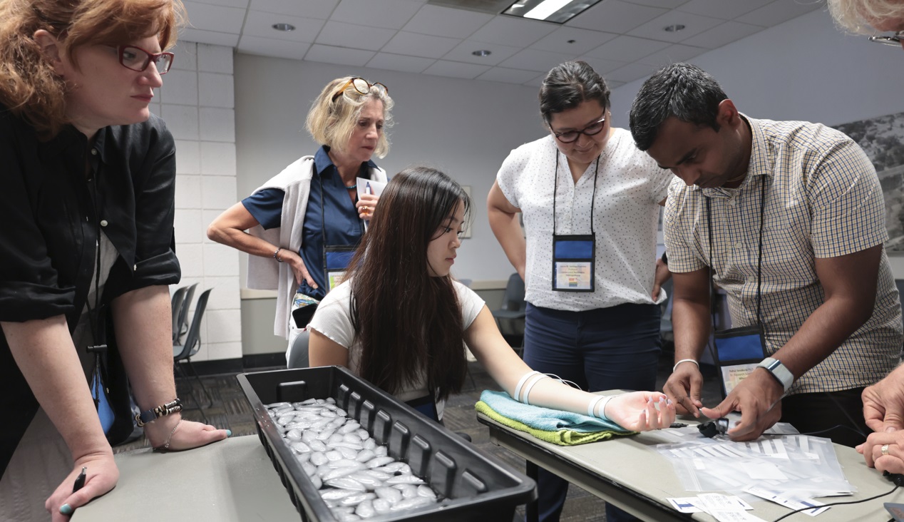
4T|Phys 2023 was the third installment in BIOPAC’s highly successful series of biennial industry conferences that began in 2017. After a two-year hiatus during the global Covid-19 pandemic, 4T came roaring back this summer with more workshops, more tracks, more sponsors, more presenters, and more countries represented than ever before. Over the course of three days, nearly 100 attendees participated in hands-on sessions with some of the top minds in physiology research, experiencing the latest tools for gathering and analyzing data.

BIOPAC’s in-house experts were joined by an array of authorities from the fields of physiology, psychology, imaging (fNIRS and fMRI), neuroscience, cardiovascular research, eye tracking, virtual reality (VR), biology, and engineering. Participants took part in workshop schedules drawn from five separate tracks that included fundamentals, which focused on the basics of data acquisition and analysis. The research track took attendees on a deeper dive into techniques and technologies, including recording evoked potentials, screen-based eye tracking, impedance cardiography using trans-radial bioimpedance velocimetry (TREV), stimulus presentation, exercise physiology, and many others. Participants were also able to tour UC Santa Barbara’s state-of-the-art Brain Imaging Lab to see up close how BIOPAC products are being used in both functional MRI (fMRI) and TREV studies.

“It was an incredible experience that left a lasting impact on me by helping me to understand more about physiological data collection and analysis,” said Dr. Parth Trivedi, a 4T|Phys attendee. “The choice of presenters and the topics covered were truly inspiring, offering valuable knowledge and fresh perspectives that enriched my understanding.”

4T|Phys 2023 Attendee MapThis year’s conference featured several new tracks such as a dedicated VR track led by partner company WorldViz. The WorldViz team showcased virtual reality solutions in a series of workshops that introduced how VR is being integrated in physiology and psychology research, explained how to create effective and immersive VR experiments, and demonstrated their latest PRISM technology for creating fully immersive projected 3D virtual environments.

Several other partner companies introduced their technologies and products both in the 4T exhibition hall and in workshop sessions. Argus Science presented their ETVision eye-tracking glasses that synch seamlessly with AcqKnowledge. Cognixion demonstrated the Cognixion One Axon-R—Research Edition wearable BCI-AR computing platform, which is being used to help individuals with neurodegenerative conditions and severe motor impairments communicate and interact with their environments assisted by augmented reality. Seenel Imaging showed off their MedelOpt Systems for whole-head EEG with fNIRS, including new VR integration. Participants also got a chance to try on the Research Ring by Senstream, a new user-friendly wearable research ring, which allows easy recording of EDA, PPG, Temperature, and ECG signals. CNSystems showed off the company’s NIBP continuous noninvasive blood pressure (NIBP) and hemodynamics devices.

If you missed the educational and networking opportunities at 4T|Phys 2023, don’t despair. We will be back again in a few years, bigger and better than ever. Watch this space for updates on our next installment. In the meantime, see our Video Tutorials and Webinars On Demand for trainings and seminars on our products and partners.


Make sure you never miss a BIOPAC event and keep up with the latest Tools, Trends, Techniques, and Technologies by contacting your local BIOPAC sales representative.

The Year in Review: Top Physiology and Neuroscience Research Stories 2022

Another calendar year has come and gone. COVID-19 remains a reality, but we continue to adapt to the ongoing pandemic and find new ways to overcome the challenges it poses at work, at school, at home, and in the lab. And speaking of research, 2022 proved to be a banner year for innovation and breakthroughs in the sciences, particularly in the physiological and neurological fields. Here is a quick round-up of some of the research stories that caught our attention.

Brain Implant Allows Locked-in Patient to Communicate

A man in his late 30s has been given the ability to communicate for the first time since being left in a completely locked-in state by amyotrophic lateral sclerosis (ALS). The patient, who was unable to move or even open his eyes, can communicate again thanks to a brain-computer interface (BCI) comprising two microelectrode arrays implanted in his motor cortex. The procedure was carried out by a team of researchers at the University of Tübingen in Germany. “This study answers a long-standing question about whether people with complete locked-in syndrome (CLIS) – who have lost all voluntary muscle control, including movement of the eyes or mouth – also lose the ability of their brain to generate commands for communication,” wrote Dr. Jonas Zimmermann, one of the co-authors of the study resulting from the procedure.

First Scan of Dying Human Brain Provides Insight into Near-death Experience

near-death experienceAn EEG recording from a dying 87-year-old patient has provided researchers with the first glimpse of the neurophysiological phenomena that occur shortly after cardiac arrest. While survivors of so-called near-death experiences have anecdotally reported intense and surreal experiences that include panoramic memory review, out-of-body experiences, and other hallucinatory phenomena, little is understood about what causes such occurrences. While experiments have been carried out on animal subjects showing a hypoactive state of brain activity under such conditions, this new data provided researchers with the first evidence “that the human brain may possess the capability to generate coordinated activity during the near-death period.”

Biomembrane Research May Advance Understanding of Memory in Computers and Humans

Drawing inspiration from the living realm, scientists at the U.S. Department of Energy’s Oak Ridge National Lab are applying biological concepts to next-generation computers with implications for both human health and cutting-edge computing. Researchers were able to demonstrate that an artificial cell membrane could imitate one of the hallmarks of biological memory and learning—long-term potentiation (LTP). The study, published in the Proceedings of the National Academy of Sciences, shows for the first time that a cell membrane by itself, without the benefit of proteins or other embedded biomolecules, can carry out LPT over several hours. The pure lipid membrane, which was formed using lipid-covered water droplets, represents the first nanoscale structure in which memory can be encoded. The discovery opens the door to neuromorphic computing, which imitates the way the human brain stores and processes information and is more efficient than traditional computing.

This is just a sampling of the exciting developments catching our attention in the sciences this year. For more explorations of the intersection of cutting-edge technology, physiology, and neuroscience, join BIOPAC at the T4 Human Physiology Conference in summer 2023. And have a happy New Year!


To learn more about BIOPAC’s role in exciting breakthroughs in neuroscience and physiology, and how we can assist you in achieving your research and teaching goals, contact your local sales representative today.

error: Content is protected !!