Hands-on Learning: How Real‑Time Physiological Data Deepens Clinical Education

Physicist Albert Einstein noted, “The only source of knowledge is experience.” This principle is particularly relevant in healthcare education, where hands-on learning is vital for cultivating the skills needed to care for patients effectively. The practice of recording and analyzing physiological signals in the learning lab serves as a powerful teaching tool, enabling students to immerse themselves in real-world scenarios and reveal autonomic responses, cardiovascular patterns, and emotional states that are often hidden from view.

For students in nursing, dentistry, and pharmacology, engaging with these signals transforms abstract concepts into concrete, measurable experiences. By analyzing heart activity, visual attention, stress markers, and pharmacodynamic effects, students not only enhance their critical thinking skills but also prepare themselves for clinical challenges with greater confidence.

Physiological signals across the health sciences teach learners to interpret subtle features embedded in physiological waveforms. Modern teaching laboratories increasingly incorporate multimodal systems to help students better understand the complexity of human physiology.

Nursing programs emphasize accurate patient monitoring and interpretation of vital signs, including electrocardiograms (ECG) and heartrate variability (HRV). A 2025 study published in Sensors evaluated the validity of Apple Watch Series 6 HRV data against laboratory‑grade ECG recordings using a BIOPAC 3‑lead ECG system to provide a comparative baseline for measurement. By synchronizing event markers between the Apple Watch and BIOPAC timeline, researchers ensured precise temporal alignment for comparing R‑R intervals and HRV indices. The results showed near-perfect agreement in R-R interval timing under resting conditions, indicating that such wearable devices can serve as a reasonable tool for certain wellness-oriented assessments. At the same time, the BIOPAC system remains the gold standard for precise physiological analysis.

For nursing students, this study demonstrated that consumer wearables must be validated against clinical-grade systems before being relied upon for assessment or decision-making. By examining the comparative data, students can explore how artifacts, sampling frequencies, and sensor placement influence the fidelity of HRV measurements. Moreover, because HRV relates directly to autonomic balance and stress physiology, the study provides a meaningful touchpoint for nursing courses in pathophysiology and patient assessment. Through hands‑on lab sessions using BIOPAC ECG amplifiers, students can visualize waveform morphology, measure beat‑to‑beat variability, and learn how cardiovascular signals reflect underlying homeostatic regulation.

Dental students should understand not only the mechanics of clinical procedures but also the emotional and perceptual experiences of their patients, especially children. A 2025 cross-sectional pilot study in the European Archives of Paediatric Dentistry used an eye tracking and  data acquisition and analysis system from BIOPAC to measure children’s gaze behavior and physiological stress when viewing images of dentists, dental chairs, and operatory designs.

Using eye-tracking metrics such as mean dwell time, fixation count, time to first fixation, and total fixation time, researchers found that study participants stared longer at dentists wearing white coats, indicating a perception of compitance and safety, but also indimidation. In addition, gender matching mattered, as children showed longer fixation on dentists of the same gender. The study also found that warm‑colored dental chairs (especially yellow) attracted greater visual attention, particularly among younger children. Simultaneously, HRV and ECG signals recorded with the BIOPAC system revealed that younger children exhibited greater autonomic arousal than older peers when exposed to dental images.

These findings offer dental students actionable insights into clinic design, caregiver attire, and emotional perception, all of which influence pediatric patient comfort. The study demonstrates how combining eye tracking with physiological monitoring can reveal otherwise hidden psychological responses. For dental educators, integrating such physiological signal analysis into coursework helps students develop empathetic, patient‑centered care strategies.
colored pillsIn pharmacology courses, students learn how drugs influence physiological systems. A study published in Brain Sciences investigated the lingering cardiovascular effects of caffeine, challenging the assumption that its effects dissipate within six hours. Using BIOPAC respiratory transducers, noninvasive blood pressure modules, bioimpedance amplifiers, and ECG amplifiers connected to a data acquisition and analysis system researchers monitored participants before and after consumption of caffeinated and decaffeinated coffee. Results showed measurable changes in heart rate, respiratory patterns, and thoracic impedance well beyond the expected metabolic window, suggesting caffeine’s physiological effects outlast its pharmacokinetic half‑life.

For pharmacy students, these findings reinforce critical distinctions between pharmacokinetics (drug metabolism) and pharmacodynamics (physiological response). By analyzing real‑world signal data, learners gain a more nuanced understanding of how stimulants affect autonomic regulation. Such insights prepare future pharmacists to counsel patients on medication timing, drug interactions, and individual responses to common agents such as caffeine.

Across nursing, dentistry, and pharmacy, physiological signals provide potent educational value. They bridge the gap between theory and clinical application by giving students concrete, measurable access to physiological processes such as autonomic reactivity, cardiovascular regulation, and perceptual attention. Using both research and education-based lab systems, learners can visualize real-time physiological data, develop analytical skills, and cultivate a deeper understanding of the human body under stress, stimulation, and clinical observation.

As healthcare grows more data‑driven, integrating hands-on experience with signal acquisition into professional training helps future clinicians interpret complex physiological information with confidence. Whether validating wearable devices, improving pediatric patient experiences, or exploring drug‑induced autonomic effects, physiological signals empower students to connect scientific principles with evidence‑based, care. Check out our webinar, Advancing Practical Learning: Biosignal Essentials, to learn more.


If you’re exploring ways to incorporate physiological signals into your curriculum or simulation lab, our team is ready to help identify the best tools and workflows for your program.

Curriculum Roadmap: Guiding Students with HAPS Learning Goals

When planning any journey, choosing the right route can mean the difference between reaching one’s destination or becoming lost in the wilderness. It helps to know the purpose of the trip; is it merely to get from point A to point B, or is the journey itself as important as reaching the destination? What stops need to be made along the way?

These questions can also resonate for instructors as they guide students along their academic journeys, as the goal of instruction is not limited to just reaching a specific destination. It is often about what occurs along the way: experiences, obstacles, and challenges encountered that shape the student and their understanding of the subject matter.

To help students and instructors on the path to better life science education, BIOPAC created a roadmap with its curriculum for the Biopac Student Lab systems (BSL) for life science instruction. The BSL curriculum comprises more than 65 lessons that serve as carefully plotted points on a path to an in-depth and invigorating understanding of general life science disciplines.

This curriculum follows a route established by the Human Anatomy and Physiology Society (HAPS). HAPS has created a set of learning goals, guidelines, and outcomes that bring together a range of topics covered in Anatomy and Physiology I & II courses at the university level for both clinical and academic degree programs.

The advantages of following such a path are fourfold. First, students are provided with an outline of course expectations. Just as every journey should begin with a clear understanding of where one is going, knowing learning goals allows students to focus their efforts on what matters most. Furthermore, instructors are encouraged to relate learning goals to real-life applications to provide meaning in a relatable context.

Secondly, with straightforward goals in mind, the instructor will find it easier to manage course design by focusing on key elements of their curriculum. These can include the following:

  • Order of topics to cover
  • Pedagogical strategies
  • Lab experiments
  • Periodic assessments that help students stay on track

Taking this a step further, by following HAPS’s goals, guidelines, and outcomes, instructors will be better equipped to plan a comprehensive program of study. Well-defined goals allow the instructor to build a solid foundation for future courses and guide learning specific to the program’s discipline. Additionally, it can reveal redundancies, overlaps, and gaps in the degree track.Goal Planner

Lastly, just as a good cartographer’s map provides evidence of where the traveler has journeyed, carefully plotted goals, guidelines, and outcomes trace the students’ track within the academic program. This provides valuable information for accreditation while ensuring students graduate with the necessary skills for their chosen field in the sciences.


To learn more about how the BSL system and curriculum can help students and instructors get the most out of anatomy and physiology courses, contact BIOPAC today.

Enhance Consistency with an Automated Lab Refresh

The global COVID-19 pandemic made in-person lab work impossible under many circumstances and difficult in the best of conditions. Now, as researchers and students begin to return to the lab, an opportunity presents itself for a fresh start. One way in which labs are refreshing their approach to research and education is by adding software and equipment that allows for the automation of procedures and analysis with the goal of increasing repeatability and reproducibility within experiments and research.

It’s been a challenging 18 months for science educators and those running research facilities. The global COVID-19 pandemic made in-person lab work impossible under many circumstances and difficult in the best of conditions. Now, as researchers and students begin to return to the lab, an opportunity presents itself for a fresh start. One way in which labs are refreshing their approach to research and education is by adding software and equipment that allows for the automation of procedures and analysis with the goal of increasing repeatability and reproducibility within experiments and research.

Crisis of reproducibility

The phrases “reproducibility crisis” and “replication crisis” became buzzwords in the scientific community in the 2010s, with that notoriety spilling over into both the scientific and mainstream media’s coverage of the sciences. Several high-profile scientific and sociological studies were called into question when researchers were unable to reproduce their results. In 2015, a group of psychology researchers under the moniker of the “Open Science Collaboration” found that they were only able to reproduce the findings of 68% of studies published in high-ranking peer-reviewed journals. A 2016 survey of 1,576 researchers by the journal Nature similarly found that 52% agreed that there was a “crisis” of reproducibility, though most said they still trusted published research.

The automated advantage

One step that researchers can take to ensure the reproducibility of their experiments and thereby boost the integrity of their data and conclusions is by automating procedures, workflow, and record keeping. These steps not only reduce the possibility for human error, but also provide a clear roadmap for future researchers to repeat processes in the lab to review and test results. The National Academies of Sciences, Engineering, and Medicine guide Reproducibility and Replicability in Science (National Academies Press, 2019) outlines several of the ways in which automation is being used to enhance reproducibility of research results, including record keeping, data analysis, data version control, and workflow management. The publication notes that “automated scientific workflows are essential in data-intensive and large-scale science missions that aim to be computationally reproducible.”

A 2019 study by researchers at the Indiana University School of Informatics, Computing, and Automated studyEngineering employed these techniques to demonstrate “how automation can be used to address many of these factors contributing to the replication crisis.” The study focused on the controlled rearing of chicks, comparing automated and non-automated research methods. The study found that the automated tracking system allowed researchers to increase the amount of data collected from each test subject, which in turn “reduced measurement error by 60% and increased the effective size of experiments by about 300%.”

An automated refresh

BIOPAC’s AcqKnowledge data acquisition and analysis software allows labs of any size to gain the advantage of automation to improve both the efficiency and effectiveness of research. Features like Batch Processing and  BIOPAC Basic Scripting help eliminate the potential for human error by consolidating multiple tasks into a single automated routine. A heightened level of standardization and consistency results in improved data quality. Adding a Basic Scripting license to AcqKnowledge 5.0.6 ensures results are reproducible regardless of who is carrying out the experiment.

Helping Students Learn Biomedical Engineering

Students entering the rigorous Biomedical Engineering (BME) discipline learn a gamut of separate physiological signals to best study healthcare applications of new technology. Simply put, as new, advanced medical devices make their way into the market, biomedical engineers are on the front lines of discovering how to best utilize these devices and corresponding software to better serve in healthcare. Well-equipped and prepared biomedical engineers study a number of signals, movements, and physiological concepts in their research, including some of the following:

Data Acquisition & Analysis Systems Respiratory System & Pulmonary Function
Spectral Analysis & Histograms Bioimpedance (Cardiac Output & Blood Flow)
Amplifiers, Transducers, and Calibration Biomechanics—Angle, Acceleration, Distance, Velocity
ECG, EDA (GSR), EEG, EGG, EMG, and EOG Physiological Control Systems
Force, Pressure, Strain, Flow, Temperature, Sound, Light Chart, Overlap, Scope, and X/Y Displays
Brain Computer Interface Blood Pressure & Heart Sounds
Signal Analysis & Processing Compartmental Modeling
Filters (FIR & IIR) Gait Analysis
Instrumentation Design

Related scientific citations are available for BME research in nearly all these areas of interest, but for students and educators, simplified practical labs can lay the groundwork for more advanced BME areas of interest, such as Gait Analysis or Brain Computer Interface (BCI). Inherent in the design of BME education is the fusion between virtual software-based analysis and physical laboratory applications—a dichotomy that favors software-based learning and research more and more. Educating students in biomechanics, biomeasurement, and biomaterials is crucial to set them up for success in the BME field, whether they go into research, education, health services, or industry. Trained biomedical engineers utilizing virtual tools in lieu of a larger laboratory instrumentation setup can save upwards of $80,000 in hardware costs for their institution. Skills taught with virtual tools are better suited to the real world “new world” of software-based analysis and signal processing.

Physiological Signal Processing Labs—colloquially known as “breadboards”—are an excellent tool to further biomedical engineering education, through both basic lessons and the physical construction of the signal processing lab device, a foray into the physical engineering side of the BME vocations. Recorded signals are analyzed and processed via Biopac Student Lab Software—a leader in BME education for university laboratories and undergraduate studies.

For example, the Biopac Student Lab Breadboard Lab Set covers lessons on constructing and running the following signal processes:

  1. Square Wave Oscillator
  2. Instrumentation Amplifier
  3. Active Filter: High Pass
  4. Active Gain Block & Low Pass
  5. Notch Filter for 50/60 Hz Rejection
  6. QRS Detection: Band Pass Filter
  7. QRS Detection: Absolute Value Circuit
  8. QRS Detection: Low Pass Filter and Overall System Test

BIOPAC also offers Biomedical Engineering curriculum for EMG-controlled robotic labs, modeling, filtering, FFT, as well as a “Best Practices for Good Data” on-demand training series on fundamentals of physiological signal processing and support for an array of BME research applications.

The benefits of this approach to BME Education help “…viscerally engage the student in an inquiry-based learning environment.”¹  Upon construction of their own Signal Processing lab, students verify signal processing models against mathematical simulations and test the models against measured signals sourced on their own bodies.

BIOPAC offers a wide array of wired and wireless equipment that can be used in your research. To find more information on solutions for recording, teaching, and analyzing Biomedical Engineering data, you can visit our product category for BME on the BIOPAC website.


¹ Alan Macy, R&D Director, BIOPAC Systems, Inc.

Panoply of Respiration and Pulmonary Measurements

The respiratory or pulmonary system performs the important functions of supplying oxygen (O2) during inhalation, removing carbon dioxide (CO2) during exhalation, and adjusting the acid-base balance (pH) of the body by removing acid-forming CO2.

All humans and animals require oxygen to carry out cellular processes of energy transformation essential for life. During cellular metabolism, oxygen is consumed when nutrients such as protein, carbohydrate and fat are oxidized, and carbon dioxide is produced as a gaseous waste product. Collectively, the processes whereby oxygen is taken up from the atmosphere, delivered to body cells and consumed, and the processes of producing carbon dioxide and delivering it to the lungs for excretion into the atmosphere constitute respiration.

Processes of respiration fall into one of three categories: external respiration, gas transport, and internal respiration. External respiration refers to mechanisms by which a person obtains oxygen from the external environment and eliminates carbon dioxide into the external environment. Gas transport refers to mechanisms used to distribute oxygen to and remove carbon dioxide from cells. Internal respiration refers to the chemical reactions of cellular metabolism in which oxygen is consumed and carbon dioxide is produced.

Gas exchange between the air in the lung and the blood is a process of simple diffusion. A gas diffuses from a region of higher concentration to a region of lower concentration, or, from an area of higher partial pressure to an area of lower partial pressure. Partial pressure is simply a way of expressing the concentration of gas molecules.

Blood transports gases to and from the body’s cells. The respiratory system supplies oxygen to the blood and removes carbon dioxide from the blood. At the beginning of inspiration, the thoracic cavity is enlarged by contraction of the diaphragm and the external intercostal muscles. Expiration begins when the inspiratory muscles relax. The diaphragm returns to its resting dome shape, decreasing thoracic and intrapulmonic volume.

What are some common measurements extracted when we measure respiration and how are these measurements acquired and analyzed?

Pulmonary Airflow — The pulmonary airflow analysis uses a calibrated airflow signal from a pneumotach transducer, of which there are many types for various airflow applications.

Compliance and Resistance Analysis is measured on a breath-by-breath basis by diving tidal volume by change in pressure between the exhale start and inhale start locations of the breath.

Respiratory Gas Analysis uses the BIOPAC O2100C and CO2100C amplifier modules for measuring and analyzing oxygen and carbon dioxide content.

Anaerobic Threshold uses AcqKnowledge software to establish a ratio of inspired volume and VO2 consumption and to integrate the air flow signal to obtain total inspired volume in real time.

Respiratory Exchange Ratio is determined by dividing VCO2 produced by VO2 consumed. An automated script for computing RER is available via download from BIOPAC.

Respiration, Gas Analysis, Pulmonary Function, and Exercise Physiology for Research applications and Education curriculum are available.

Respiratory Cycle and Exercise Physiology are supported by the new BSL HOME Practical Lab Kit, enabling students to keep abreast of curriculum demands while safely distanced.

We invite you to sign up for BIOPAC’s free How to Record Great Respiration Data webinar on demand. You may also watch archived webinar events on demand by visiting our Webinars page.


BIOPAC offers a wide array of wired and wireless equipment that can be used in your research. To find more information on solutions for recording and analyzing signals such as ECG, heart rate, respiration, and more using any platforms mentioned in this blog post, you can visit the individual application pages on the BIOPAC website.

Photo by Victor Garcia on Unsplash

Libraries in 2020: From Curbside Pickup to Practical Lab Kits

Libraries can support remote students with distance learning tools like BSL Home practical lab kits, accessible wifi or hotspot loaners, and more.

By: Jasmine Anderson, Biopac Student Lab Product Manager

People call the library the heart of the university campus, which speaks for the memories students and alumni associate with it. It’s the essential college stomping ground and uniquely serves as both a quiet refuge for studying and a meeting place for free-flowing ideas. At the library, peers mingle, study groups collaborate, and you can find solace from the stress of student life between the book stacks. Well, at least that was true before COVID-19…

Now facing the pandemic, libraries are performing their own open-heart surgery to maintain the flow of shared knowledge and support a thriving academic experience. They are redesigning ways to safely provide information and tools to students, facilitating inter-disciplinary course reserves and collaboration, and helping to maximize resource funding and logistics across campus.

“Quiet, please” doesn’t apply these days…talk to your librarian to discover new funding options!

To maintain their role in the college experience while also evolving with the recent changes to academia, libraries are adopting electronic print collections, virtual technology, and self-service stations. Adding these services—along with the infrastructure for self-checkout, curbside pickup, and the ability to loan technology such as laptops, anatomical models, and lab kits—helps support the transition to distance learning at universities.

Libraries can also help students in the sciences by providing funding for portable lab kits to fill the gap left by practical lab closures. In the fall, students can reserve BSL Home kits on loan from the library to participate in hands-on physiology experiments from anywhere. With BSL Home, students record and analyze data from their own bodies and receive the same authentic, open-ended, and complex learning framework that’s true of the traditional lab.

To champion this, the Association of College & Research Libraries (ACRL) put forth guidelines for instruction programs stating that libraries should demonstrate a “commitment to purchasing and providing support for classroom technologies.” Alongside the University of Wisconsin, several other institutions are following suit by providing resources and funding to support classroom technologies. A handful of these universities are listed below with their library action plan (Table 1).

Institution/University Library Action Plan
Auburn University Auburn’s subject librarians offer services to help faculty by purchasing books, journals, and electronic resources as well as supporting teaching and research in specific disciplines.
Case Western University Case Western’s Library equipment borrowing program takes suggestions for what they should be carrying online.
Georgia Southern University Georgia Southern provides a materials request form that faculty can complete online to request the purchase of teaching and research materials.
Michigan State University Michigan State allows instructors to request item purchases to place them on course reserve, similar to the University of Wisconsin MERIT Library.
University of California, Santa Barbara UCSB allows similar reserve requests by directing faculty to make requests to their subject librarians.
University of California, Los Angeles UCLA utilizes CCLE (Common Collaboration and Learning Environment), a campus-wide service that makes collaboration possible across schools, divisions, and departments and helps to advance remote teaching, learning and research.
University of Nevada, Las Vegas UNLV takes faculty purchase recommendations through a form on their website which can specifically be recommended to their healthcare library.

Table 1 – A list of Institution/University Libraries paired with their action plan to support classroom technologies.

Even though libraries will not operate in their normal manner this fall, they still remain at the heart of campus life, working to support the student body in stimulating and innovative ways. The students of 2020 won’t have the same memories in the library as classes before them, but with new technology available online and kits to take home, it’s encouraging to see the library continue to be a place of shared knowledge and scholarship. For more information on how your school can accommodate practical labs with BSL Home, talk to your librarian and reach us at info@biopac.com.

Don’t Let COVID-19 Lock Down Your Labs

By: Jasmine Anderson, Biopac Student Lab Product Manager 

The COVID-19 pandemic turned higher education upside-down when stay-at-home orders forced educational institutions to shift to distance learning this Spring. In hindsight, no one could have predicted what 2020 would entail – and now, while facing the pandemic, the future seems even more impossible to plan. 

When the novel coronavirus put a lockdown on our higher education institutions, instructors were required to make swift decisions on how they would continue. In the news, institutions are slowly releasing their course of action on an individual basis and plans appear to vary across the board. Some schools are announcing that they’ll forge ahead this fall while others are opting to hold off on deciding or have already closed their campus. Regardless of the plan, a common theme shines through – the fact that everyone’s trying to do the best that they can, given these complicated and un–chartered circumstances.  

To help save practical labs, BIOPAC is releasing a new teaching platform, called BSL Home, that brings the lab experience directly to your students. With BSL Home students can learn physiology by recording from their own body and can run the hands-on lessons from their own home, in the dorm, at the library, wherever. The objective of Biopac Student Lab is to learn-by-doing and we’re here to help you navigate this new normal. To find out more about our distance learning solutions and regional events for the Biopac Student Lab community, please contact your local sales representative.

How are you navigating this new normal? 

To start the conversation, use the comment box to share your school’s strategy for saving the practical lab this fall. 


The Eyes Have It—Mobile Eye Tracking

Eye tracking has been a consistent player in the physiological research field for a number of years, and more recently a “mobile” variation of this technology has become increasingly widespread. But just what is mobile eye tracking and how does it work? Unlike eye tracking studies tethered to a lab, where a participant visually follows an object on a computer screen, mobile eye tracking data can be acquired outside of the lab. All that is required, in fact, is a pair of glasses. Not just any glasses, but ETVision glasses, capable of recording not only multiple parameters of a participant’s “eye image” but also the scene the participant is gazing at and responding to. Let’s look briefly on how eye tracking glasses work. The lightweight ETVision glasses are equipped with two eye-facing cameras combined with a pair of LEDs for recording the various components of the “eye image,” such as pupil size/position, gaze path, fixation (when the participant’s eyes lock onto an area of interest), internal and external data markers, vergence angle (angle between the eyes), and corneal reflections (CRs). The cameras also look at the contrast between the pupil and the surrounding iris. This constant real-time feedback is recorded every 5.5 milliseconds and the eye images are rendered at 180 frames per second.

In addition to eye cameras, a built-in forward-facing 720p scene camera records the viewable area in front of the participant. This enables the landscape or scenery the participant is looking at to be combined and synchronized with the eye image data (say, if the participant is driving a car or riding a mountain bike).

Did I say riding a mountain bike? Yes. Full eye tracking data can be collected and stored in a 256 GB SD card and the logged data exported to a .*csv file, which in turn can be imported into AcqKnowledge software along with other logged physiological data. The glasses also have a coated visor for reducing glare. (Glare causes squinting and the eye tracking cameras do not perform as well on eyes that are squinting or closing due to excessive light or reflections.)

In addition, a small microphone built into the eyeglasses enables two-way audio between the investigator and the participant. Already wear glasses? No problem. Eye tracking glasses can be worn over corrective lenses without issue. However, contact lenses should be cleaned before wearing them under eye tracking glasses. The glasses are connected to a small controller via a standard HDMI cable, and the controller houses a 256 GB SD card. In addition to the SD card used for logging remote data, the controller can also be connected to a host computer via WiFi or LAN. The lithium-polymer battery will run the eye tracking glasses for 5.5 hours on a full charge.

Using the AcqKnowledge software’s Media feature, the eye tracking video data can be replayed with a tight correlation to other physiological data collected during the experiment by the BioNomadix Logger. (Other data acquisition hardware types, such as MP160 or MP36R, can be used within the lab.) The eye tracking video and graph data can be displayed side-by-side on the screen. Placing the I-beam cursor at any point in the AcqKnowledge graph takes you to the corresponding point in the eye-tracking video (say, during activities such as driving a car). Eye tracking data for vertical position, horizontal position, pupil diameter, and more can be imported as channels into the AcqKnowledge graph. This data will be synchronized with the eye tracking video, along with any added event markers.

Interested in more details about ETVision Eye Tracking Glasses and AcqKnowledge software? To see it all in action, we invite you to view our free on-demand Mobile Eye Tracking Webinar.


BIOPAC offers a wide array of wired and wireless equipment that can be used in your research. To find more information on solutions for recording and analyzing signals such as ECG, heart rate, respiration, and more using any platforms mentioned in this blog post, you can visit the individual application pages on the BIOPAC website.

Photo by Amanda Dalbjörn on Unsplash

Top Signals for Life Science Instruction

Experience has shown that beginning physiology students are more engaged when acquiring and viewing signals from their own (or a lab partner’s) bodies in real time, particularly signals that are noninvasive to capture and easy to acquire. Watching a waveform trace across the screen and respond immediately to physical input is a sure-fire way to spark wonder and curiosity in a student. The following is a list (in no particular order) of the most common signals gathered in a beginning lab environment using the Biopac Student Lab System.

Electromyogram (EMG)

EMG is probably the top signal as far as simplicity, as the EMG signal can be consciously and directly controlled by the student, whether it be by simply making a fist to contract the forearm muscle or squeezing a hand dynamometer to measure grip strength. In addition to a raw EMG signal, a second channel showing integrated EMG can be simultaneously displayed. (Biopac Student Lab Lesson 1 and Lesson 2.)

Electroencephalogram (EEG)

EEG is another easy-to-acquire signal. Although not consciously controllable as EMG, the student can observe differences in the four simple periodic rhythms present in brain activity (alpha = 8-13 Hz, beta = 13-30 Hz, delta = 1-5 Hz, and theta = 4-8 Hz) while engaged in activities such as opening/closing eyes, solving simple mental math problems, or hyperventilating. (Biopac Student Lab Lesson 3 and Lesson 4.)

Electrocardiogram (ECG)

The ECG represents electrical events of the cardiac cycle whereas Ventricular Systole and Ventricular Diastole represent mechanical events (contraction and relaxation of cardiac muscle, passive opening and closing of intracardiac valves, etc.). Because the ECG reflects electrical activity, it is a useful “picture” of heart activity and presents several interesting components under various conditions, such as while sitting, standing, lying down, and after deep breathing or exercise. Various lead configurations highlight observable differences in the QRS complex which become apparent in analysis. (Biopac Student Lab Lessons 5, 6, and 7.)

Respiration (RSP) cycle and pulmonary function

As easy as breathing, measuring RSP signals under a variety of circumstances is another common study. Using a respiratory transducer (SS5LB) and an airflow transducer (SS11LB), we can record and analyze chest expansion and contraction during the breathing cycle, as well as pulmonary volumes/capacities and the effects of aerobic exercise. (Biopac Student Lab Lessons 8, 12, 13, and 15.)

Electrooculogram (EOG)

Large and small movements of the eye can be tracked and studied under various conditions by attaching simple adhesive electrodes to the forehead and temples in conjunction with the same SS2L electrode lead set used for recording EMG and ECG signals. (Biopac Student Lab Lesson 10.)

Reaction Time

Possibly the most fun signal to record. The participant holds a hand switch (SS10L) and pushes the button when an aural (click sound) or visual (light flash) stimulus is presented. The reaction time interval between the presentation of the stimulus and the participant’s voluntary reaction (button push) are recorded and analyzed. A variety of stimuli are presented, such as reaction to a fixed stimulus (e.g., every four seconds) opposed to a random stimulus (one to 10 seconds). Reactions are recorded holding the hand switch alternately in both the right and the left hand. It’s interesting to compare the difference in reaction time elicited from the dominant vs. non-dominant hand in response to the aural or visual stimulus. (BSL Lessons 11 and 11A.)

Other signals and studies commonly acquired in Biopac Student Lab are:

We also invite you to watch a free on-demand webinar detailing Biopac Student Lab Lessons.


BIOPAC offers a wide array of wired and wireless equipment that can be used in your research. To find more information on solutions for recording and analyzing signals such as ECG, heart rate, respiration and more using any platforms mentioned in this blog post, you can visit the individual application pages on the BIOPAC website.

Photo by JESHOOTS.COM on Unsplash


T4 Conference Wrap-up

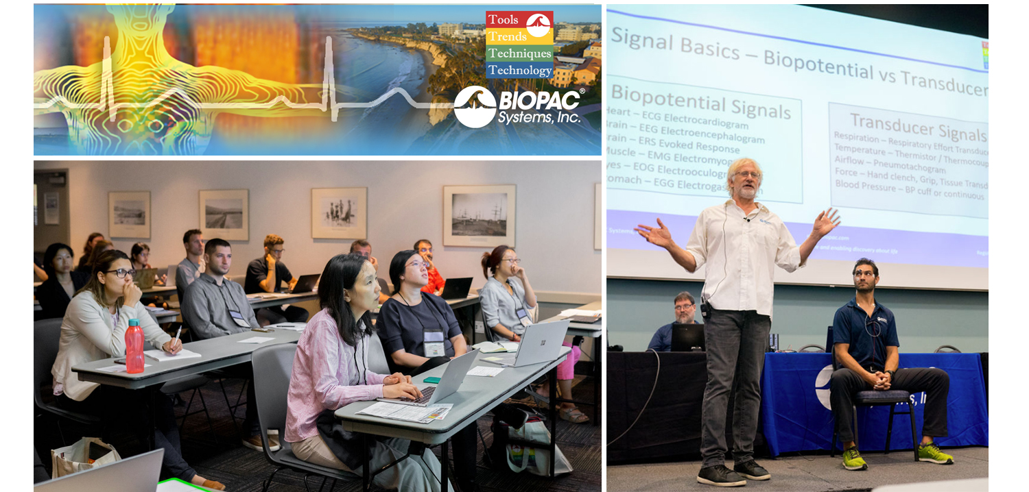
August is a month of barbecues, baseball, and last-minute vacations against the backdrop of a fading summer, and the eighth month of 2019 proved to be an equally exciting time at BIOPAC, with something of a pleasant vacation thrown in. On August 12-14, BIOPAC hosted its 2nd Biennial “Tools, Trends, Techniques, and Technology” Human Physiology Conference, known as “T4” to our friends and partners around the world. This three-day event, first held in 2017, featured lively, informative sessions hosted by BIOPAC staff, partners, noted academics, and industry experts, and covered a wide range of physiological techniques and disciplines. Over 150 attendees joined BIOPAC at the 2019 T4 Conference staged once again at the beautiful University of California at Santa Barbara campus. Participants and instructors winged in from China, Australia, Germany, Sweden, Italy, Spain, U.K., Ireland, and Egypt, as well as from wide regions of the U.S and Canada to soak up the idyllic surroundings of this ocean-side campus.

The focus of BIOPAC’s T4 Conference was to provide a fun, relaxed environment while hosting nearly 40 hands-on, human physiology training workshops chaired by over 20 distinguished instructors from around the globe. One of the many conference highlights was the “Mindfulness, Meditation, and Measurement: Mindfulness Exercises in Physiological Monitoring” session, hosted by Dr. Allan Weiss of the University of Southern California and  BIOPAC Co-founder and Director of R & D Alan Macy. These sessions, conducted in two parts over a two day period were booked beyond capacity, even though the early morning fog hadn’t yet lifted from the campus skyline.

The varied additional sessions covered a range from the basics, “Setting Up Your Lab, Getting Great Data and Troubleshooting,” (hosted by Dr. Terry Blumenthal of Wake Forest University, and BIOPAC’s Alan Macy and Tim Cook), and “Creating Your Own Lessons Using BSL PRO,” (hosted by Dr. Taffeta Elliot of the New Mexico Institute of Mining and Technology), to exciting new technologies such as “Integrating Screen-based Eye Trackers with AcqKnowledge,” with Keith Jackson of BIOPAC partner company EyeTech, BIOPAC CEO Frazer Findlay, and BIOPAC Engineering Manager Ed Peterlin. Additional wide-ranging physiological disciplines were also covered, including MRI techniques, stimulation, and exercise physiology. All sessions were conducted using BIOPAC hardware and software systems, as well as platforms provided by BIOPAC partners such as Cedrus, fNIR, WorldViz, Psychology Software Tools, and CNSystems. At the end of each day, it was time to relax and unwind with a beverage of choice, a a walk along the beach, and some good conversation. Each evening featured excellent meals and camaraderie in the relaxed yet elegant environment of some of Santa Barbara and Goleta’s most stunning surroundings. After three great days of immersive sessions, it came time for everyone to part ways, having forged new collaborative partnerships and an enhanced knowledge about the full potential of BIOPAC and BIOPAC partner products.

You don’t have to wait for T4 2021 to learn more about life science data—watch Fundamentals of Physiological Data Recording now!

For a closer look at T4, and a preview of what is sure to be an exciting 2021 event, we invite you to visit our T4 Human Physiology Conference page.

Looking for additional resources, tips, tricks, and expert advice on BIOPAC and BIOPAC Partner products? Please also visit our Webinars and Video Tutorial pages.


BIOPAC offers a wide array of wired and wireless equipment that can be used in your research. To find more information on solutions for recording and analyzing signals such as ECG, heart rate, respiration and more using any platforms mentioned in this blog post, you can visit the individual application pages on the BIOPAC website.

error: Content is protected !!