Aesthetically Pleasing: Exploring How the Brain Perceives the Arts

Beauty is in the eye of the beholder—a mostly subjective experience, so the saying implies. While we may share many common ideals about beauty and aesthetics, our reactions to art, music, and literature can diverge, converge, and overlap to a myriad of degrees. Neuroaesthetics explores how the brain perceives and responds to art, music, and other aesthetically evocative experiences. Combining neuroscience with psychology and the arts, researchers seek to understand why humans find certain images, sounds, or designs appealing and how these perceptions influence our emotions, cognition, and behavior. This interdisciplinary science has practical applications across a wide range of fields, including design, marketing, education, and mental health therapy, where understanding aesthetic experience can inform and enhance user engagement, learning, and emotional well-being.

Biometric signals provide insight into emotional and cognitive processing, allowing researchers to gain valuable data on how the brain creates and responds to aesthetically engaging stimuli. Commonly used techniques include electrodermal activity (EDA), which measures changes in skin conductance linked to emotional arousal; electroencephalography (EEG), which tracks brainwave patterns associated with attention and engagement; functional near-infrared spectroscopy (fNIRS), which monitors blood flow changes in the brain; and functional magnetic resonance imaging (fMRI), which provides high-resolution images of neural activity.

Additionally, the use of virtual reality (VR) and augmented reality (AR) is expanding in neuroaesthetics research, allowing scientists to create controlled, immersive environments that mimic real-world experiences. These technologies offer new ways to investigate how individuals engage with art and design in dynamic, interactive settings, pushing the boundaries of how we understand and harness aesthetic experiences.

Researchers in Shanghai, China, for example, investigated how different visual modes—2D and 3D VR—affect emotional arousal, demonstrating how immersive environments influence emotional experiences. The research team utilized EEG to monitor brain activity and EDA to measure skin conductance responses, both indicators of emotional arousal. EDA and EEG data were recorded while study participants wore VR headsets to view 2D and 3D environments. Data was collected and analyzed using a BIOPAC MP system with AcqKnowledge software. The findings revealed that 3D VR environments elicited more intense emotional stimulation than 2D settings, as evidenced by higher beta (21–30 Hz) EEG power and increased EDA responses. According to the published findings, results suggest that enhanced perception in 3D environments leads to greater emotional engagement, highlighting the potential of 3D VR in applications such as immersive therapy, virtual training, and interactive entertainment.

MusicA team of German researchers recently examined the intersection of neurology, psychology, music, and literature. Their findings, published in the journal NeuroImage, explored how melodic features influence the neural processing of spoken poems and songs, examining the interplay between language and music. Researchers employed fMRI to investigate the neural mechanisms underlying the processing of musical and nonmusical verse. The fMRI data revealed that both spoken poems and songs activated bilateral temporal auditory areas of the brain. One aspect of the study was the measurement of participants’ emotional responses to the stimulus, i.e., whether they liked the song or poem to which they were listening while the fMRI scan was conducted. To gather this data, participants interacted with a BIOPAC differential pressure transducer connected to the participants’ right and left index fingers to record the degree to which they enjoyed the music. The researchers stated the findings suggest that melodic properties are central to the aesthetic experience of both speech and music.

A study published by Britain’s Royal Society looked at the connection between aesthetic appreciation and emotional states, specifically how exposure to art or music can shift feelings of anxiety to curiosity. To understand how the brain perceives beauty, the researchers used EDA to assess participants’ physiological arousal during exposure to aesthetically pleasing stimuli, such as music. EDA data was gathered using a BIOPAC EDA amplifier connected to a data acquisition and analysis system while participants were administered anxiety-inducing electrical tactile stimulation during music listening sessions. The study demonstrated that aesthetic appreciation can reduce anxiety and promote curiosity by analyzing the EDA data alongside self-reported measures. The research team concluded that their results “might have potential applications across different human activities such as learning and therapy. The idea that aesthetic appreciation might affect anxious states and that expecting aesthetic rewards can transform anxiety into arousal and curiosity plays a central role in some psychotherapeutic models of human change.”

These examples provide a rough outline of a bigger picture, illustrating the many ways researchers use physiological signals to better understand the human mind’s response to art, music, and literature. For more information on the tools BIOPAC provides to assist in neurological and physiological research, see our online training and webinars.


Are you planning a research project to investigate the intersection of aesthetics and neuroscience? Contact a BIOPAC representative in your area to identify the right resources and equipment for a successful study.

The Year in Review: Top Physiology and Neuroscience Research Stories 2022

Another calendar year has come and gone. COVID-19 remains a reality, but we continue to adapt to the ongoing pandemic and find new ways to overcome the challenges it poses at work, at school, at home, and in the lab. And speaking of research, 2022 proved to be a banner year for innovation and breakthroughs in the sciences, particularly in the physiological and neurological fields. Here is a quick round-up of some of the research stories that caught our attention.

Brain Implant Allows Locked-in Patient to Communicate

A man in his late 30s has been given the ability to communicate for the first time since being left in a completely locked-in state by amyotrophic lateral sclerosis (ALS). The patient, who was unable to move or even open his eyes, can communicate again thanks to a brain-computer interface (BCI) comprising two microelectrode arrays implanted in his motor cortex. The procedure was carried out by a team of researchers at the University of Tübingen in Germany. “This study answers a long-standing question about whether people with complete locked-in syndrome (CLIS) – who have lost all voluntary muscle control, including movement of the eyes or mouth – also lose the ability of their brain to generate commands for communication,” wrote Dr. Jonas Zimmermann, one of the co-authors of the study resulting from the procedure.

First Scan of Dying Human Brain Provides Insight into Near-death Experience

near-death experienceAn EEG recording from a dying 87-year-old patient has provided researchers with the first glimpse of the neurophysiological phenomena that occur shortly after cardiac arrest. While survivors of so-called near-death experiences have anecdotally reported intense and surreal experiences that include panoramic memory review, out-of-body experiences, and other hallucinatory phenomena, little is understood about what causes such occurrences. While experiments have been carried out on animal subjects showing a hypoactive state of brain activity under such conditions, this new data provided researchers with the first evidence “that the human brain may possess the capability to generate coordinated activity during the near-death period.”

Biomembrane Research May Advance Understanding of Memory in Computers and Humans

Drawing inspiration from the living realm, scientists at the U.S. Department of Energy’s Oak Ridge National Lab are applying biological concepts to next-generation computers with implications for both human health and cutting-edge computing. Researchers were able to demonstrate that an artificial cell membrane could imitate one of the hallmarks of biological memory and learning—long-term potentiation (LTP). The study, published in the Proceedings of the National Academy of Sciences, shows for the first time that a cell membrane by itself, without the benefit of proteins or other embedded biomolecules, can carry out LPT over several hours. The pure lipid membrane, which was formed using lipid-covered water droplets, represents the first nanoscale structure in which memory can be encoded. The discovery opens the door to neuromorphic computing, which imitates the way the human brain stores and processes information and is more efficient than traditional computing.

This is just a sampling of the exciting developments catching our attention in the sciences this year. For more explorations of the intersection of cutting-edge technology, physiology, and neuroscience, join BIOPAC at the T4 Human Physiology Conference in summer 2023. And have a happy New Year!


To learn more about BIOPAC’s role in exciting breakthroughs in neuroscience and physiology, and how we can assist you in achieving your research and teaching goals, contact your local sales representative today.

fNIRS Systems—a NIR miracle

At the dawn of the 19th century, pioneering astronomer Sir William Herschel discovered the existence of infrared radiation, which is electromagnetic radiation invisible to the human eye but identifiable via its ability to heat objects. Infrared radiation is commonly divided into three categories, near, mid, or far infrared waves. In ensuing years, infrared radiation was found to be useful for industrial, scientific, military, and law enforcement applications, such as heat signature tracking and night vision goggles. Over two decades ago, a new technology called near infrared spectroscopy (NIRS) was developed for non-invasively measuring specific aspects of human physiology using near infra-red waves. While far infrared waves lie closest to the microwave region, near infrared waves are closest in length to the visible light part of the electromagnetic spectrum, and are microscopic in size. Unlike far infrared waves, near infrared waves do not produce any heat. In fact, we can’t feel them at all. They are the small waves used by common devices such as a
remote control.

Today, near infrared waves are commonly used  for studying many application areas of human physiology, notably by the fNIRS Imaging Systems to gather precise, high-density brain function and muscle oxygenation data.

How does fNIRS work with regard to human physiology? It’s an optical technology which sends light into the tissue and records the light that scatters back via a photo-diode and sensor attached to the subject. The fNIRS continuous wave technology uses an optical window in the near infrared spectrum that enables light to pass through. For example, when you hold your hand in front of a strong white light, the tissue can appear red and almost translucent. fNIRS shines light into the skull or muscle tissue, and using special detectors, records the resulting light scatter that returns to the surface.  The light penetrates skin, bone, and alights on the brain before its return trip to the surface detectors, and accomplishes this painlessly without heat. In fact, the subject does not even feel the near infrared energy and the light is harmless. The result is startling images and functional data of the brain at work. The fNIRS Systems (ranging from Educational to High-Density versions) include fNIRsoft software for processing, analyzing and visualizing functional near infrared (fNIR) spectroscopy signals through a graphical user interface. Additionally, the fNIRS Systems work with the BIOPAC MP160 Research System and AcqKnowledge software in conjunction with other physiological signals such as ECG, respiration, cardiac output, blood pressure, electrodermal activity, and stimulus response markers. AcqKnowledge software also provides automated analysis tools for event related potentials and ensemble averaging.

fNIRS brain function application areas include workload assignment, VR,  stress, attention, geriatrics, economics, ergonomics, learning and performance, sports science, HCI, pain perception, and cognitive workload – low or high.

fNIRS Systems can also be used to measure muscle oxygenation data, including oxidative capacity, muscle load, lateral imbalances (left vs. right muscle group) oxygenated hemoglobin and de-oxygenated hemoglobin in muscle activity.

We invite you to enjoy a comprehensive free fNIRS webinar and to visit our fNIRS products page.

BIOPAC offers a wide array of wired and wireless equipment that can be used in your research. To find more information on solutions for recording and analyzing signals such as ECG, heart rate, respiration and more using any platforms mentioned in this blog post, you can visit the individual application pages on the BIOPAC website.

Dark Current Measurement as a Noise Management Tool

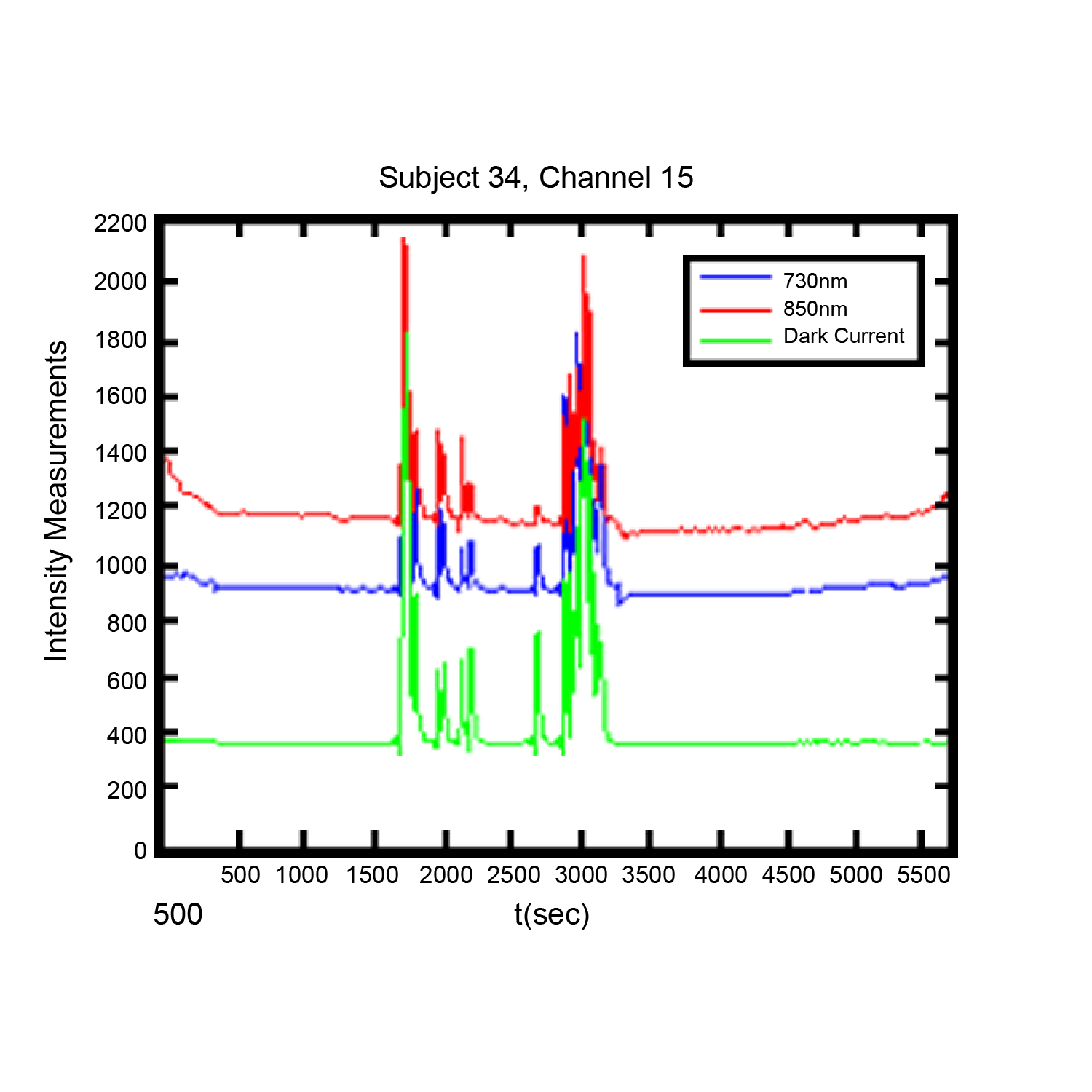
Noise in functional near infrared (FNIR) imaging data utilized for neurophysiological brain functioning research is a challenge to researchers, The use of dark current measurement in functional near infrared (fNIR) brain imaging can provide a valuable tool in identifying and managing certain type of noise in recordings.

By Davood Tashayyod, FNIR Devices

The use of dark current measurement in functional near infrared (fNIR) brain imaging (measurements taken not only when LED type light sources shine light at wavelengths 730 and 850 nm but also when they do not shine light (dark current condition)) can provide a valuable tool in identifying and managing certain types of noise in recordings.

Noise in functional near infrared (fNIR) imaging data utilized for neurophysiological brain functioning research is a challenge to researchers, in that excessive or unmanaged noise may pose a risk to the quality of gathered data and hence in the interpretation of the results. This noise arises from two general categories of sources, biological sources and non-biological sources. It should be noted that sources of noise are routinely present, so that noise cannot be eliminated but rather must be managed. Biological sources of noise include the respiratory and cardiac cycles and slow vasomotor oscillations. Non-biological sources of noise may include contamination from other external devices and noise induced by improper sensor application (light leakage, in the case of fNIR sensors), cable movements, or instruments or equipment used in the course of the experiment. These sources, independently or in combination, can mask the signals of interest.

Close attention to good laboratory practices, to include proper sensor placement, trial runs to establish baselines, and other techniques can help to reduce noise. Close attention to experiment protocols during the experiment can also be of assistance. However, it is in post-experiment data processing and analysis phases that the researcher has the greatest number of tools to manage noise. These tools include intuitive or visual analysis, filtering, and specialized analysis. Each method has strengths and weaknesses, but used in combination they offer a reliable method of managing noise and improving data quality.

Visual or intuitive analysis depends upon researcher experience and qualification, and some artifacts may be too small to be readily apparent. Filtering is useful, but in some range of measurements noisy data segments may be so great or may not contain any signal originated from the brain such as in the case when the sensor loses contact with the skin and captures data only from the ambient that it may not make sense to remove noise, rather such data segments should be identified and discarded.

Specialized analysis, such as dark current measurement, provide a very useful method for both identifying the noise not readily apparent to the naked eye and identifying data segments where filtering is likely to not be useful. A discussion of the use of dark current measurement can be found here.

For more information on optical braining imaging systems, visit BIOPAC’s fNIR page. BIOPAC also provides a wide range of tools for recording, displaying, classifying, and analyzing psychophysiological measurements—including EEG, ECG, EDA, PPG, and emotional states—visit BIOPAC’sapplications pages and view BIOPAC’s full line of physiology data acquisition and analysis systems, electrodes,amplifiers, and wearable, wireless transmitters and loggers.

BIOPAC Systems, Inc. provides life science researchers and educators with data acquisition and analysis systems that inspire people and enable greater discovery about life. Visit us at www.biopac.com.

error: Content is protected !!