
No Pain, All Gain: How EMG and Other Physiological Signals Aid Injury Avoidance

It can happen anywhere, anytime: on the pickleball court, during a pristine nature hike, or even while stepping off the front porch on the way to work. An ankle turns, a knee buckles, and suddenly you’re on the ground wondering what just happened. While such injuries often seem random, researchers who study human movement know they are rarely without warning.

Kinesiologists, scientists who study the mechanics and physiology of movement, have found that subtle changes in muscle activation, fatigue, coordination, balance, and neuromuscular control often emerge long before a strain, sprain, or overuse injury becomes apparent. To better understand these hidden danger signs, researchers increasingly rely on physiological measurements that reveal how the body’s muscular, nervous, cardiovascular, and biomechanical systems respond to activity. Such insights help identify injury risk factors and guide strategies designed to keep people moving safely and effectively.

Among the many tools used in kinesiology research, electromyography (EMG) is one of the most common because it reveals when and how muscles activate during movement. However, EMG is only one part of the story. Researchers also study force production, joint motion, movement acceleration, cardiovascular activity, respiration, muscle oxygenation, and neural activity to better understand how the body adapts to physical demands. Together, these measures provide a more complete picture of movement and help uncover patterns that observation alone may miss.

The applications of this work extend far beyond sports. Physiological monitoring helps clinicians evaluate rehabilitation progress, guide return-to-activity decisions, support fall-prevention efforts among older adults, and help design safer workplaces and training programs. Increasingly, researchers are shifting from simply treating injuries to identifying and addressing the factors that contribute to them before they occur.

Researchers in India conducted a cross-sectional study comparing elite and recreational university badminton athletes to explore whether physiological and neuromuscular characteristics could serve as injury-risk markers. The investigators collected a range of performance and neuromuscular measurements, including surface EMG recordings, to examine differences between competitive levels and establish preliminary benchmarks for injury-risk stratification. Surface muscle activity was recorded using a BIOPAC two-channel data acquisition system as part of the neuromuscular assessment protocol. The study found that elite athletes exhibited distinct physiological and neuromuscular profiles compared to recreational players, suggesting that objective physiological markers may help identify movement patterns and performance characteristics associated with increased injury vulnerability. These conclusions support the growing use of physiological profiling to identify athletes who may benefit from targeted injury-prevention strategies before injuries occur.

A team of researchers in Tunisia investigated how fatigue of the ankle evertor muscles affects force perception and neuromuscular function in healthy adults. Ankle injuries are among the most common musculoskeletal injuries in both athletic and recreational populations, making ankle stability an important area of study. Using surface EMG to measure muscle activity before and after a fatigue protocol, the researchers examined how localized fatigue affected participants’ ability to accurately reproduce force levels. Researchers used a BIOPAC EMG amplifier and data acquisition system to record electrical activity from muscles involved in ankle stabilization. The researchers found that fatigue impaired force perception and altered neuromuscular responses, suggesting that fatigue may reduce the body’s ability to control ankle movements accurately. The study results add to growing evidence that fatigue-related physiological changes can increase injury risk, particularly during prolonged exercise or competition when neuromuscular control begins to decline.

A multi-institutional team of researchers from the United States examined neuromuscular function in individuals who had undergone anterior cruciate ligament (ACL) reconstruction, comparing them with healthy controls. The researchers wanted to better understand whether the relationships between muscle performance, muscle activation, and nervous system function change after ACL surgery. Participants completed a series of strength and neuromuscular assessments, with quadriceps muscle activity recorded using a BIOPAC data acquisition system with AcqKnowledge software and an EMG amplifier. BIOPAC isolated stimulators were also used during testing to help determine how fully participants could activate their quadriceps muscles. The study found that several relationships observed in healthy knees were no longer present after ACL reconstruction, suggesting that changes in neuromuscular control can persist long after surgery and rehabilitation are complete. These findings highlight how physiological monitoring can uncover hidden deficits that may not be apparent during routine recovery assessments, helping clinicians make more informed return-to-sport decisions and potentially reducing the risk of future injury.

As these examples demonstrate, physiological measurements give researchers powerful tools to identify injury risk before injuries occur, understand how fatigue alters movement control, and evaluate recovery following musculoskeletal injury. By revealing the physiological mechanisms that influence human movement, signals such as EMG and related neuromuscular measures help bridge the gap between laboratory research and practical injury-prevention strategies. As kinesiology research continues to evolve, physiological monitoring will likely play an increasingly important role in supporting safer movement, more effective rehabilitation, and improved long-term physical performance.


Are you planning a kinesiology-focused study? BIOPAC local sales representatives have the information you need to design and equip your research project.
Muscles in Motion: Electromyography in Physiological Research

When we think about the human muscular system, movement often comes to mind. Activities such as running, lifting, and maintaining posture all rely on coordinated skeletal muscle activity, while cardiac muscle drives circulation and smooth muscle supports essential functions like digestion. Yet muscles do more than move us from point A to B; they also reflect what is happening inside the body. Patterns of muscle activation can reveal information about physical effort, fatigue, coordination, and recovery, as well as emotional expression, stress, and cognitive engagement. Electromyography (EMG) is a widely used technique for measuring the electrical signals generated by muscles during contraction, providing researchers with a direct and objective means to study both physiological function and the links among the nervous system, behavior, and mental state. By capturing muscle activity that may be too subtle to observe visually, EMG enables precise investigation of processes ranging from motor control and performance to emotional and stress-related responses.

There are several types of EMG, each suited to different research needs. Surface EMG (sEMG) uses small sensors placed on the skin to detect activity from nearby muscles and is the most common approach in behavioral and human performance studies. Intramuscular EMG involves inserting a fine-needle electrode directly into a muscle to record activity from specific muscle fibers, often used in clinical or specialized research settings. Facial EMG (fEMG), a form of surface EMG, focuses on facial muscles. By measuring muscle activity associated with smiling or frowning, fEMG allows researchers to study emotional responses, even when those responses are subtle or not consciously expressed.

One of the key strengths of EMG is its ease of integration with other physiological measurements. In many studies, EMG is recorded alongside electrocardiography (ECG), which measures heart activity; electroencephalography (EEG), which records electrical activity in the brain; functional near-infrared spectroscopy (fNIRS), which tracks changes in brain oxygenation; and eye tracking, which monitors visual attention and gaze patterns. By synchronizing these signals in time, researchers can better understand how different systems in the body work together. For example, combining EMG with EEG can reveal how brain activity leads to muscle movement, while pairing EMG with ECG can show how physical effort relates to heart responses during stress or exercise. This multimodal approach is central to advancing research in areas such as emotion, cognition, and human performance.

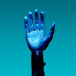
EMG also has important real-world applications. In rehabilitation, it helps clinicians monitor muscle recovery after injury. In ergonomics, it provides objective data about muscle strain in the workplace. In sports science, EMG helps optimize training and performance by identifying how and when muscles are activated. Increasingly, EMG is also used in human–machine interfaces, such as systems that allow people to control prosthetic limbs using their muscle signals. Its ability to detect fine-grained changes in muscle activity makes EMG a valuable tool across both research and applied settings.

Best practices for EMG data quality begin with careful electrode placement and attention to potential sources of noise. For sEMG and fEMG, electrodes should be placed over the “belly” of the target muscle, aligned with the muscle fibers, and spaced consistently to capture clean, interpretable signals. Gently cleaning the skin and, when appropriate, lightly abrading the surface helps reduce impedance and improve signal quality. In fEMG studies, precise and repeatable placement over well-defined facial muscles is critical, as these muscles are small and closely spaced. To minimize noise and artifacts, researchers should secure electrodes and cables to prevent movement, ask participants to limit unnecessary body motion, and ensure proper grounding of the recording system. Electrical interference from nearby equipment, power lines, or poor cable management can also contaminate EMG signals, so shielded cables and a controlled recording environment are recommended. Finally, synchronizing EMG with other physiological signals using a common acquisition system helps prevent timing errors and reduces the need for later corrections, resulting in cleaner data and more reliable conclusions.

hand mechanicsRecent studies demonstrate how EMG continues to expand into new and innovative areas. In a study published in Results in Engineering, a team of researchers from China developed a low-computational method for recognizing hand gestures from EMG signals to improve prosthetic device control. The researchers extracted key features from EMG recordings and used a lightweight machine-learning model, a multilayer perceptron (MLP), to classify gestures in real time. EMG data were collected using a four-channel BIOPAC data acquisition system running Biopac Student Lab (BSL) software. Surface Ag/AgCl electrodes were placed on four forearm muscles to capture relevant muscular activity. The study showed that accurate gesture recognition can be achieved without heavy computational requirements, making the system more practical for everyday use. This work highlights how EMG can support the development of more responsive, accessible assistive technologies.

Another study, conducted by a team of researchers from Mahidol University in Thailand and presented in the Institute of Electrical and Electronics Engineers (IEEE) proceedings, explored how combining EMG with motion sensors can improve ergonomic assessments. Focusing on occupational health, the study integrated surface EMG with data from an inertial measurement unit (IMU), a sensor that tracks movement and orientation. The researchers used the IMU to calculate Rapid Upper Limb Assessment (RULA) scores—a common method for evaluating postural risk—while EMG data collected via a BIOPAC four-channel research system provided insight into muscle activity. By combining these measures, the team found that posture alone did not always reflect actual muscle strain. The inclusion of EMG revealed additional muscle stress that might otherwise go unnoticed, suggesting that multimodal approaches can lead to more accurate workplace assessments and better injury-prevention strategies.

Researchers from Louisiana State University and the Pennington Biomedical Research Center in Baton Rouge, Louisiana, examined how training experience influences muscle activation and fatigue during resistance exercise performed with and without blood flow restriction (BFR) in young men. The study, published in Physiological Reports, compared trained and untrained participants while they completed knee extension exercises under low-, medium-, and high-load conditions. The team used sEMG to measure muscle excitation and total muscle activation in the vastus lateralis, with participants fitted with a portable BIOPAC BioNomadix Wireless EMG system. EMG signals were sampled at 2000 Hz and analyzed using a data acquisition system with AcqKnowledge software, allowing the researchers to quantify both peak muscle excitation and overall activation across repetitions. The results showed that low-load exercise with blood flow restriction produced lower peak muscle excitation but substantially greater total muscle activation compared to traditional high-load exercise, regardless of training status. These findings suggest that low-load BFR training may provide an effective alternative for stimulating muscle adaptations while reducing mechanical stress, with potential applications in rehabilitation and strength training programs.

Taken together, these examples illustrate the versatility and effectiveness of EMG in neuroscience, psychophysiological research, and beyond. Whether used to study emotion, improve physical training and workplace safety, or develop assistive technologies, EMG provides a direct link between muscle activity and human behavior. When combined with other physiological measures, it offers a more comprehensive understanding of how the brain and body interact. As research tools and techniques continue to advance, EMG will remain an essential method for exploring brain-body connections. For additional information on how to design studies around EMG, best practices for gathering EMG data, and how to integrate EMG with other signals, check out our webinar on EMG acquisition and analysis.


If EMG will be part of your next physiological or psychophysiological research project, reach out to our knowledgeable staff, who are standing by to help you with each stage of the planning and design process.

Boom! Pow! Clang! Exploring Stress Resilience with Startle Response and PPI

We have all felt that electrical jolt that comes with hearing a sudden, unexpected loud noise—the clang of a dropped cooking pan, the blast of a car horn, or the pop of a balloon. The reaction is instantaneous and nearly uncontrollable as muscles tense, eyes blink, pupils dilate, and one’s heart seems to leap from the chest. The startle response—a rapid, automatic reaction to sudden stimuli like loud noises—has a practical purpose, helping protect us from outside threats. It can also pose a danger if the response occurs while engaging in precision activities, such as driving a vehicle or operating machinery.

Within the psychophysiological field, the startle response can reveal how our brains and bodies process stress, attention, and emotional regulation. In pursuit of ways to mitigate its detrimental effects, researchers often study this reflex alongside prepulse inhibition (PPI). In this latter process, a weaker sound presented just before the startling noise reduces the strength of the response. To capture data when studying these phenomena, scientists use physiological signals such as electromyography (EMG) to measure the tiny muscle contractions around the eye during a blink, electrocardiography (ECG) for heart rate changes, electrodermal activity (EDA) for skin conductance, and respiration to track stress responses. In addition, researchers often pair these physiological measures with brain imaging techniques, like functional magnetic resonance imaging (fMRI) or electroencephalography (EEG), as well as cognitive tasks to build a complete picture of how startle responses connect with brain function and behavior.

Studies of startle response and PPI intersect with a wide range of fields, from neuroscience and psychiatry to aviation, defense, and occupational health. Clinical investigations of altered startle and PPI patterns are helping scientists uncover biomarkers of vulnerability or resilience. These studies are providing new insight into potential treatments for conditions like schizophrenia, PTSD, anxiety, and neurodegenerative disorders.

pilotIn applied contexts, like pilot training or high-stress professions, startle measures are used to understand how sudden stress impacts performance and decision-making. A study conducted by researchers at the University of Zagreb in Croatia with U.S. partners aimed to determine whether stress resilience markers, including startle response and PPI, could predict pilot performance under sudden startle events. Seventeen military pilot cadets completed baseline testing of physiological and cognitive features—including startle reflex habituation, PPI, heart rate variability (HRV), and multitasking ability—followed by flight simulator sessions with induced startle events. Physiological signals such as ECG, EMG (eye-blink startle reflex), respiration, and EDA were recorded via their respective BIOPAC amplifiers connected to a data acquisition and analysis system. Regression modeling showed that startle habituation, autonomic recovery, and cognitive task performance provided an accurate prediction of flight performance. The findings suggest that psychophysiological measures can help identify pilots who are more resistant to startle-induced performance degradation. Such insights could offer practical applications in pilot screening, training design, and aviation safety.

A team of Spanish researchers wanted to see how short-term (acute) stress affects both PPI and higher-level thinking (working memory) in healthy young adults. They used the Maastricht Acute Stress Test (MAST) to induce stress in one group, while another group did a non-stressful control task. After this, all participants heard loud noises that triggered the eye-blink startle response. In some trials, a prepulse immediately preceded the loud noise, which typically reduces the blink reflex. The startle (eye-blink) was measured from the orbicularis oculi muscle using BIOPAC Ag/AgCl electrodes connected to an Electromyography amplifier that fed data to a data acquisition system running AcqKnowledge software. This allowed the researchers to precisely record the intensity of a blink and the degree to which prepulse inhibited it. The team also tested working memory using tasks like digit span backwards and letter-number sequencing, in addition to other cognitive tests. They found that stress made startle responses stronger and reduced PPI, thereby degrading the participants’ ability to filter out the loud noise when it was preceded by a quieter one. Stress also impaired performance on working memory tasks compared to the non-stressed group. In practical terms, these results suggest that acute stress doesn’t just make one more reactive; it can interfere with basic sensory gating and cognitive control. This has implications for stressful jobs or situations (e.g., emergency responders or pilots) and possibly for understanding disorders where PPI or working memory is impaired.

These are just a few examples of how research into startle response and PPI can improve our understanding of the brain’s basic filtering and stress-response systems. The knowledge these studies provide may eventually translate into practical tools for diagnosis, prevention, and performance optimization. For additional information on research applications, check out our webinar on Startle and PPI.


Are you struggling to find the right tools to plan your startle response and PPI study? Our team of customer service experts will put you on the right path to gathering the best data possible for your research project.

Speak Up: Physiological Signals for Speech Research

In the symphony of human communication, speech is the principal instrument. Behind every uttered word lies a complex interplay of physiological and psychological signals, each telling a unique story about the speaker’s state of mind and physical well-being. The critical data provided by these signals has become a pivotal focus in both speech research and therapy, offering useful insights into the mechanisms of verbal communication and its disorders while enhancing speech ability and overall health.

Electromyography (EMG), a technique used to measure muscle activity, plays an invaluable role in speech research by revealing the interaction of facial muscles involved in articulation. By monitoring EMG signals from muscles such as the lips, tongue, and jaw, researchers can elucidate the precise movements and coordination required for speech production, shedding light on the underlying mechanisms of speech disorders like dysarthria or apraxia.

A French study published in the 2024 edition of Science Reports used EMG to establish the role tongue reflex plays in speech posture control. The presence of a tongue reflex in maintaining tongue posture, the proper positioning of the tongue in the mouth, has been a matter of debate among speech researchers. The study team employed an unorthodox recording technique that involved attaching EMG electrodes to the base of the tongue “to record activation of the anterior genioglossus, which is responsible for the control of the anterior part of the tongue.” A robotic arm was used to rapidly stretch the tongue while researchers measured muscular reflex EMG data during the formation of various vowel sounds, which were fed to a BIOPAC EMG amplifier for signal amplification and filtering. According to the research team, the study results “represent the first physiological demonstration in humans that the tongue is driven by reflex in response to a sudden stretch.”

Electrocardiography (ECG), which measures the electrical activity of the heart, offers insights into the intricate relationship between emotion and speech. Research has shown that changes in heart rate variability (HRV) correlate with emotional arousal and cognitive load during speech tasks. By integrating ECG data with speech analysis, researchers can explore the complex connection between emotional states and linguistic expression, paving the way for emotion-aware speech synthesis systems or interventions for social communication disorders.microphone

A 2023 study by researchers at Vanderbilt University used ECG to examine the relationship between respiratory sinus arrhythmia (RSA) on the persistence of stuttering and, specifically, the role emotional regulation plays in stuttering. Researchers measured potential differences in RSA in preschool-age children with persistent stuttering, children who recovered from stuttering, and children who do not stutter. RSA, an index of parasympathetic nervous system activity, was measured during the viewing of both neutral (baseline) and emotionally stimulating video clips and during subsequent speaking tasks. To obtain RSA for each study participant, ECG data was gathered via a BIOPAC data collection and analysis system running AcqKnowledge. A low RSA baseline is often associated with difficulty regulating emotion and, according to the study’s main hypothesis, plays a role in speech disorders like stuttering. The study’s findings supported this hypothesis, with some caveats, but the overall result demonstrated that emotional regulation could provide a promising avenue for speech therapy.

Electroencephalography (EEG), a noninvasive method for recording brain activity, offers insight into neural underpinnings of speech processing and comprehension. By analyzing EEG signals, researchers can map the spatiotemporal dynamics of neural networks involved in language processing, from phonological encoding to semantic integration. EEG-based studies offer insights into the neural signatures of speech disorders like aphasia or developmental language disorders, informing targeted therapeutic interventions and neurorehabilitation strategies. EEG has presented challenges with regard to artifacts created by participant movement during speech, but recent studies have shown promise in EEG as a speech research tool.

This is just an introduction to the signals that are being used to provide researchers with a better understanding of speech and speech pathology. For additional information on how BIOPAC products are being integrated into the study of speech, see our application note on Speech Motor Control.


Are you planning a research project focusing on speech? Our regional sales representatives are ready to help you find the right tools for your study.

2023 in Review: The Best of Research Using BIOPAC

As another year winds to a close, it is a holiday tradition to look back on the last 12 months and take stock. ‘Tis the season of “top 10” lists, “best of” articles, and “year in review” segments in the media. So, not to be left out, we here at BIOPAC have been assessing 2023 from our own unique vantage point. We have put together a brief list of some of our favorite research studies from the year that relied on BIOPAC products to gather and analyze data. We have nervous passengers in driverless cars, killer clowns, and individuals giving us conspicuous side-eye, all for your reading pleasure.

Backseat Drivers

While the advent of self-driving vehicles offers considerable potential for increasing the convenience of transportation and travel, this developing technology is not without controversy. Both passengers and those who would potentially share the road with autonomous vehicles worry about safety issues. A recent study funded by the French National Research Agency examined how passengers in self-driving vehicles reacted when navigating shared-space areas with large concentrations of pedestrians. The study defined shared spaces as “areas where all forms of segregation between pedestrians and vehicles (e.g., road markings, traffic signals, signs, curbs, and barriers) are minimized or eliminated.” Study participants were seated in front of a computer-based driving simulator. EDA data was gathered via a BioNomadix wireless  EDA transmitter and amplifier connected to a BIOPAC data acquisition and analysis system. Data was gathered across a variety of variables including changes in vehicle speed, concentrations of pedestrians, and proximity. Among the study’s conclusions was the suggestion that cognitive processes involved in collision risk assessments may lead to physiological adaptations that allow passengers to cope with stress related to vehicle-pedestrian interactions in shared spaces.

Send in the (Demonic) Clowns

VR HMDAnother study that caught our attention in 2023 examined how stressors created in a virtual reality (VR) environment are manifested in measurable physiological stress reactions. VR has seen a growth of applications beyond entertainment in recent years. It is being used for a wide range of applications such as training in simulated environments where it is necessary to create stressful conditions that mimic real-world experiences. A team of German researchers examined a variety of physiological signals, including electroencephalography (EEG), electrodermal activity (EDA), and peripheral skin temperature (ST) to determine which are most reliable for accurately measuring the stress levels of participants while they experienced cinematic horror scenarios in a VR environment. Study participants wore a VR headset that displayed four-minute episodic horror movies featuring demonic clowns. EEG data was gathered with an SS2L electrode lead set and general-purpose electrodes connected to the left temporal lobe. ECG recording was conducted with a second lead using an SS2L electrode lead set. The research team used an SS3LA EDA transducer to collect EDA data. All data was fed to an MP36 system running Biopac Student Lab (BSL) software. The study successfully demonstrated that stress can be triggered in VR and leads to a physiological and measurable stress reaction.

Are You Looking at Me?

The third study that sparked our interest in 2023 looked at how individuals react to the gaze of another person. A team of researchers from Finland and China used facial electromyography (EMG), skin conductance response (SCR), and heart rate (HR) to measure affective and attentional responses to faces with a direct and averted gaze. The study sought to measure both vicarious responses to watching two individuals who were looking at each other as well as individuals looking directly at the participant. SCR was recorded using a BIOPAC EDA100C amplifier connected to an MP data acquisition system. The participant’s facial muscle activity was measured and amplified with an EMG100C amplifier. The electrocardiogram (ECG) signal was measured with electrodes placed on the left and right inner forearm and amplified with an ECG100C amplifier. While the study did not find significant vicarious eye contact response, it did measure considerable affective response from direct gaze from one or more individuals. The study’s results could provide a better understanding of the role physiology plays in non-verbal communication between individuals and groups.

This is just a small sampling of the many studies that relied on BIOPAC equipment to gather and analyze data in 2023. For more ideas on how BIOPAC is leading the way in physiology, psychophysiology, neurology, and other areas of research, check out our webinars or watch one of our many online training videos. See you in 2024!


Start 2024 right by leveraging BIOPAC products in your next research study. Reach out to your regional sales representative to learn more.

Mind and Body: Signals to Enhance Your Psychophysiology Research

The human body and mind share an intrinsic link, both influencing and reflecting the health and general well-being of the other. Within the field of psychophysiology, researchers gain a wealth of insight into mental processes and disorders by studying physiological signals recorded from both the brain and body. Signals such as electrodermal activity (EDA), skin temperature (SKT), electroencephalogram (EEG), heart rate variability (HRV), and functional near-infrared spectroscopy (fNIRS) provide just a few of the tools helping us to better understand these mind-body interactions.

A wide range of research technologies has made these signals increasingly accessible in the lab as well as in everyday environments, allowing studies to place participants in increasingly naturalistic settings while gathering critical physiological data. In this way, researchers can provide a more accurate picture of how study participants react to stress and arousal, experience emotion, or interact within group environments.

One recent study used EDA and heart rate (HR) to examine the role of emotion in student success within computer-based learning environments (CBLE). As discussed in previous posts, a range of signals can be used to measure the body’s physiological response to emotion, otherwise known as “affect.” By gaining a better understanding of learners’ emotional responses to CBLE stimuli, researchers in this study hope to apply their data to improvements in CBLE design. EDA data was gathered from participants via SS57LA EDA leads while HR signals were collected with SS2LB shielded lead sets, both of which were connected to a BSL MP36 data acquisition and analysis system. Participants were shown imagery designed to evoke a negative emotional reaction before being asked to read a complex academic paper. The study was able to establish a link between the participants’ emotions and their comprehension of the reading material.

head with brain exposedIn another study, researchers used EDA and skin conductance response (SCR) to measure participants’ reactions to conditioned stimuli (CS) and unconditioned stimuli (US). The goal was to determine if fear-inducing memories could be modified either pharmacologically or through behavior modification to overcome fear. An MP36R System with AcqKnowledge software allowed analysis of SCR signals gathered via SS3LA finger transducers across a range of experiments designed to trigger fear-inducing memories. While the overall results were inconclusive, the SCR signals provided valuable feedback regarding the participants’ response to stimuli.

While EDA is a common thread through many of these psychophysiology studies, other signals provide researchers with a more complete and nuanced data set from which to draw their conclusions. Researchers exploring the factors influencing musical preferences combined EDA data with ECG and facial electromyography (fEMG) signals to assess how listeners reacted to familiar and unfamiliar musical stimuli. Data from all three signals were fed to an MP36 data acquisition and analysis system. The study found a strong correlation between musical preferences and familiarity with material as reflected by the subjects’ emotional responses.

These examples only scratch the surface of how psychophysiological research is being aided by an array of signals, all made accessible with available technology. To learn more about how to implement these signals in your next study, see BIOPAC’s Psychophysiology Applications webpage.


Contact your BIOPAC sales representative to learn more about the wide range of data-gathering tools available to help you construct your next psychophysiology study.

error: Content is protected !!
