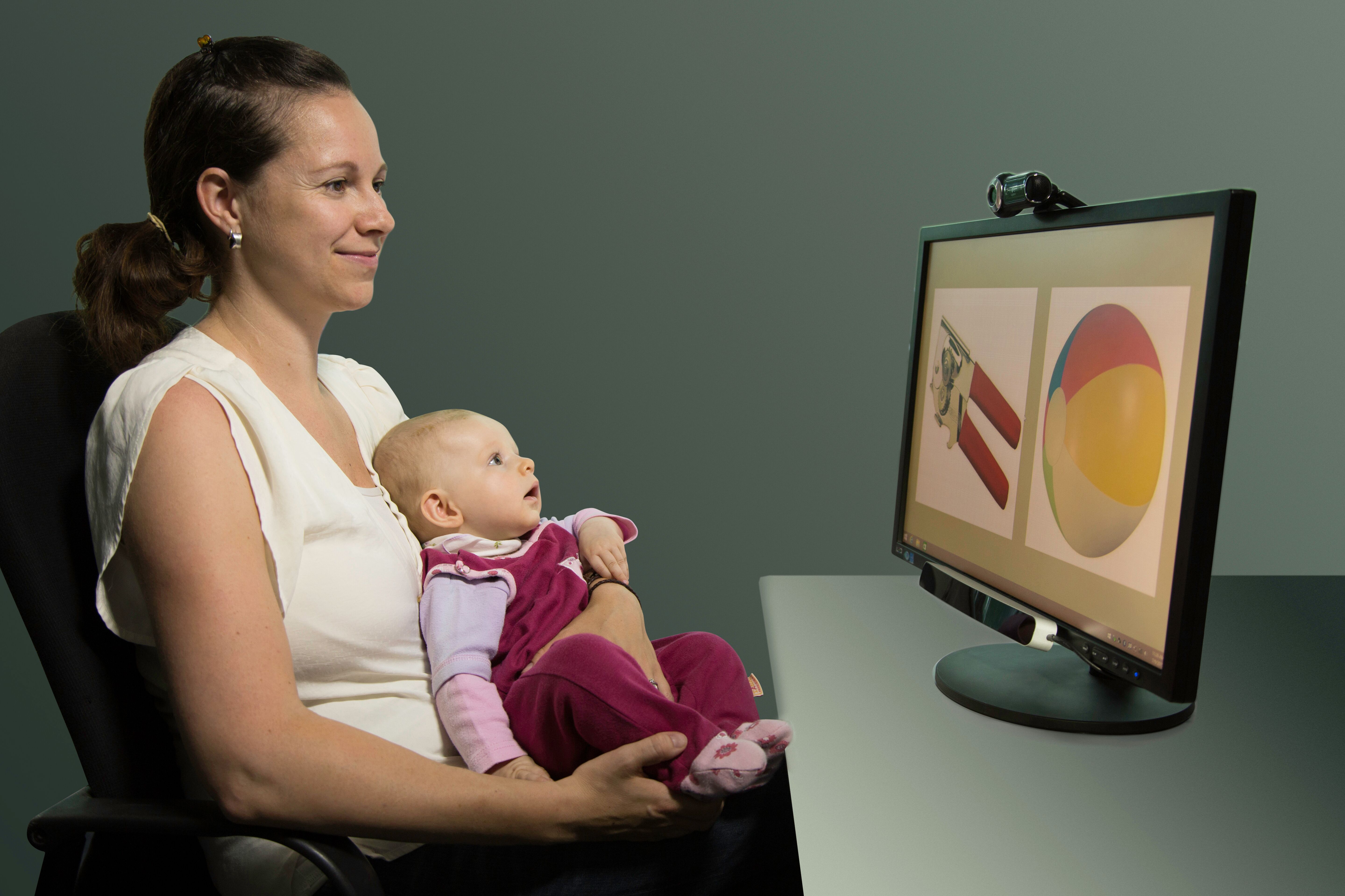
Eye-Catching: Mobile Tech Enhances Immersive Experiments

Freed from the limitations of stationary eye tracking technology, mobile eye tracking enables researchers to study their subjects while they perform a wide range of immersive tasks in natural settings.

Sixteenth-century pediatrician Thomas Phaer mused that “the eyes are the windows to the soul.” These sensory organs and their movements certainly provide a wealth of information to researchers studying the interaction of mind and body. Eye tracking has long been a key tool of physiological and psychophysiological research, and recent developments in mobile eye tracking take this valuable data resource out of the confines of the lab and into the real world.

Freed from the limitations of stationary eye tracking technology, mobile eye tracking enables researchers to study participants while they perform a wide range of immersive tasks in natural settings. These self-contained devices come in a variety of form factors including wearable glasses that provide untethered eye tracking data acquisition in real time that can be combined with other physiological data collected during live experiments.

Today’s lightweight mobile devices can record a broad spectrum of eye tracking data, including pupil size and position, gaze path, vergence angle, and areas of interest (AOI). The latter connects stimuli to eye-movement metrics, providing data on the participant’s psychophysiological response to stimuli within their field of vision. In previous blog posts and webinars, we have explored how mobile eye tracking technology gathers this data. Numerous studies have been published that demonstrate its potential in physiological and psychophysiological research. Settings and applications include user experience, sports and physical therapy, public safety and law enforcement, driving and equipment operation, and work environments from the office to the factory floor.

Mobile eye tracking technology allowed researchers at The University of Sheffield to take their experiments to the city streets for a 2020 study into how different lighting affects cyclists navigating urban environments at night. Mobile eye tracking provided data on the gaze behavior of nocturnal cyclists in parallel with skin conductance response (SCR) to better understand how obstacle detection is influenced by variations in type, location, and level of lighting. Bicycles and their riders were equipped with a BIOPAC data acquisition unit and audio stimulation units along with a BioNomadix EDA transmitter and electrodes to record the participants’ SCR. Eye tracking data, including AOI, were synchronized with SCR data and analyzed using AcqKnowledge software to determine how the riders reacted to a variety of lighting conditions. Researchers concluded that lighting plays a significant role in riders’ comfort levels at night and that improved lighting could increase urban bicycle use.

VGM playerLess intrusive by design, mobile eye tracking can provide indoor experiments with a heightened degree of immersion. A study at the University of British Columbia used mobile eye tracking glasses to create a more natural environment to investigate how the immersive effects of electronic gaming machines (EGM) impact gambling addiction. The study looked at how machine design triggered psychophysiological responses in participants by assigning various AOI to EGM screens. ECG data were recorded along with respiration effort via a BIOPAC respiration effort transducer and amplifier, which were fed to a data acquisition unit and synchronized with AOI data recorded via mobile eye tracking glasses. The study was able to successfully identify several key markers for game immersion using AOI that suggest behavioral links between immersion and gambling addiction.

Possible applications for mobile eye tracking in research continue to expand with the evolution of the technology. To learn more about recent developments in mobile eye tracking, check out our latest webinar on mobile eye tracking glasses with AcqKnowledge integration.


Learn more about how to integrate mobile eye tracking into your next research project by contacting your local BIOPAC sales representative.

Mind and Body: Signals to Enhance Your Psychophysiology Research

The human body and mind share an intrinsic link, both influencing and reflecting the health and general well-being of the other. Within the field of psychophysiology, researchers gain a wealth of insight into mental processes and disorders by studying physiological signals recorded from both the brain and body. Signals such as electrodermal activity (EDA), skin temperature (SKT), electroencephalogram (EEG), heart rate variability (HRV), and functional near-infrared spectroscopy (fNIRS) provide just a few of the tools helping us to better understand these mind-body interactions.

A wide range of research technologies has made these signals increasingly accessible in the lab as well as in everyday environments, allowing studies to place participants in increasingly naturalistic settings while gathering critical physiological data. In this way, researchers can provide a more accurate picture of how study participants react to stress and arousal, experience emotion, or interact within group environments.

One recent study used EDA and heart rate (HR) to examine the role of emotion in student success within computer-based learning environments (CBLE). As discussed in previous posts, a range of signals can be used to measure the body’s physiological response to emotion, otherwise known as “affect.” By gaining a better understanding of learners’ emotional responses to CBLE stimuli, researchers in this study hope to apply their data to improvements in CBLE design. EDA data was gathered from participants via SS57LA EDA leads while HR signals were collected with SS2LB shielded lead sets, both of which were connected to a BSL MP36 data acquisition and analysis system. Participants were shown imagery designed to evoke a negative emotional reaction before being asked to read a complex academic paper. The study was able to establish a link between the participants’ emotions and their comprehension of the reading material.

head with brain exposedIn another study, researchers used EDA and skin conductance response (SCR) to measure participants’ reactions to conditioned stimuli (CS) and unconditioned stimuli (US). The goal was to determine if fear-inducing memories could be modified either pharmacologically or through behavior modification to overcome fear. An MP36R System with AcqKnowledge software allowed analysis of SCR signals gathered via SS3LA finger transducers across a range of experiments designed to trigger fear-inducing memories. While the overall results were inconclusive, the SCR signals provided valuable feedback regarding the participants’ response to stimuli.

While EDA is a common thread through many of these psychophysiology studies, other signals provide researchers with a more complete and nuanced data set from which to draw their conclusions. Researchers exploring the factors influencing musical preferences combined EDA data with ECG and facial electromyography (fEMG) signals to assess how listeners reacted to familiar and unfamiliar musical stimuli. Data from all three signals were fed to an MP36 data acquisition and analysis system. The study found a strong correlation between musical preferences and familiarity with material as reflected by the subjects’ emotional responses.

These examples only scratch the surface of how psychophysiological research is being aided by an array of signals, all made accessible with available technology. To learn more about how to implement these signals in your next study, see BIOPAC’s Psychophysiology Applications webpage.


Contact your BIOPAC sales representative to learn more about the wide range of data-gathering tools available to help you construct your next psychophysiology study.

The Year in Review: Top Physiology and Neuroscience Research Stories 2022

Another calendar year has come and gone. COVID-19 remains a reality, but we continue to adapt to the ongoing pandemic and find new ways to overcome the challenges it poses at work, at school, at home, and in the lab. And speaking of research, 2022 proved to be a banner year for innovation and breakthroughs in the sciences, particularly in the physiological and neurological fields. Here is a quick round-up of some of the research stories that caught our attention.

Brain Implant Allows Locked-in Patient to Communicate

A man in his late 30s has been given the ability to communicate for the first time since being left in a completely locked-in state by amyotrophic lateral sclerosis (ALS). The patient, who was unable to move or even open his eyes, can communicate again thanks to a brain-computer interface (BCI) comprising two microelectrode arrays implanted in his motor cortex. The procedure was carried out by a team of researchers at the University of Tübingen in Germany. “This study answers a long-standing question about whether people with complete locked-in syndrome (CLIS) – who have lost all voluntary muscle control, including movement of the eyes or mouth – also lose the ability of their brain to generate commands for communication,” wrote Dr. Jonas Zimmermann, one of the co-authors of the study resulting from the procedure.

First Scan of Dying Human Brain Provides Insight into Near-death Experience

near-death experienceAn EEG recording from a dying 87-year-old patient has provided researchers with the first glimpse of the neurophysiological phenomena that occur shortly after cardiac arrest. While survivors of so-called near-death experiences have anecdotally reported intense and surreal experiences that include panoramic memory review, out-of-body experiences, and other hallucinatory phenomena, little is understood about what causes such occurrences. While experiments have been carried out on animal subjects showing a hypoactive state of brain activity under such conditions, this new data provided researchers with the first evidence “that the human brain may possess the capability to generate coordinated activity during the near-death period.”

Biomembrane Research May Advance Understanding of Memory in Computers and Humans

Drawing inspiration from the living realm, scientists at the U.S. Department of Energy’s Oak Ridge National Lab are applying biological concepts to next-generation computers with implications for both human health and cutting-edge computing. Researchers were able to demonstrate that an artificial cell membrane could imitate one of the hallmarks of biological memory and learning—long-term potentiation (LTP). The study, published in the Proceedings of the National Academy of Sciences, shows for the first time that a cell membrane by itself, without the benefit of proteins or other embedded biomolecules, can carry out LPT over several hours. The pure lipid membrane, which was formed using lipid-covered water droplets, represents the first nanoscale structure in which memory can be encoded. The discovery opens the door to neuromorphic computing, which imitates the way the human brain stores and processes information and is more efficient than traditional computing.

This is just a sampling of the exciting developments catching our attention in the sciences this year. For more explorations of the intersection of cutting-edge technology, physiology, and neuroscience, join BIOPAC at the T4 Human Physiology Conference in summer 2023. And have a happy New Year!


To learn more about BIOPAC’s role in exciting breakthroughs in neuroscience and physiology, and how we can assist you in achieving your research and teaching goals, contact your local sales representative today.

Human Factors: Physiological Signals Enhance Ergonomics Research

Ergonomics plays a role in almost every aspect of human interaction with the world around us—how we work, entertain ourselves, rest, and relax. Simply put, ergonomics (sometimes termed “human factors and ergonomics” or simply “human factors”) refers to the way in which physiological and psychological principles are applied to design, particularly in ways that benefit a user’s physical and psychological well-being and productivity.

In practice, ergonomics addresses more than our physical interactions but also encompasses our mental, emotional, and interpersonal states. What unites these different applications of ergonomic theory is that they can be studied and better understood with many of the same tools and signals applied in other areas of physiological and psychophysiological research. These can include ECG, EOG, fNIRS, EEG, and EMG, to name just a few.

Ergonomics can be broken down into three primary domains. Physical ergonomics relates to how the body interacts with the physical environment, from the design of tools we use every day to the physical environments in which we live and work. Cognitive ergonomics focuses on how our mental abilities are influenced by the systems and tasks with which we are presented. Organizational ergonomics (also known as macroergonomics) explores how to optimize organizational structures, policies, and processes within various social settings, including work environments.

Eye TrackingOne example of how physiological signals are being applied in physical ergonomics research involves the application of electrooculogram (EOG) signals to analyze eye movements, particularly saccades, as a measurement of eye fatigue caused by exposure to digital screens. The ubiquity of such screens in both our work and recreational environments makes such research critical to maintaining user well-being and productivity. This 2018 study utilized EOG electrodes connected via SS2LB leads to an MP36 data acquisition unit running BSL software to record and analyze EOG signals. Researchers were able to determine the optimal reading and viewing distance to minimize eye fatigue and discomfort, improving user health and productivity.

Cognitive ergonomics come into play when discussing the ease-of-use for a product. This is particularly important when dealing with how less technologically savvy groups interact with new technology, a challenge is explored in a 2019 study into how to develop a home heart monitor that would be easy for older users to operate. The study compared user interaction with device hardware, a mobile app, and a cloud-based software platform while using a BIOPAC research-level system as a control. Researchers created the signal baseline with an ECG amplifier connected to a data acquisition and analysis unit, comparing these results to the quality of the signal the user was able to obtain using the self-administered home devices and applications. This approach accurately assesses both the user-friendliness and quality of data obtained by several telemedicine systems.

These examples offer just a small sample of the ways in which physiological signals are being applied in the study of ergonomics. For an in-depth exploration of the topic, watch our webinar series on Ergonomics and Fatigue.


To learn more about using BIOPAC hardware and software to enhance your ergonomics study or design research, contact your local sales representative.

Evoked Potentials: Low-amplitude Signals for High-value Data

Researchers studying the nervous system have a host of physiological signals at their disposal to provide critical data on neurological function. Various signals present advantages and disadvantages, depending on the demands of the research being conducted. Evoked potentials offer a particularly effective means of measuring the nervous system’s response to certain stimuli, notably visual and auditory cues.

Evoked potentials are employed in both research and clinical settings to measure electrical activity in the central nervous system. Stimulation of specific sensory neural pathways produces this electrical activity, allowing researchers to see the speed and thoroughness with which these signals reach the brain. Evoked potentials provide researchers with heightened precision when isolating and localizing a participant’s response to stimuli.

Because evoked potential amplitudes tend to be low, from less than one to just a few microvolts, these signals can be difficult to differentiate from other higher amplitude signals and background noise. This is resolved by using signal averaging to separate the low-amplitude signals from the noise. Multiple readings are taken with evoked potential signals time-locked to the stimulus. Averaging these measurements allows the evoked potential to be filtered from the random signal noise.

Along with providing useful data for research, evoked potential tests are commonly used in clinical settings to diagnose and explore treatments for a range of disorders such as multiple sclerosis (MS), epilepsy, glaucoma, neuropathies, Alzheimer’s, and others. In both research and clinical applications, evoked potential tests provide several advantages over using other approaches since they are noninvasive, painless, and provide results more quickly than other neurological scans, such as MRI. Types of evoked potentials can be categorized by the stimuli with which they are associated. Visual evoked potentials (VEP) are triggered by visual stimuli such as light flashes or by participants viewing certain patterns. Auditory evoked potentials (AEP) measure responses to sound stimuli such as clicks. Somatosensory evoked potentials (SEP) are triggered by electrical stimulation of peripheral nerves.

A 2022 study by researchers at the Autonomous University of Aguascalientes in Mexico evaluated the effectiveness of using VEP waveforms embedded in electroencephalographic (EEG) signals to determine the health of the optic nerve. The team employed an MP36 data acquisition system connected to an SS2LB lead set to record EEG signals from study participants. eyeThe signal data were used to create a database of VEPs that could be processed with analysis software for the early detection of glaucoma and other optic nerve-related illnesses.

A similar study combined the use of EEG signals with VEP to examine how participants recognize emotion in others. Researchers from two universities in China relied on a variation of VEP known as steady-state visual evoked potential (SSVEP), which exhibits distinctive frequency and phase properties. EEG signals were recorded via a BIOPAC data acquisition unit while participants were shown flickering images representing three emotional states—delight, sadness, and anger. The study found a positive correlation between SSVEP amplitude, alpha-frequency entrainment, and accurate identification of emotion.

These are just a few examples of how evoked potentials provide researchers with valuable insight into the central nervous system’s response to stimuli and its relationship to a wide range of physiological states. For additional information on how to use BIOPAC hardware and AcqKnowledge to stimulate a range of senses using electrical, auditory, visual, haptic, scent, and thermal stimuli and record evoked potentials, check out our webinar on Exogenous and Stimulus-related Evoked Potentials.


To learn more about how your study can benefit from using BIOPAC hardware and software to record and analyze evoked potential data, contact your local sales representative.

Workflow: Research Smarter with Automated Routines

You have probably heard the saying “why work harder when you can work smarter?” While this may seem like a cliché, there is no denying that smart work, or what might be more accurately termed “efficient effort,” not only alleviates worker stress but in doing so minimizes human error while creating opportunities for increased productivity and creativity.

In an earlier post on lab automation, we discussed how increased workloads and human error have contributed to a crisis of reproducibility and repeatability in the research community. To summarize, repeatability and reproducibility provide the foundation upon which the scientific method is built. The results of academic research only retain value for the wider scientific community when the conditions that produce such results can be repeated and shared amongst other researchers.

There are several best practices that can contribute to ensuring repeatability and reproducibility of research by maintaining the conditions under which lab work is conducted. These include, among other factors, consistency in the location or research environments, the tools used for measurement as well as other equipment used in experiments, and the routines and processes used to gather and analyze data.

As discussed in the previous blog post, automation of data gathering and analysis has been one of the best ways to ensure consistency and repeatability while helping to eliminate human error, as outlined in a 2019 article on improving reproducibility in synthetic biology through automation:

“The successful implementation of automation technology can help to improve both the throughput and reproducibility of experiments, although the high cost and lack of flexibility of traditional lab automation has so far hindered their wide-spread adoption in academia. Recently though, there have been advances in automation technology that, when combined with new protocol sharing methods and protocol management systems, may allow for researchers to gain the benefits of automation.”

—Jessop-Fabre and Sonnenschein

Before when discussing how to automate routines for data acquisition and analysis, the focus has been on the use of scripts to string together processes. While this achieves the goal of creating laboratory conditions that are more readily repeatable, scripting requires a specific skill set. This brings us back to the notion of working smarter, not harder.Plan Smart

Previous versions of BIOPAC’s own AcqKnowledge data gathering and analysis software offered a powerful Scripting tool as an add-on licensed feature. The latest version of AcqKnowledge supports a new licensed feature with Scripting called Workflow. Workflow takes all the functionality of scripts and places it in an easy-to-use, drag-and-drop interface that requires no prior scripting knowledge.

Workflow is the embodiment of the “work smarter, not harder” philosophy, allowing users to create automated routines to process and analyze data under consistent and repeatable conditions. By speeding signal conditioning and preparation for analysis, Workflow boosts efficiency. The ability to save and share workflows makes it easier for colleagues and reviewers to replicate research conditions. BIOPAC CEO, Frazer Findlay hosted a recent webinar in which he provided a detailed introduction to creating automated routines with Workflow.


For additional information on the latest version of BIOPAC’s AcqKnowledge software as well as other research tools and how they can help streamline your lab work, contact your local sales representative.

Biopac Student Lab | What I Learned This Summer

A Student’s Perspective

Guest post by Ashley F.

Like many of my university peers, I wrapped up my freshman year of college working a summer internship in hopes of gaining valuable professional experience. Rather than relaxing on the beach, I occupied my time between June and August with electronic breadboards and ice bucket plunges. All this took place at BIOPAC where I was tasked with reviewing the BSL PRO Lessons, helping the company continue to refine and improve these teaching aids.

Let me start by confessing that I am not a physiology or anatomy major, but rather a development and psychology major. Given this difference, I did not expect to find that so many of the BSL PRO Lessons connect to what I am studying. BSL PRO Lessons stand out compared to your average textbook for multiple reasons. For me, the primary difference is that with BSL PRO Lessons, you get to watch the data take shape in real-time. And it’s not just random data, but data from your own body—your heart beating, your finger twitching, your eyes tracking a pendulum. Because the data is from you and not a textbook, the lessons are more interesting to analyze.

Throughout the summer, I worked through each “human” BSL Lesson, gaining experience with the software, equipment, and workspace. Each lesson includes a ‘background’ section that provides context to the lesson and connects each one to different concepts. This context makes the data from each lesson relevant to more disciplinary studies than just physiology and anatomy, which in turn broadens the student’s knowledge. Because BIOPAC wanted to test the materials under realistic student lab conditions, I wasn’t given much guidance on how to approach individual lessons. Rather, I would simply read the manual and follow the instructions, which turned out to be surprisingly easy as I figured out how to do most of it on my own.

With a total of 39 BSL PRO lessons, you would expect at least a few to be quite similar. And yet, BIOPAC understood the importance of unique lessons and ensured that each one had a different background section, used different equipment, and covered a different topic.

woman submerging head in cold waterEach lesson includes fun concepts to keep you engaged, like dunking your head in cold water to see how your heart rate changes in Lesson H08: Dive Reflex, using equipment to make your finger twitch in Lesson H06: Finger Twitch, or testing and timing your reflexes in H16: Reflexes & Reaction Time.

While each lesson is unique and covers a different topic in physiology and anatomy, the lessons do follow a clear progression as you move from BSL PRO Lesson 1 to 40. They start shorter, utilizing more basic concepts and equipment. Then, as you progress through the lessons, you begin to work through increasingly complex physiological concepts until you end on a lesson involving the use of a breadboard. This last lesson involves nine separate labs, each as involving as the earlier individual lessons. This amount of work may sound daunting at first, but the skills picked up while completing the previous lessons leave you well-prepared to complete the final lesson and analyze clear data.

Working through BSL PRO Lessons is a unique and interactive experience that allows students to learn, connect, and analyze personalized data from a broad range of physiological and anatomy concepts, leaving you with a well-rounded and contextualized education experience.


Learn more about BIOPAC’s education solutions, including complete teaching systems for a variety of life science disciplines. BIOPAC also provides a wide range of research systems for recording, displaying, classifying, and analyzing physiological signals & measurements.

BIOPAC Systems, Inc. helps life science researchers and educators
measure physiology anywhere, any time, from any subject.
Visit us at www.biopac.com.

Skin Deep: Using EDA to Measure Affective Response

We say something “gets under our skin” when it elicits an unpleasant or irritating emotional response. This connection between our skin and emotion runs deeper than a simple idiomatic expression, however. Along with performing several critical functions—protective, regulatory, and sensory—our skin provides a window into our physiological and emotional states as we respond to a range of stimuli. Electrodermal Activity (EDA) is the mechanism providing these insights.

EDA, also known as galvanic skin response (GSR), measures eccrine activity resulting from the sympathetic nervous system’s response to environmental stimulation. As a function of the autonomic nervous system, it is an involuntary response to a range of sensory inputs, including touch, sight, sound, odor, and taste. EDA regulates the rate of sweat produced by sweat glands in the extremities, particularly in the fingertips, palms, and soles of the feet.

EDA increases in conjunction with the level of stress or nervousness experienced by an individual. For this reason, it provides researchers with a valuable means of measuring affective response to external stimuli. As we explored in a previous post, affect is the measurable physiological expression of emotion. While there are numerous physiological signals that researchers can employ to measure affect, EDA in particular functions as an indicator of arousal. For this reason, EDA has become one of the most relied upon signals employed by researchers exploring psychophysiological phenomena.

Violence StudyA recent Slovakian study published in Adaptive Human Behavior and Physiology illustrates how EDA is commonly employed in such research. Researchers studying the influence of gender differences on reaction to violent imagery used skin conductivity response (SCR), the mechanism behind EDA, to measure levels of arousal when participants were shown images of war and sports. The goal of the study was to test the “male warrior hypothesis” (MWH). The theory suggests that men have evolved psychological mechanisms to form aggressive alliances with the goal of acquiring or protecting reproductive resources in war and that team sports stimulate that same impulse. EDA was measured using BIOPAC electrodes and leads feeding data to an MP36 data acquisition unit. While the study found that visual perception of team sports and war imagery stimulated different levels of excitement as measured by EDA in men and women, it was inconclusive in showing that sports provide a substitute for war in the sample of participants.

A study conducted at the University of Ottawa and published in the Journal of Behavior Therapy and Experimental Psychiatry used EDA to examine whether response-focused emotion regulation (RF-ER) strategies provided an effective way to control evoked emotions—specifically, does smiling make one feel better in times of heightened stress? EDA data was gathered from participants using a BIOPAC skin conductance transducer and amplifier connected to a BIOPAC data acquisition unit while participants were shown images designed to evoke both arousal and negative responses. Participants were asked to either suppress their emotional response to images or employ expressive dissonance (e.g., smile when disturbed). The results of the study shed doubt on previously held assumptions that RF-ER strategies were effective in controlling emotion.

These are just a sampling of the ways in which EDA is being used in psychophysiological research. For additional information on the background, tools, and techniques for using EDA in your studies, visit our EDA Resource Directory, read our EDA Introductory Guide. and watch Electrodermal Activity: Intro to Recording and Analysis or any of our EDA webinars.


For additional information on how BIOPAC can assist you with your research using EDA and a range of other physiological signals, contact your local sales representative.

Guilt Trips: Taking Measure of Our Moral Compass

“So full of artless jealousy is guilt, It spills itself in fearing to be spilt”
—Gertrude, William Shakespeare’s “Hamlet”

In Act 4 of Shakespeare’s famous tragedy, Hamlet’s mother Gertrude points out that the harder one tries to conceal guilt, the easier it is to expose. Little does she realize that she may have hit upon how future researchers approach the study of guilt in the lab. For practical purposes, it is a primary motivator in moral action and impacts how an individual responds to certain stimuli.

Psychologists have developed a myriad of tests to gauge guilt in a clinical setting such as the Test of Self-Conscious Affect (TOSCA), a multiple-choice test that tracks responses to various moral scenarios. While such techniques have been useful in linking guilt with conditions such as depression and anxiety, they have also shown their limitations in providing a deeper understanding of guilt’s influence on behavior, development, and psychological health.

In a recent BIOPAC webinar, UCSB professor Dr. Hongbo Yu addressed these limitations. Scenario-based tasks, he points out, are non-committal in examining the causes of guilt and do a poor job of measuring behavioral tendencies linked to guilt. Interaction-based research methods, however, allow participants to experience emotions in real-time, allow direct control of the causes of guilt in the lab, and allow for direct measurement of responses to stimuli.

As discussed in prior posts on affect, the body’s physiological manifestation of emotion, there is a range of methods and physiological signals available to researchers for measuring affect and gaining a better understanding of emotional states like guilt. Dr. Yu utilized eye-tracking, electrodermal activity (EDA), and functional magnetic resonance imaging (fMRI) in his examples. These are just a few of the signals currently being used in the lab to measure guilt.

One of the practical applications of guilt research is detecting when a subject is concealing the truth, as one would with a polygraph test. A recent study conducted by researchers at the University of Wisconsin-Madison’s Department of Psychology sought to gauge deception among research participants taking the Concealed Information Test (CIT). The goal was to improve upon traditional polygraph test signals such as heart rate and respiratory rate. Respiration data was gathered using a respiration transducer belt while ECG signals were collected with BIOPAC electrodes. In addition, researchers gathered EDA data using a transducer and finger electrodes. The study found that thGuilty Girle EDA signal was significantly more effective in accurately measuring physiological responses during the test procedure.

A 2018 study by researchers at Liverpool Hope University looked at the link between guilt and aggression in children by examining activity in the autonomic nervous system during so-called “bad behavior.” The study measured respiratory sinus arrhythmia (RSA) and skin conductance in children 4–8 years old while they imagined engaging in antisocial activities. EDA and ECG signals were gathered via wireless amplifiers and fed to an MP150 data acquisition unit (superseded by the MP160) running AcqKnowledge software. The study found a linkage between physiological responses to aggression and guilt that may provide a better understanding of the mechanisms behind guilt and related behavior in early and middle childhood.

These are just a few examples of how physiological signals can be applied to measuring guilt in research. BIOPAC offers a wide range of data acquisition, stimulus presentation, and VR options suitable for creating custom guilt research environments in the lab.


For additional information, contact your BIOPAC representative to learn more about the range of signals that can be utilized to gather data on guilt and other emotional states in the lab.

Baby Blues: Neonatal, Infant, and Toddler Research Challenges

The first five years of a child’s life are marked by considerable change—both physiological and psychological. Young children undergo incremental development in their ability to think and solve problems, their emotions, and their linguistic abilities, to name just a few developmental changes. Over time, the rapidity and extent of this development vary by individual, even among children of the same age. For these reasons, research techniques and tools must be adopted that are suitable for the participant’s age group. In this constantly evolving segment of the physiological and psychophysiological field, new approaches and technologies are being implemented with the needs of our youngest participants in mind.

Different Needs, New Approaches

A recent article published by researchers at the University of California Irvine outlined some of the challenges in conducting research within the neonatal to toddler age groups. Chief among these was the higher levels of attrition among these groups compared to older test subjects. Much of this can be linked to the general characteristics of the test group—reduced ability to sit still for long periods of time, shorter attention span, and lack of motivation to participate. While these factors may not surprise an experienced parent or childcare provider, they can throw a wrench into any study that has not taken such challenges into account.

Tools for Toddlers

Choosing the right tools and applying them appropriately in the lab setting can not only significantly improve attrition rates, but also the quality of data. Receiving a clear, uninterrupted signal when gathering data from a research participant is challenging under the best of conditions and can seem insurmountable when it involves a potentially fussy, fidgety young child or infant. Thankfully, research tools and technologies have been developed with our youngest participants in mind. These include electrodes for infants that are not only sized appropriately but also designed not to irritate sensitive skin, as well as infant-sized EEG caps that are comfortable enough to be worn for extended periods and secure to prevent excess movement and signal loss.

Combining Technique with Technology

Dyadic ResearchSome researchers have found the best approach to tackling the challenges presented by these youngest participants is a combination of age-appropriate strategies and specialized equipment. A 2020 dyadic study published in the Journal of Cognitive Neuroscience examined the Effects of Maternal Singing Style on Mother-Infant Arousal and Behavior by using skin conductance (SC) levels as a measure of arousal in both the mother and infant participants. Both participants were fitted with pre-gelled electrodes attached to BioNomadix wireless EDA transmitters, which are less cumbersome than a fully wired solution. While the mother’s electrodes were attached at the neck, the infants were attached to the foot and secured in place with paper medical tape and covered with a sock. This ensured the electrodes would be held securely in place and reduce movement that could negatively impact signal quality.

We have only scratched the surface of the tools and strategies available for conducting lab research on very young participants. For additional information, watch our webinar on Research and Best Practices for Recording from Newborns, Infants, and Toddlers.


For additional information on research tools for young participants, contact your local BIOPAC sales representative.

error: Content is protected !!