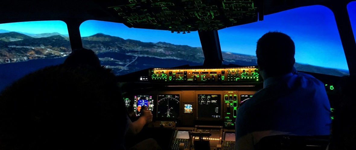
Neuroergonomics: Measuring Cognitive Workload in Real-World Tasks

In some high-stakes jobs, the pressure can feel overwhelming. From air traffic controllers tracking dozens of aircraft simultaneously to surgeons guiding robotic instruments through delicate procedures, many modern occupations require people to process extraordinary amounts of information while maintaining accuracy and focus. As workplaces become more automated and technologically sophisticated, understanding how the brain and body respond to these demands has become a central challenge for ergonomics researchers. A growing area of study, known as neuroergonomics, combines neuroscience, physiology, and human factors research to better understand how people perform in complex real-world environments.

In human factors research, neuroergonomics conintues to gain traction by offering a more comprehensive picture of cognitive workload than traditional measures. Two individuals may perform a task equally well while experiencing dramatically different levels of mental effort. Excessive cognitive workload can contribute to fatigue, stress, errors, and reduced situational awareness, while insufficient workload can lead to disengagement and decreased vigilance. Researchers are therefore concentrating attention on measuring not only what people do but also how their brains and bodies respond as they do it.

Among the most valuable tools for studying cognitive workload are physiological signals that provide objective measurements of mental state. Electroencephalography (EEG) can reveal changes in neural activity associated with attention and cognitive effort, while functional near-infrared spectroscopy (fNIRS) measures changes in cortical blood oxygenation linked to mental workload. Electrocardiography (ECG) and heart rate variability (HRV) offer insight into autonomic nervous system activity, and eye-tracking measures such as pupil diameter and gaze behavior can indicate shifts in attention and cognitive demand. More often, researchers are combining multiple synchronized signals to create a more complete view of how people interact with demanding tasks and environments.

A team of researchers from Drexel University and Lockheed Martin investigated how multiple physiological signals can be combined to measure cognitive workload across a range of mental activities. Participants completed tasks involving working memory, vigilance, risk assessment, attention shifting, situation awareness, and inhibitory control while researchers simultaneously monitored EEG, fNIRS, ECG, photoplethysmography (PPG), electrooculography (EOG), and eye tracking. Brain activity was measured using an fNIR Devices system alongside other physiological monitoring technologies. The researchers found that different physiological measures varied in their sensitivity to specific task demands, with neuroimaging measures providing particularly valuable insight into changes in workload. The findings demonstrate the value of combining signals from the brain, heart, and eyes to create a broader perspective of cognitive effort in complex environments.

Researchers at Loughborough University in the United Kingdom examined how rising task complexity influences cognitive workload in modern manufacturing environments. Participants performed a series of assembly tasks while researchers recorded pupil diameter with Tobii eye-tracking and measured HRV with wearable ECG sensors. By comparing physiological responses across low-, medium-, and high-complexity tasks, the team identified significant changes in both pupillometry and cardiac indicators of cognitive load. Their findings suggest that physiological monitoring can provide an objective means of evaluating cognitive ergonomics in highly digitalized manufacturing environments and may help support the design of more sustainable, human-centered workplaces.
flight simAnother important challenge in neuroergonomics is determining which physiological measurements provide the most reliable indicators of mental workload. A collaborative team of researchers from France investigated how physiological measurements can be used to establish standardized levels of mental workload during aviation-inspired tasks. Using the Multi-Attribute Task Battery (MATB-II), participants performed simulated flight-related tasks under varying levels of demand while researchers measured EEG, eye-tracking, electrodermal activity, and cardiac activity. ECG and EDA signals were acquired using a BIOPAC data acquisition and analysis system running AcqKnowledge software. The study found that several cardiac, ocular, and electroencephalographic measures successfully distinguished high-workload conditions and correlated with subjective workload ratings. The researchers found that several physiological measures, particularly EEG, eye tracking, and cardiovascular metrics, reliably distinguished between different workload levels and closely tracked participants’ subjective assessments of task difficulty. Their findings suggest that multimodal physiological monitoring could support the development of adaptive systems that detect cognitive overload in real time, improving both performance and safety in demanding operational environments.

These studies highlight the increasingly multimodal nature of neuroergonomics research. Eye tracking provides insight into attention and visual processing, cardiovascular measures reveal how the autonomic nervous system responds to mental demands, and brain-imaging techniques such as EEG and fNIRS offer direct windows into neural workload. By combining these complementary perspectives, researchers are moving beyond simple measures of task performance toward a deeper understanding of how people think, adapt, and perform in complex environments. As workplaces continue to evolve alongside automation and intelligent technologies, neuroergonomics is helping researchers design systems that better support human capabilities, improve safety, and reduce the hidden costs of cognitive overload.


Are you planning a multimodal neuroergonomics study? Our team of experts can help you plan your research and identify the tools that will provide the best possible data.

Hands-on Learning: How Real‑Time Physiological Data Deepens Clinical Education

Physicist Albert Einstein noted, “The only source of knowledge is experience.” This principle is particularly relevant in healthcare education, where hands-on learning is vital for cultivating the skills needed to care for patients effectively. The practice of recording and analyzing physiological signals in the learning lab serves as a powerful teaching tool, enabling students to immerse themselves in real-world scenarios and reveal autonomic responses, cardiovascular patterns, and emotional states that are often hidden from view.

For students in nursing, dentistry, and pharmacology, engaging with these signals transforms abstract concepts into concrete, measurable experiences. By analyzing heart activity, visual attention, stress markers, and pharmacodynamic effects, students not only enhance their critical thinking skills but also prepare themselves for clinical challenges with greater confidence.

Physiological signals across the health sciences teach learners to interpret subtle features embedded in physiological waveforms. Modern teaching laboratories increasingly incorporate multimodal systems to help students better understand the complexity of human physiology.

Nursing programs emphasize accurate patient monitoring and interpretation of vital signs, including electrocardiograms (ECG) and heartrate variability (HRV). A 2025 study published in Sensors evaluated the validity of Apple Watch Series 6 HRV data against laboratory‑grade ECG recordings using a BIOPAC 3‑lead ECG system to provide a comparative baseline for measurement. By synchronizing event markers between the Apple Watch and BIOPAC timeline, researchers ensured precise temporal alignment for comparing R‑R intervals and HRV indices. The results showed near-perfect agreement in R-R interval timing under resting conditions, indicating that such wearable devices can serve as a reasonable tool for certain wellness-oriented assessments. At the same time, the BIOPAC system remains the gold standard for precise physiological analysis.

For nursing students, this study demonstrated that consumer wearables must be validated against clinical-grade systems before being relied upon for assessment or decision-making. By examining the comparative data, students can explore how artifacts, sampling frequencies, and sensor placement influence the fidelity of HRV measurements. Moreover, because HRV relates directly to autonomic balance and stress physiology, the study provides a meaningful touchpoint for nursing courses in pathophysiology and patient assessment. Through hands‑on lab sessions using BIOPAC ECG amplifiers, students can visualize waveform morphology, measure beat‑to‑beat variability, and learn how cardiovascular signals reflect underlying homeostatic regulation.

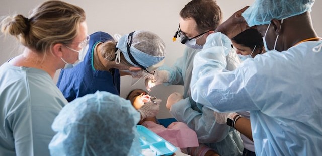
Dental students should understand not only the mechanics of clinical procedures but also the emotional and perceptual experiences of their patients, especially children. A 2025 cross-sectional pilot study in the European Archives of Paediatric Dentistry used an eye tracking and  data acquisition and analysis system from BIOPAC to measure children’s gaze behavior and physiological stress when viewing images of dentists, dental chairs, and operatory designs.

Using eye-tracking metrics such as mean dwell time, fixation count, time to first fixation, and total fixation time, researchers found that study participants stared longer at dentists wearing white coats, indicating a perception of compitance and safety, but also indimidation. In addition, gender matching mattered, as children showed longer fixation on dentists of the same gender. The study also found that warm‑colored dental chairs (especially yellow) attracted greater visual attention, particularly among younger children. Simultaneously, HRV and ECG signals recorded with the BIOPAC system revealed that younger children exhibited greater autonomic arousal than older peers when exposed to dental images.

These findings offer dental students actionable insights into clinic design, caregiver attire, and emotional perception, all of which influence pediatric patient comfort. The study demonstrates how combining eye tracking with physiological monitoring can reveal otherwise hidden psychological responses. For dental educators, integrating such physiological signal analysis into coursework helps students develop empathetic, patient‑centered care strategies.

colored pillsIn pharmacology courses, students learn how drugs influence physiological systems. A study published in Brain Sciences investigated the lingering cardiovascular effects of caffeine, challenging the assumption that its effects dissipate within six hours. Using BIOPAC respiratory transducers, noninvasive blood pressure modules, bioimpedance amplifiers, and ECG amplifiers connected to a data acquisition and analysis system researchers monitored participants before and after consumption of caffeinated and decaffeinated coffee. Results showed measurable changes in heart rate, respiratory patterns, and thoracic impedance well beyond the expected metabolic window, suggesting caffeine’s physiological effects outlast its pharmacokinetic half‑life.
For pharmacy students, these findings reinforce critical distinctions between pharmacokinetics (drug metabolism) and pharmacodynamics (physiological response). By analyzing real‑world signal data, learners gain a more nuanced understanding of how stimulants affect autonomic regulation. Such insights prepare future pharmacists to counsel patients on medication timing, drug interactions, and individual responses to common agents such as caffeine.

Across nursing, dentistry, and pharmacy, physiological signals provide potent educational value. They bridge the gap between theory and clinical application by giving students concrete, measurable access to physiological processes such as autonomic reactivity, cardiovascular regulation, and perceptual attention. Using both research and education-based lab systems, learners can visualize real-time physiological data, develop analytical skills, and cultivate a deeper understanding of the human body under stress, stimulation, and clinical observation.

As healthcare grows more data‑driven, integrating hands-on experience with signal acquisition into professional training helps future clinicians interpret complex physiological information with confidence. Whether validating wearable devices, improving pediatric patient experiences, or exploring drug‑induced autonomic effects, physiological signals empower students to connect scientific principles with evidence‑based, care. Check out our webinar, Advancing Practical Learning: Biosignal Essentials, to learn more.


If you’re exploring ways to incorporate physiological signals into your curriculum or simulation lab, our team is ready to help identify the best tools and workflows for your program.

Brain and Body: SPA-fNIRS Solves Physiological Signal Noise

Functional near-infrared spectroscopy (fNIRS) offers researchers a powerful noninvasive tool for studying brain activity by measuring changes in blood oxygen levels. These changes reflect how various brain regions respond to stimuli such as tasks that involve thinking, emotion, movement, and social interaction. These are often in natural, real-world settings that are not possible with other brain imaging methods. However, fNIRS is not without its own limitations. Systemic fluctuations in the body, such as breathing patterns, cardiovascular changes, and autonomic responses, can result in signal noise that is misinterpreted as brain activity by traditional fNIRS. The solution: a visit to the SPA.

Systemic Physiology Augmented fNIRS (SPA-fNIRS) is an advanced approach to fNIRS that goes beyond measuring local changes in cerebral oxygenation. While standard fNIRS measures cortical hemodynamics by tracking oxygenated (HbO) and deoxygenated (HbR) hemoglobin, noise originating from systemic physiological changes can corrupt those signals. Combining data from both neural and systemic physiological signals, SPA-fNIRS provides researchers with a more diverse and accurate view of how the brain and body interact in real time.

The method is being applied across various fields, including psychophysiology, cognitive neuroscience, exercise physiology, mental health, and human performance research. Studies have utilized SPA-fNIRS to investigate a range of topics, including meditation and emotional regulation, movement control, and social interaction. The broader goal is to move beyond studying the brain in isolation and to understand it as part of an integrated mind-body system. This holistic view is transforming how scientists interpret brain-function data, opening new paths for applied research in education, rehabilitation, and human-machine interface design.

soundResearchers in India investigated the impact of a traditional sound-based meditation technique, known as the Mind-Sound Resonance Technique (MSRT), on both brain and heart activity. Using fNIRS to track changes in oxygen levels in the prefrontal cortex, and a BIOPAC data acquisition and analysis system with AcqKnowledge software to record and analyze heart rate variability (HRV), the study employed a SPA-fNIRS design, integrating neural and physiological data in a single, synchronized dataset. HRV analysis allowed the researchers to quantify shifts in autonomic balance during and after meditation. The results showed increased oxygenation in the right prefrontal cortex and enhanced low-frequency HRV components, reflecting greater parasympathetic regulation. Together, these findings suggest that sound-based meditation not only improves brain oxygenation but also promotes calmer, more balanced heart-brain dynamics, illustrating how SPA-fNIRS methods can reveal the comprehensive physiological impact of mindfulness practices.

In her doctoral research at the Université de Lille in France, neuroscientist Aurore Guérin examined how the brain controls movement speed using a multimodal combination of fNIRS and physiological data recording systems. Participants performed repetitive arm movements at fast, natural, and slow tempos. At the same time, the study captured oxygenation in the prefrontal and motor cortices using fNIRS, alongside heart rate and respiration signals collected with a BIOPAC data acquisition system. A respiratory effort transducer measured breathing patterns, while a BioNomadix ECG module recorded cardiac activity for HRV analysis. All physiological signals were synchronized and analyzed in AcqKnowledge software, allowing the team to identify and remove systemic influences (e.g., heartbeat and breathing rhythms) from the fNIRS signals, following a SPA-fNIRS approach. The results revealed that faster movements primarily engaged motor regions, while slower, more deliberate pacing resulted in higher prefrontal activation, reflecting greater cognitive control. By integrating physiological monitoring with fNIRS, the study provided a clearer picture of how the brain and body coordinate timing and control during movement.

These studies illustrate how SPA-fNIRS is reshaping the way scientists investigate the interaction between brain and body. By combining neural and physiological data in a single synchronized system, researchers can separate brain activity from systemic noise and uncover the interplay between cognition, emotion, and autonomic regulation. For more information on this topic, watch our webinar on Multimodal SPA-fNIRS and check our our latest multimoldal fNIRS research products.


Are you planning a study that blends fNIRS with other physiological signals? Contact your local BIOPAC customer service representative to learn more about how SPA-fNIRS can benefit your research.

Boom! Pow! Clang! Exploring Stress Resilience with Startle Response and PPI

We have all felt that electrical jolt that comes with hearing a sudden, unexpected loud noise—the clang of a dropped cooking pan, the blast of a car horn, or the pop of a balloon. The reaction is instantaneous and nearly uncontrollable as muscles tense, eyes blink, pupils dilate, and one’s heart seems to leap from the chest. The startle response—a rapid, automatic reaction to sudden stimuli like loud noises—has a practical purpose, helping protect us from outside threats. It can also pose a danger if the response occurs while engaging in precision activities, such as driving a vehicle or operating machinery.

Within the psychophysiological field, the startle response can reveal how our brains and bodies process stress, attention, and emotional regulation. In pursuit of ways to mitigate its detrimental effects, researchers often study this reflex alongside prepulse inhibition (PPI). In this latter process, a weaker sound presented just before the startling noise reduces the strength of the response. To capture data when studying these phenomena, scientists use physiological signals such as electromyography (EMG) to measure the tiny muscle contractions around the eye during a blink, electrocardiography (ECG) for heart rate changes, electrodermal activity (EDA) for skin conductance, and respiration to track stress responses. In addition, researchers often pair these physiological measures with brain imaging techniques, like functional magnetic resonance imaging (fMRI) or electroencephalography (EEG), as well as cognitive tasks to build a complete picture of how startle responses connect with brain function and behavior.

Studies of startle response and PPI intersect with a wide range of fields, from neuroscience and psychiatry to aviation, defense, and occupational health. Clinical investigations of altered startle and PPI patterns are helping scientists uncover biomarkers of vulnerability or resilience. These studies are providing new insight into potential treatments for conditions like schizophrenia, PTSD, anxiety, and neurodegenerative disorders.

pilotIn applied contexts, like pilot training or high-stress professions, startle measures are used to understand how sudden stress impacts performance and decision-making. A study conducted by researchers at the University of Zagreb in Croatia with U.S. partners aimed to determine whether stress resilience markers, including startle response and PPI, could predict pilot performance under sudden startle events. Seventeen military pilot cadets completed baseline testing of physiological and cognitive features—including startle reflex habituation, PPI, heart rate variability (HRV), and multitasking ability—followed by flight simulator sessions with induced startle events. Physiological signals such as ECG, EMG (eye-blink startle reflex), respiration, and EDA were recorded via their respective BIOPAC amplifiers connected to a data acquisition and analysis system. Regression modeling showed that startle habituation, autonomic recovery, and cognitive task performance provided an accurate prediction of flight performance. The findings suggest that psychophysiological measures can help identify pilots who are more resistant to startle-induced performance degradation. Such insights could offer practical applications in pilot screening, training design, and aviation safety.

A team of Spanish researchers wanted to see how short-term (acute) stress affects both PPI and higher-level thinking (working memory) in healthy young adults. They used the Maastricht Acute Stress Test (MAST) to induce stress in one group, while another group did a non-stressful control task. After this, all participants heard loud noises that triggered the eye-blink startle response. In some trials, a prepulse immediately preceded the loud noise, which typically reduces the blink reflex. The startle (eye-blink) was measured from the orbicularis oculi muscle using BIOPAC Ag/AgCl electrodes connected to an Electromyography amplifier that fed data to a data acquisition system running AcqKnowledge software. This allowed the researchers to precisely record the intensity of a blink and the degree to which prepulse inhibited it. The team also tested working memory using tasks like digit span backwards and letter-number sequencing, in addition to other cognitive tests. They found that stress made startle responses stronger and reduced PPI, thereby degrading the participants’ ability to filter out the loud noise when it was preceded by a quieter one. Stress also impaired performance on working memory tasks compared to the non-stressed group. In practical terms, these results suggest that acute stress doesn’t just make one more reactive; it can interfere with basic sensory gating and cognitive control. This has implications for stressful jobs or situations (e.g., emergency responders or pilots) and possibly for understanding disorders where PPI or working memory is impaired.

These are just a few examples of how research into startle response and PPI can improve our understanding of the brain’s basic filtering and stress-response systems. The knowledge these studies provide may eventually translate into practical tools for diagnosis, prevention, and performance optimization. For additional information on research applications, check out our webinar on Startle and PPI.


Are you struggling to find the right tools to plan your startle response and PPI study? Our team of customer service experts will put you on the right path to gathering the best data possible for your research project.

Mind Over Matter: Cognitive Neuroscience and Aging

“Age is an issue of mind over matter. If you don’t mind, it doesn’t matter.”  —  Mark Twain.

While American author and humorist Mark Twain suggested that mental attitude could make aging less daunting, modern science shows that the changes our bodies and brains undergo over time are very real. Yet researchers are discovering that aging doesn’t have to mean inevitable cognitive decline. In fact, the growing field of cognitive neuroscience is revealing ways to track, understand, and even influence how our brains change with age, offering hope that we can preserve mental acuity, independence, and quality of life well into our later years.

When studying how aging affects the brain, cognitive neuroscience researchers use a variety of tools. In addition to standard behavioral tests like the Stroop test, n-back test, and verbal fluency exercises, which track reaction times, accuracy, and problem-solving strategies, they also rely on neuroimaging and physiological measurements to gather deeper insights. Neuroimaging techniques include electroencephalography (EEG), which monitors real-time electrical activity in the brain; functional near-infrared spectroscopy (fNIRS), which measures blood oxygen levels in specific cortical regions during cognitive tasks; and magnetic resonance imaging (MRI), which provides detailed structural and functional maps of the brain.

Other physiological signals play an essential role in this field, including heart rate variability (HRV), a key indicator of the autonomic nervous system’s ability to balance various physiological functions and handle stress. Electrodermal activity (EDA) allows researchers to track arousal among participants undergoing cognitive exercises. Electromyography (EMG) measures muscle activation during fine and gross motor tasks. Eye tracking and electrooculography (EOG) provide researchers with data on how aging impacts attention and the ability to process visual cues. Respiration and blood pressure provide vital information on vascular health, which is often linked to cognitive performance.

Taiwanese researchers from National Yang Ming Chiao Tung University in Taipei turned to HRV and fNIRS to uncover how aging and, more specifically, mild cognitive impairment (MCI), affect both brain and heart responses during cognitive tasks. Healthy young adults, cognitively healthy older adults, and older adults with MCI were asked to perform cognitive function and executive tests. Researchers recorded the participants’ prefrontal cortex (PFC) activation through changes in oxyhemoglobin using fNIRS and autonomic responses via HRV. The team gathered ECG data using a BIOPAC data acquisition module running AcqKnowledge data acquisition and analysis software, and equipped with an ECG amplifier. The study found that older adults who did not exhibit MCI showed lower global and executive cognitive performance, reduced HRV, but increased prefrontal activation. The research team interpreted this to be the result of compensatory neural effort. However, older adults with MCI showed even greater cognitive decline, but no further differences in HRV or prefrontal activation. The findings suggest that HRV and PFC activation patterns could serve as biomarkers for distinguishing healthy aging from early pathological decline.

To better understand how learning processes develop and change with age, researchers from Tilburg University in the Netherlands investigated how younger adults and older adults differ in their brain and body responses while learning a visuomotor sequence task. To capture these responses, the team measured EEG, skin conductance (EDA), heart rate, HRV, breathing rate, and eye movements during the task. Physiological data were collected using BIOPAC’s BioNomadix wireless dual ECG/respiration amplifier and PPG/EDA amplifier, which continuously recorded three-lead ECG, respiration, and EDA at 2,000 Hz, with signals processed and analyzed in AcqKnowledge software. This setup allowed the researchers to precisely synchronize physiological activity with learning performance. Both younger and older adults improved with practice, but older adults needed more trials to reach the same precision. Brainwave activity showed apparent age differences, while measures like HRV, respiration, and skin conductance did not. This suggests that EEG is a more sensitive marker of age-related learning changes and could help guide training programs for older adults.

readingA long-term study by researchers from Radboud University Medical Center in the Netherlands, together with UK collaborators, tracked a group of healthy older adults over nearly 11 years to see how their brain blood-flow regulation changed by their 80s. Using transcranial Doppler ultrasound to measure blood velocity in the middle cerebral arteries, the team also monitored control signals including heart rate, blood pressure, and breathing gases. Physiological data were recorded with a BIOPAC data acquisition and analysis system, with end-tidal CO₂ (EtCO₂) measured using capnography via a BIOPAC carbon dioxide measurement amplifier. The researchers found a slight decline in CO₂ reactivity but, surprisingly, an improvement in dynamic cerebral autoregulation (the brain’s ability to maintain steady blood flow despite blood pressure changes). These effects were most apparent in participants who later reported memory complaints, suggesting that while aging may reduce vascular responsiveness, the autoregulatory system may help compensate for early cognitive challenges. The findings show how vascular health in the brain relates to aging and memory, providing valuable insight for detecting early risks of dementia and shaping preventive strategies.
These studies highlight how combining brain imaging with physiological measurements is helping scientists reveal the complex relationship between aging, cognition, and overall health. Through cognitive neuroscience, researchers are identifying early signs of decline and gaining a better understanding of how the brain and body adapt over time. Research is paving the way to more effective screening, personalized interventions, and lifestyle strategies that may help people maintain mental sharpness and independence well into older age. For more information on the tools and techniques being applied to cognitive neuroscience research, see our Webinars and visit our page on Cognitive State Analysis.


Are you planning a study on how aging affects the brain or just looking to expand your cognitive neuroscience toolbox? Our customer service team is ready to help you find what you need to obtain the best possible data.

A System of Systems: Network Physiology Connects the Dots

In the field of physiology, it is tempting to see the human body as a collection of independent systems performing a range of functions—converting food to energy, protecting us from harm and disease, and helping us react to and interact with our environments, to name a few. It is easy to overlook that these systems often work together and are interconnected. The field of network physiology does just that—look at how these systems (nervous, cardiovascular, muscular, etc.) work together, seeing them as part of a dynamic network rather than a collection of independent parts.

The author of the article linked above, Boston University research professor of physics Plamen Ivanov, first coined the term “network physiology” in 2006. Through his work as director of the Keck Laboratory of Network Physiology, Ivanov and his team pioneered research in this new and rapidly growing field. The latest research methods, breakthroughs, and ideas in network physiology will take center stage this summer at the Fourth International Summer Institute on Network Physiology (ININP) hosted at the Lake Como School of Advanced Studies in Italy.

Researchers are exploring various ways to apply the concepts of network physiology, including how stress affects both the brain and the body. Medical researchers are looking into how to identify early warning signs of future health problems in how various physiological systems interact when developing treatments that address multiple bodily systems. While this may sound like the multimodal approach to physiology—collecting data simultaneously and synchronously from multiple systems—the two terms are not synonymous. Network physiology uses multimodal methods to collect data, but goes further by connecting the dots between the various systems.

A study by a team of researchers from the University of New York and the University of Houston demonstrated how combining data from different body systems can give a more complete picture of how the brain and body work together, specifically in response to music and mental effort. The study explored how different types of music—either calming or exciting—affect a person’s working memory (the ability to retain and use information for short periods) and overall brain and body arousal. During the experiment, participants completed memory tasks while listening to either calming or exciting music. Researchers collected detailed data from the body using a set of BIOPAC sensors and electrodes, including general-purpose electrodes for gathering electrocardiogram (ECG) signals, a respiratory effort transducer, a wireless skin temperature sensor, a wireless pulse transducer for photoplethysmogram (PPG) data, and electrodes to measure electrodermal activity (EDA), blood flow in the finger, and muscle activity in the shoulders. These multimodal signals were gathered using a BIOPAC data acquisition and analysis system. The data was collected at 2 kHz to capture even small changes in the body’s responses. The research team found that while the difficulty of the memory tasks didn’t significantly impact arousal levels, the type of music did. Exciting music helped participants feel more alert and even improved their performance on some tasks.

connected manA 2024 South Korean study investigated how the autonomic nervous system (ANS) responds differently to social pain (such as emotional distress from losing a loved one) versus physical pain (like discomfort from a pressure cuff). Researchers used a BIOPAC 16-channel data acquisition and analysis system running AcqKnowledge software to collect a range of signals from 73 healthy young adult participants. The following signals were gathered synchronously using a variety of sensors and transducers: ECG electrodes placed on both wrists and the left ankle to monitor heart activity, EDA sensors attached to the index and middle fingers to assess skin conductance (indicating stress levels), PPG sensor on the thumb to measure blood volume changes, chest-wrapped sensor to track breathing patterns, and a temperature sensor on the ring finger to monitor peripheral temperature changes. Participants experienced social pain via a 60-second grief-themed film clip and physical pain through a blood pressure cuff inflated to 250 mmHg. Social pain increased heart rate and skin conductance, while lowering blood volume pulse, pulse transit time, breathing rate, and finger temperature, reflecting a mix of sympathetic and parasympathetic nervous system activity. Physical pain, on the other hand, raised heart rate variability and skin conductance, reduced blood volume pulse and pulse transit time, but left finger temperature unchanged, suggesting activation of the body’s stress response system. These distinct patterns show how different types of pain trigger unique responses across interconnected body systems.

A multimodal approach was employed in a study by researchers from several institutions including the University of Southern California, University of California, Irvine, Rutgers University, Vanderbilt University, and Wayne State University School of Medicine to examine how daily heart rate variability (HRV) biofeedback training influences brain function, emotional regulation, and physiological responses in both younger and older adults. In the study, 193 participants were randomly assigned to either increase or decrease their heart rate oscillations through daily biofeedback sessions over five weeks. Before and after the intervention, participants underwent various assessments, including MRI scans, psychological evaluations, and sleep monitoring using wristband sensors. During the MRI sessions, physiological data such as blood pressure, pulse, respiration (using a BIOPAC MRI-safe respiration transducer and module), and end-tidal CO₂ were continuously recorded. During stress tasks, blood pressure and hemodynamic measurements were taken using a noninvasive blood pressure monitor and transferred to a BIOPAC data acquisition and analysis system. The study’s findings suggest that HRV biofeedback can positively impact both brain function and physiological health, highlighting the interconnected nature of bodily systems, a central concept in network physiology.

These are just a few examples of how network physiology is being applied in research today. For more information on the topic, including links to related webinars, citations, and events, see our Network Physiology applications page.


Are you planning to integrate a range of signals for your next network physiology study? Our customer service specialists can help identify the best equipment and strategies for the synchronous collection of high-quality multimodal data.

Mind-Body Connection: Biofeedback in Research

The connection between the mind and body has long intrigued scientists, driving research into how our thoughts, emotions, and mental states can influence physical health and well-being. Biofeedback has become a recognized approach for helping people manage stress, reduce pain, and even enhance cognitive and physical performance. Biofeedback involves using sensors and real-time data to monitor physiological functions that are typically automatic, such as heart rate, breathing, muscle tension, and skin conductance. With this information, individuals can learn to gain greater awareness and, in some cases, voluntarily control these bodily processes.

Common physiological signals used in biofeedback include electrodermal activity (EDA), which reflects sweat gland activity and emotional arousal; electroencephalogram (EEG), which measures electrical activity in the brain; electrocardiogram (ECG), which measures heart electrical activity; heart rate variability (HRV), an index of autonomic nervous system flexibility; and respiration rate, among others. These signals offer researchers a window into the body’s stress responses, regulatory mechanisms, and even aspects of cognition and emotional control.

To complement physiological monitoring, researchers are increasingly integrating tools like stimulus presentation systems, virtual reality (VR) environments, and eye tracking technology into biofeedback studies. These tools allow scientists to study how people react to controlled stimuli, immerse participants in realistic scenarios, and track where and how long someone focuses visually, all while collecting detailed data on their physiological responses. This combination of immersive experiences and real-time monitoring helps researchers investigate how the body responds to stress or cognitive challenges and whether individuals can be trained to influence those responses.

Swiss researchers at ETH Zurich investigated whether individuals could voluntarily control their arousal states using real-time feedback on their pupil size. Pupil size is a physiological marker closely linked to the brain’s arousal systems, particularly the locus coeruleus. Study participants employed mental strategies to adjust their pupil diameter, receiving visual feedback through a dynamically changing circle that reflected their pupil size. The participants’ cardiovascular data, including ECG and respiration, were recorded to provide insights into the physiological effects of pupil self-regulation. The research team employed a BIOPAC data acquisition and analysis system running AcqKnowledge software to record ECG data continuously at a sampling rate of 1,000 Hz. Additionally, a BIOPAC respiratory effort transducer was used to measure respiration, although these respiratory data were not reported in the publication. The study’s findings demonstrated that pupil-based biofeedback enabled participants to modulate not only their pupil size but also associated neural activity and cardiovascular responses, such as heart rate. The results suggest a non-invasive method for individuals to control their autonomic functions, with potential applications in managing stress and anxiety disorders.

mind-bodyVagally mediated heart rate variability (vmHRV) refers to the natural changes in the time between heartbeats that are controlled by the vagus nerve, which is closely tied to how well a person can adapt to stress and regulate their body. Researchers from Université Paris Cité explored whether a brief session of vmHRV biofeedback could help people better control their memory, specifically whether it could help them tell the difference between real and false memories. In the study, 71 healthy young adults completed a VR memory task after receiving either vmHRV biofeedback or a simple breathing exercise. To track their physiological responses, the team used a BIOPAC data acquisition and analysis system and recorded heart activity via a three-lead ECG configured according to Einthoven’s triangle using Lead II. The results showed that this short biofeedback session had no immediate effect on memory accuracy or on vmHRV levels during the task. This suggests that a single session of HRV biofeedback may not be enough to influence memory-related brain functions, underlining the complex relationship between the body’s stress responses and cognitive control.

​A study published in the Journal of Human Kinetics looked at whether just one 10-minute session of HRV biofeedback, using a special breathing technique called resonance frequency breathing, could affect the level of activity of certain nerve cells. Researchers measured this by looking at a reflex in the lower leg muscle (the soleus) called the Hoffman reflex (H-reflex). Twelve healthy men took part in both the HRV biofeedback session and a regular breathing session in a random order. To measure muscle activity, researchers used a BIOPAC surface electrode on the leg and a reference electrode on the hand. The signals were recorded with a BIOPAC data acquisition system and analyzed with AcqKnowledge software, which helped line up the timing of muscle activity with electrical stimulation. The results showed that HRV biofeedback lowered the strength of the reflex and a related ratio (Hmax/Mmax), suggesting the spinal nerve cells were less excitable. These findings point to the potential of short HRV biofeedback sessions to affect the nervous system in ways that could be useful for physical therapy or improving movement control.

To learn more about how to integrate biofeedback techniques into your next research project, check out BIOPAC’s webinar, Biofeedback: Theory, Techniques, and Tools for Research.


Are you planning a research project that will utilize biofeedback data? Our expert customer service representatives can help you find the right tools for all your biofeedback needs.


Only the Lonely: The Human Impact of Social Isolation

Humans by their nature are social creatures. A sense of community, daily interactions, and shared experiences are tied to our overall mental and physical well-being. Few events in modern history have shut down such interactions more thoroughly than the outbreak of COVID-19, which forced millions into prolonged isolation. As lockdowns and social distancing became the norm, an increasing number of people reported heightened feelings of loneliness, anxiety, and depression. Beyond the pandemic, other modern social factors contribute to isolation, including the growing reliance on social media and virtual interactions in place of face-to-face connections. While technology offers new ways to stay in touch, excessive screen time and digital communication can sometimes deepen feelings of loneliness by reducing meaningful, in-person social engagement. In the wake of these societal shifts, researchers are working to understand the long-term consequences of social isolation on both mental and physical health.

Studies suggest that loneliness isn’t just an emotional experience—it has measurable physiological effects on the body. Using advanced biometric signals, researchers are trying to uncover how isolation influences our nervous system, stress responses, and overall wellness. Heart rate variability (HRV) provides insights into autonomic nervous system balance, while electrodermal activity (EDA) reflects changes in stress and emotional arousal. Respiration patterns, electromyography (EMG) readings, and other physiological markers help paint a complete picture of how isolation affects both mind and body. By combining psychological assessments with physiological data, scientists aim to better understand how chronic loneliness can contribute to conditions such as cardiovascular disease, weakened immune function, and mental health disorders.

In one such study published in Britain’s Royal Society Open Science journal in 2024, researchers looked at the effect of social isolation on human adolescents, specifically their ability to learn to perceive threats. Study participants between the ages of 16 and 19 underwent sessions of complete isolation and isolation combined with virtual social interaction, as well as baseline sessions. Following each session, study participants reported their psychological state and completed a threat learning task (participants learned to associate neutral shapes with an aversive sound) to measure physiological response to learned threats and safety cues. During these sessions, researchers recorded participants’ EDA signals using BIOPAC EDA finger transducers connected to an MP36R data acquisition and analysis system running AcqKnowledge software. Results showed heightened EDA levels during threat extinction following isolation compared to the baseline. Researchers reported that their findings suggest that isolation and loneliness among adolescents might increase vulnerability and increase the instances of emotional disorders such as phobias, anxiety, and obsessive-compulsive disorder (OCD).

A team of researchers in Brazil compared how giving and receiving of social touch as well as heart rate variability serve as accurate predictors of loneliness in college-age adults. Participant loneliness levels were measured using the UCLA Loneliness Scale. Participants completed a psychiatric screening questionnaire that reported loneliness, social support, and social touch levels. Participant HRV measurements were taken via electrocardiographic (ECG) signal processing using a BIOPAC ECG amplifier connected to a 16-channel MP data acquisition and analysis system running AcqKnowledge. According to the researchers’ published findings, “social support and social touch, specifically receiving touch, accounted for a significant proportion of the variance in loneliness. However, neither giving touch nor resting HRV was a significant predictor of loneliness.”alone

A study by researchers from the University of Bonn examined the link between loneliness and how males and females process trauma and react to fear-inducing stimuli. Over three days, participants were exposed to experimental trauma and evoked intrusive thoughts. Researchers employed functional magnetic resonance imaging (fMRI) scans to assess neural habituation to fearful faces and fear learning (conditioning and extinction) before trauma exposure. Fear conditioning and extinction tasks used a BIOPAC MP system to deliver stimulus in the form of electric impulses. EDA and respiration data were recorded using the BIOPAC data acquisition and analysis system. Study results revealed “a significant interaction between loneliness and sex such that loneliness is associated with more intrusions in men, but not in women.” The implication is, according to the study’s authors, “that loneliness may confer vulnerability to intrusive memories after trauma exposure in healthy men and that this phenotype relates to altered limbic processing of fear signals.”

As the physiological and psychological effects of the COVID-19 pandemic continue to resonate and we grapple with the isolating influence of technology, increasing numbers of research studies are being launched to measure these impacts. Studies into human isolation and loneliness represent just one small corner of a broad spectrum of research on the topic. For information on how BIOPAC equipment is being used in such research areas, check out our on-demand webinars for additional ideas on how to deploy BIOPAC resources in your next study.


Are you planning a research project investigating the effects of isolation, loneliness, or other related subjects? Reach out to our team of experts to help you get started with the right resources and equipment.


Pour One Out: The Physiology of Alcohol Consumption

“Dry January,” the cutting out of alcohol consumption during the first month of the calendar year, entered the public vernacular a little over a decade ago. The idea was first promoted in 2013 by a British charity to encourage alcohol abstinence, or at the very least, healthy moderation. The idea took off as a counterbalance to the excesses of indulgence that often characterize the end-of-year holiday season. As if on cue, the U.S. surgeon general released a report earlier this month linking increased cancer risk to even moderate alcohol use and called for alcohol products to carry a warning label like those found on tobacco products.

Since the early 1990s, the prevailing belief among some in the medical community held that moderate amounts of alcohol may not necessarily be detrimental to overall health and may even have beneficial effects. Despite the surgeon general’s report, the consensus in the benefits versus risks debate remains elusive, a fact summarized well in a recent article in The Atlantic and an accompanying podcast by journalist Derek Thompson. Without wading further into the controversy, it is worth pointing out that there is a rich and growing body of research into the effects of alcohol on human physiology, cognitive and psychological well-being, and longevity, all of which rely on some form of life science metrics to measure these effects.

Physiological signals offer a powerful window into the biological and neurological effects of alcohol consumption, providing objective, real-time insights into the body’s and brain’s response. By measuring electrical, chemical, and physical signals generated by the body, researchers can examine changes in heart rate, brain activity, muscle tone, and more. Commonly used signals in alcohol studies include electroencephalography (EEG) to monitor brain activity, electromyography (EMG) for muscle function, electrocardiography (ECG) for heart rhythms, and electrodermal activity (EDA) and galvanic skin response (GSR) for stress and arousal levels. Each tool captures aspects of how alcohol influences the human system, from cognitive processing impairments to autonomic regulation shifts. In research settings, these signals are applied to explore both short- and long-term effects of alcohol. For example, EEG studies can track alterations in brainwave patterns, shedding light on reduced attention, memory disruption, or impaired decision-making during intoxication. ECG and GSR data, meanwhile, reveal how alcohol modulates cardiovascular activity and stress responses, highlighting its impact on the autonomic nervous system (ANS).

A 2021 study by researchers at Wake Forest University and John Hopkins University School of Medicine used heart rate variability (HRV) to examine the relationship between alcohol abstinence, ANS function, and whole brain functional brain networks among everyday drinkers. Whole brain functional brain networks demonstrate how different parts of the brain work together as a connected system. This study combined functional brain wave mapping generated with resting-state MRI scans during three-day periods of alcohol consumption and abstinence. The effects of abstinence were measured using HRV. Alcohol craving in patients with an alcohol use disorder (AUD) has been associated with high-frequency heart rate variability (HF-HRV) with acute consumption being related to decreasing HF-HRV. During the study, ECG data was collected via a BIOPAC data collection and analysis system. The study concluded that “while brain networks do differ across drinking states in risky drinkers, the change is primarily driven by HRV.”woman drinking

A current study by a team of German researchers explores the effectiveness of Virtual Reality (VR) in Cue Exposure Therapy (CET), a strategy in Cognitive Behavioral Therapy (CBT) used for treating AUD. Research participants are exposed to VR scenarios with alcohol-related cues. VR with integrated eye tracking offers advantages over real-life scenarios when developing new treatment options, according to the study authors. Participants wore head-mounted displays (HMD) to immerse them in three different scenarios while heart rate, HRV, pupillometry, and EDA were measured. Respiration and ECG data are gathered via a wireless BioNomadix BN-RSPEC while PPG and EDA are gathered with a BN-PPGED. Physiological signal data is then fed to a BIOPAC data acquisition system running AcqKnowledge software for analysis. While the study is ongoing, researchers believe it will lead to a better understanding of the induction of craving in VR cue exposure…” and that it could offer “new diagnostic and therapeutic perspectives…providing a basis for future biofeedback training in VR exposure therapy.”

These are a few of the ways physiological signals are being applied in alcohol-related research. For more information on integrating HRV analysis into your next research study, see our webinar on the topic. Better yet, get hands-on training on a full range of research equipment and techniques at our biannual conference on Tools, Trends, Techniques, and Technologies in Human Physiology, 4T|Phys.


Are you planning an alcohol or addiction-related study? BIOPAC has the tools and our regional sales staff is ready to help you with everything you need.

Facing Fear: Measuring What Scares Us

In many countries, October ushers in the season of ghosts, goblins, and all manner of frightening things that inhabit our deepest fears. While the popularity of Halloween reflects the thrills many derive from a good scare, facing one’s fears is not always fun and games. Our fears can protect us from danger, but they can also hamper individuals from performing the simplest tasks and negatively impact one’s quality of life and mental health.

The study of fear conditioning and fear learning sheds light on how humans and animals develop responses to threatening stimuli. Research often relies on psychophysiological measurements—techniques that capture the body’s physical responses to fear. Key among these measurements are skin conductance response (SCR), electrodermal activity (EDA), heart rate variability (HRV), respiration, and electromyography (EMG).

SCR and EDA track changes in sweat gland activity, providing a direct measure of emotional arousal when a person experiences fear. HRV and respiration rate reveal the autonomic nervous system’s reaction to stress, indicating shifts between sympathetic (fight-or-flight) and parasympathetic (rest-and-digest) states. EMG measures muscle activity, especially facial muscles, to detect subtle expressions or physical tension associated with fear. Understanding these mechanisms has practical applications in understanding and treating anxiety disorders, phobias, and post-traumatic stress disorder (PTSD). By analyzing the body’s automatic fear responses, scientists can develop targeted therapies to help individuals recondition or dampen these reactions.

A study published in the Journal of Behavior Therapy and Experimental Psychiatry used a range of indices to assess learned fear (unconditioned stimulus expectancy or US-expectancy) in individuals. These included both self-reporting and measurements of autonomic response such as skin conductance. Researchers using E-Prime Stimulus Presentation Systems, paired various neutral visual stimuli with unconditioned stimuli in the form of electrical shocks delivered via a constant voltage stimulator coupled with a BIOPAC STM100C amplifier. After a brief break, both neutral stimuli were administered without shock. Skin conductance response data was gathered and analyzed during each phase with a BIOPAC data acquisition and analysis system and software. Results showed a positive correlation between US-expectancy ratings and skin conductance response.

facing fearResearchers at the University of Bologna in Italy took a different approach to measuring the psychophysiological mechanisms of fear learning by focusing on HRV. In their study, published in the journal Psychophysiology, they explained that “understanding transient dynamics of the autonomic nervous system during fear learning remains a critical step to translate basic research into treatment of fear-related disorders.” Researchers gathered SCR and electrocardiogram (ECG) data. As with the previous study, mild electrical shocks were applied as unconditioned stimuli and paired with neutral or conditioned stimuli (CS). SCR signals were acquired using a skin conductance transducer attached to the participants’ fingertips connected to a BIOPAC EDA amplifier. ECG data was collected using disposable surface electrodes connected to an ECG100C amplifier. EDA and ECG signals were processed with a BIOPAC data acquisition and analysis system running AcqKnowledge software, which calculated HRV from the ECG data. Researchers concluded that “learned fear elicited a transient heart rate deceleration in anticipation of noxious stimuli.” They added that the study’s findings “provide a proximal measure of the involvement of cardiac vagal dynamics into the psychophysiology of fear learning and extinction, thus offering new insights for the characterization of fear in mental health and illness.”

Another study published in the journal Psychophysiology examines fear learning by applying a broad range of physiological signals and responses to determine which provides the “optimal measurement of human trace fear conditioning.” For this study, researchers compared SCR, pupil size response, heart period response (via ECG), respiration amplitude response, and electromyography (EMG) to measure participants’ responses to electrical current, audio, and visual stimulation. EDA data was gathered via a BIOPAC EDA amplifier, while ECG, EMG, and respiration data were fed to BIOPAC ECG100C, EMG100C, and RSP100C amplifiers, respectively. All data was processed using BIOPAC data acquisition and analysis systems and software. Results of the experiment suggested that startle eye-blink response through EMG provided the most sensitive metric for measuring fear conditioning.

Fear is just one of the emotions that can be studied through the body’s affective response to stimuli. Read more about the relationship between affect and emotion or watch our webinar on Emotion and Objective Physiological Measures.


Are you facing the reality of putting together your own fear-based study? Don’t be afraid to contact your local BIOPAC customer service representative for help finding the right tools for the job.

error: Content is protected !!