
Behind the Wall of Slumber: Multimodal Physiology Reveals Sleep’s Impact on Body and Mind

Most of us know what a restless night feels like the next day: foggy thinking, a shorter fuse, and that sensation of running on empty. For many people, that feeling never fully goes away. An estimated 50 to 70 million Americans live with a chronic sleep or wakefulness disorder, according to the National Heart, Lung, and Blood Institute, and about 10% have chronic insomnia. Poor sleep is also linked to anxiety, depression, and long-term cognitive decline, and its effects change as we age. To understand how, researchers are taking a multimodal approach, recording multiple physiological signals at once to capture what sleep does to the brain, heart, and nervous system.

Sleep touches nearly every system in the body. During sleep, the brain clears metabolic waste through cerebrospinal fluid, a process that may weaken as sleep becomes lighter and more fragmented with age, potentially setting the stage for neurodegenerative disease. Sleep loss also slows reaction times, dulls memory, and makes emotions harder to regulate. For healthcare workers, pilots, military personnel, and shift workers, a tired mind can lead to costly errors, injuries, or worse. No single measurement captures all of this, so researchers combine signals from across the body.

Electrodermal activity (EDA), or skin conductance, tracks changes in sweat gland activity driven by the sympathetic nervous system, the body’s fight-or-flight response, making it a sensitive marker of emotional arousal. Electrocardiography (ECG) records the heart’s electrical activity, from which researchers calculate heart rate variability (HRV): the beat-to-beat variation that reflects the balance between the sympathetic and parasympathetic (rest-and-digest) systems. HRV shifts with sleep deprivation and has been tied to cognitive performance. Photoplethysmography (PPG) and respiration sensors track pulse and breathing, helping researchers separate the body’s rhythms from brain activity in neuroimaging data.

A study on the aging brain focused on waste clearance. A doctoral researcher at Boston University’s College of Engineering, working with collaborators at Massachusetts General Hospital and Harvard Medical School, recorded young (18–39) and older (60–85) adults during nighttime rest with simultaneous EEG and fast fMRI, and measured their cerebrospinal fluid (CSF) flow. Pulse oximetry, skin conductance, and respiration were monitored with BIOPAC sensors, including an MRI-safe pneumatic respiration belt, and acquired in AcqKnowledge software at 2,000 Hz, synchronized with the MRI data through scanner triggers. Older adults showed significantly less CSF flow during sleep, a decline tied to both neural and vascular factors, which could limit the brain’s ability to clear waste. Their scans also revealed altered activity in arousal-regulating brain regions, which may help explain why sleep becomes more fragmented with age.

Sleep also shapes how we process fear. Researchers at New York University’s Tandon School of Engineering, working with Massachusetts General Hospital and Harvard Medical School, compared 24 adults with insomnia disorder and 24 good sleepers during a fear conditioning and extinction task in an fMRI scanner. Participants learned to associate an image with a mild shock, then learned that the image was safe. Skin conductance was recorded with a BIOPAC data acquisition system with AcqKnowledge software, then separated into rapid responses and slower baseline shifts to estimate each participant’s arousal over time. Those with insomnia stayed more physiologically aroused after the threat was gone, a sign of impaired fear extinction that may have implications for anxiety and PTSD.

Sleep DeprivedChronic sleep problems build over months and years, but one sleepless night is enough to measure a decline. Biomedical engineers at the University of Connecticut, with collaborators at Bucknell University and the Naval Submarine Medical Research Laboratory, kept 19 participants awake for 26 hours while they completed cognitive tests every two hours, including a flight-simulation task. During each round, the team recorded ECG for HRV with a wearable BIOPAC BioNomadix wireless ECG transmitter. As the hours passed, reaction times slowed, accuracy dropped in the overnight rounds, and sleepiness climbed. A brain-stimulation technique tested midway did not reverse these effects. The researchers see HRV as a promising way to track fatigue continuously and objectively in safety-critical jobs.

These studies just touch on the myriad of ways sleep impacts the brain and body, from the aging brain’s nightly maintenance to the emotional cost of insomnia to how quickly alertness erodes without rest. A multimodal approach lets researchers see effects that self-reports alone can’t show, bringing them closer to understanding why sleep matters for lifelong health.


Planning a study on sleep and health? Reach out to our experts to find the right tools to record and analyze multimodal physiological data.
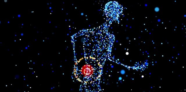

Everybody Hurts: Studying the Physiological and Neurological Components of Pain

A Buddhist saying states, “Pain is inevitable, but suffering is optional.” Pain’s dual nature has presented a conundrum for philosophy and the sciences. As a warning system, it protects us from harm, but it can also be the source of profound psychological and emotional damage. Pain is a complex and deeply subjective experience, yet it is one of the most critical areas of study in neuroscience.

Understanding pain involves not only exploring its physical origins but also decoding the neural circuits and physiological signals that contribute to its perception and regulation. Neuroscientific research into pain aims to answer fundamental questions about how pain is processed in the brain, how it varies from person to person, and how we can develop better treatments for chronic conditions.

This research uses various physiological signals to provide insights into pain mechanisms. Electroencephalography (EEG) captures brainwave activity, revealing how different regions of the brain respond to painful stimuli. Photoplethysmography (PPG) tracks blood flow changes, while electrocardiography (ECG) and skin conductance response (SCR) measure autonomic nervous system activity, offering clues about how the body reacts to stress and discomfort. Respiration patterns, too, are key indicators, as breathing can change in response to pain. Together, these signals help scientists map the body’s response to pain, shedding light on the underlying neural pathways and guiding the development of more effective pain management strategies. Understanding these physiological markers is a critical step toward personalized pain care and improving the quality of life for millions worldwide.

Pain research commonly focuses on the neurological mechanisms of pain to better understand its causes and how it can be treated. Researchers publishing their findings in the Journal of Biomedical and Health Informatics developed a device for studying pain perception biomarkers using thermal stimulation. Their device generates minutely controlled thermal stimuli using a computer-driven Peltier cell while simultaneously capturing EEG and PPG signals. Researchers identified the heat pain threshold (HPT) for each subject, defined as the maximum the subject can withstand when the Peltier cell gradually increases the temperature. The team used two-dimensional spectral entropy (SE) obtained from both the EEG and PPG signals to differentiate the condition of pain. EEG and PPG signals were fed to a BIOPAC MP36 data acquisition and analysis system running Biopac Student Lab (BSL) software. The study concluded that hemodynamics, brain dynamics, and their interactions can be used to discriminate thermal pain perception, which could have practical applications in monitoring pain perception. Possible applications include the study and treatment of “pathologies that affect the peripheral nervous system, such as small fiber neuropathies, fibromyalgia, or painful diabetic neuropathy.”

The pain mechanism associated specifically with fibromyalgia, one of the most common chronic pain disorders, was the focus of a study at the University of Jae´n in Spain. Despite its prevalence, much is still unknown about the underlying cause of this condition. Clinical and experimental studies of individuals diagnosed with fibromyalgia syndrome (FMS) have revealed autonomic nervous system (ANS) deficiencies. While both the parasympathetic and sympathetic components of the ANS have been studied, less is known about the sympathetic component as it relates to FMS. In this study, researchers used SCR as an indirect measure of the sweat response, which is controlled by the sympathetic mechanisms of the ANS. Researchers used a BIOPAC MP36 running AcqKnowledge to collect SCR data via an EDA lead set connected to participants’ fingers. The study found reduced skin conductance levels (SCL) and SCR in FMS patients compared to the non-FMS control group, indicating lower levels of sweating and reduced sympathetic activity in the skin. These factors provided the research team with a better understanding of the sympathetic nervous system’s role in FMS, with the potential for revealing new routes to better pain management and treatment.

For several decades, researchers and clinicians have studied the use of hypnosis to treat chronic pain. More recently, new technologies such as virtualpills reality have been incorporated into these techniques. A team of researchers from the University of Strasbourg in France studied the application of virtual reality hypnosis (VRH) to regulate HPT in healthy adult participants. Researchers used several physiological signals to measure autonomic function during the experiment, including ECG, respiration, SCR, and EEG. ECG signals were recorded using the BIOPAC ECG amplifier, while respiration rate was measured by a thermistor transducer that determined the difference in temperature between inhaled and exhaled air. SCRs were measured from the index and middle fingers of the dominant hand with a BIOPAC skin conductance transducer. Physiological signals were fed into a BIOPAC data acquisition system for analysis. A computer-generated immersive environment was created in VR and combined with a hypnotic auditory script. As with classical hypnosis treatments, the VRH scheme in this study began with an “induction period” followed by a “dissociation” period (separation between the mental state and the environment), followed by suggestions of pain reduction as analgesia. The study results supported a positive correlation between VRH and an increase in the participants’ pain threshold with promising implications for new non-pharmacological approaches to pain management and treatment.

These are just some of the ways that BIOPAC equipment is being used to explore the frontier of pain research and help ease the suffering of millions who deal with chronic pain and its physical and psychological effects. See our webinars page for additional information on how physiological signals can be applied to your next study.


Are you planning a study on pain or any related neurological or physiological area of research? Our team of experts is ready to help you find the tools to ensure success.

error: Content is protected !!