Muscles in Motion: Electromyography in Physiological Research

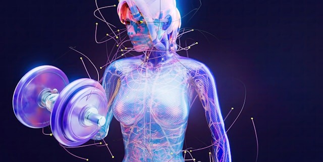
When we think about the human muscular system, movement often comes to mind. Activities such as running, lifting, and maintaining posture all rely on coordinated skeletal muscle activity, while cardiac muscle drives circulation and smooth muscle supports essential functions like digestion. Yet muscles do more than move us from point A to B; they also reflect what is happening inside the body. Patterns of muscle activation can reveal information about physical effort, fatigue, coordination, and recovery, as well as emotional expression, stress, and cognitive engagement. Electromyography (EMG) is a widely used technique for measuring the electrical signals generated by muscles during contraction, providing researchers with a direct and objective means to study both physiological function and the links among the nervous system, behavior, and mental state. By capturing muscle activity that may be too subtle to observe visually, EMG enables precise investigation of processes ranging from motor control and performance to emotional and stress-related responses.

There are several types of EMG, each suited to different research needs. Surface EMG (sEMG) uses small sensors placed on the skin to detect activity from nearby muscles and is the most common approach in behavioral and human performance studies. Intramuscular EMG involves inserting a fine-needle electrode directly into a muscle to record activity from specific muscle fibers, often used in clinical or specialized research settings. Facial EMG (fEMG), a form of surface EMG, focuses on facial muscles. By measuring muscle activity associated with smiling or frowning, fEMG allows researchers to study emotional responses, even when those responses are subtle or not consciously expressed.

One of the key strengths of EMG is its ease of integration with other physiological measurements. In many studies, EMG is recorded alongside electrocardiography (ECG), which measures heart activity; electroencephalography (EEG), which records electrical activity in the brain; functional near-infrared spectroscopy (fNIRS), which tracks changes in brain oxygenation; and eye tracking, which monitors visual attention and gaze patterns. By synchronizing these signals in time, researchers can better understand how different systems in the body work together. For example, combining EMG with EEG can reveal how brain activity leads to muscle movement, while pairing EMG with ECG can show how physical effort relates to heart responses during stress or exercise. This multimodal approach is central to advancing research in areas such as emotion, cognition, and human performance.

EMG also has important real-world applications. In rehabilitation, it helps clinicians monitor muscle recovery after injury. In ergonomics, it provides objective data about muscle strain in the workplace. In sports science, EMG helps optimize training and performance by identifying how and when muscles are activated. Increasingly, EMG is also used in human–machine interfaces, such as systems that allow people to control prosthetic limbs using their muscle signals. Its ability to detect fine-grained changes in muscle activity makes EMG a valuable tool across both research and applied settings.

Best practices for EMG data quality begin with careful electrode placement and attention to potential sources of noise. For sEMG and fEMG, electrodes should be placed over the “belly” of the target muscle, aligned with the muscle fibers, and spaced consistently to capture clean, interpretable signals. Gently cleaning the skin and, when appropriate, lightly abrading the surface helps reduce impedance and improve signal quality. In fEMG studies, precise and repeatable placement over well-defined facial muscles is critical, as these muscles are small and closely spaced. To minimize noise and artifacts, researchers should secure electrodes and cables to prevent movement, ask participants to limit unnecessary body motion, and ensure proper grounding of the recording system. Electrical interference from nearby equipment, power lines, or poor cable management can also contaminate EMG signals, so shielded cables and a controlled recording environment are recommended. Finally, synchronizing EMG with other physiological signals using a common acquisition system helps prevent timing errors and reduces the need for later corrections, resulting in cleaner data and more reliable conclusions.
hand mechanicsRecent studies demonstrate how EMG continues to expand into new and innovative areas. In a study published in Results in Engineering, a team of researchers from China developed a low-computational method for recognizing hand gestures from EMG signals to improve prosthetic device control. The researchers extracted key features from EMG recordings and used a lightweight machine-learning model, a multilayer perceptron (MLP), to classify gestures in real time. EMG data were collected using a four-channel BIOPAC data acquisition system running Biopac Student Lab (BSL) software. Surface Ag/AgCl electrodes were placed on four forearm muscles to capture relevant muscular activity. The study showed that accurate gesture recognition can be achieved without heavy computational requirements, making the system more practical for everyday use. This work highlights how EMG can support the development of more responsive, accessible assistive technologies.

Another study, conducted by a team of researchers from Mahidol University in Thailand and presented in the Institute of Electrical and Electronics Engineers (IEEE) proceedings, explored how combining EMG with motion sensors can improve ergonomic assessments. Focusing on occupational health, the study integrated surface EMG with data from an inertial measurement unit (IMU), a sensor that tracks movement and orientation. The researchers used the IMU to calculate Rapid Upper Limb Assessment (RULA) scores—a common method for evaluating postural risk—while EMG data collected via a BIOPAC four-channel research system provided insight into muscle activity. By combining these measures, the team found that posture alone did not always reflect actual muscle strain. The inclusion of EMG revealed additional muscle stress that might otherwise go unnoticed, suggesting that multimodal approaches can lead to more accurate workplace assessments and better injury-prevention strategies.

Researchers from Louisiana State University and the Pennington Biomedical Research Center in Baton Rouge, Louisiana, examined how training experience influences muscle activation and fatigue during resistance exercise performed with and without blood flow restriction (BFR) in young men. The study, published in Physiological Reports, compared trained and untrained participants while they completed knee extension exercises under low-, medium-, and high-load conditions. The team used sEMG to measure muscle excitation and total muscle activation in the vastus lateralis, with participants fitted with a portable BIOPAC BioNomadix Wireless EMG system. EMG signals were sampled at 2000 Hz and analyzed using a data acquisition system with AcqKnowledge software, allowing the researchers to quantify both peak muscle excitation and overall activation across repetitions. The results showed that low-load exercise with blood flow restriction produced lower peak muscle excitation but substantially greater total muscle activation compared to traditional high-load exercise, regardless of training status. These findings suggest that low-load BFR training may provide an effective alternative for stimulating muscle adaptations while reducing mechanical stress, with potential applications in rehabilitation and strength training programs.

Taken together, these examples illustrate the versatility and effectiveness of EMG in neuroscience, psychophysiological research, and beyond. Whether used to study emotion, improve physical training and workplace safety, or develop assistive technologies, EMG provides a direct link between muscle activity and human behavior. When combined with other physiological measures, it offers a more comprehensive understanding of how the brain and body interact. As research tools and techniques continue to advance, EMG will remain an essential method for exploring brain-body connections. For additional information on how to design studies around EMG, best practices for gathering EMG data, and how to integrate EMG with other signals, check out our webinar on EMG acquisition and analysis.


If EMG will be part of your next physiological or psychophysiological research project, reach out to our knowledgeable staff, who are standing by to help you with each stage of the planning and design process.

Going for Gold: Physiology Research Powers Olympic Athletes

As the world’s top athletes gather for the 2024 Olympic Games in Paris, they hope that years of rigorous preparation and training to ensure peak performance will pay off with a trip to the winner’s podium. Physiology research plays a critical role in these preparations, providing athletes and their coaches with valuable insights into how to best optimize training regimens and enhance athletic performance. With the introduction of new sports in the competition, understanding the specific physiological demands of each sport is critical. Advanced tools and techniques, like those developed by BIOPAC, allow researchers to study the body’s responses to different types of movement, providing tailored training programs that cater to the unique needs of each athlete.

A range of physiological signals is pivotal in sports and exercise research. Electromyography (EMG) is used to analyze muscle activation patterns, helping athletes refine their movements for efficiency and power. Electrodermal activity (EDA) provides insights into stress and arousal levels, which are essential for mental preparation and focus. Monitoring respiration rates and patterns can optimize breathing techniques, while electrocardiography (ECG) and heart rate variability (HRV) assess heart health and endurance capacity. By integrating these physiological signals, researchers and coaches can develop comprehensive training strategies that improve performance and minimize injury risk, giving athletes the best chance at Olympic gold.

One of the newest sports introduced at the Paris Olympics is breaking or competitive breakdancing. Olympic breaking competitions will feature teams of male and female competitors facing off with opposing teams in one-on-one dance battles. Dance, whether competitive or recreational, has been the subject of extensive study as researchers have examined this intersection of music and rhythmic bodily movement. A study published in 2021 in the journal Nature examined how younger and older adults timed bodily movement to both metronomic rhythms and music. The study compared participants’ responses to auditory cues from a metronome and music with finger tapping, toe tapping, and dance steps. The pace of whole-body stepping movements was recorded using BIOPAC heal-toe strike sensors attached to participants’ shoes with data fed to a data acquisition and analysis system running AcqKnowledge software. The study found that whole-body movement with musical accompaniment positively influenced participants’ ability to time movements accurately with encouraging implications for the treatment of rhythmically impaired individuals, such as those with developmental disorders.

Paris 2024Surfing debuted at the 2020 Olympics in Tokyo and is returning for the Paris games where it will be joined by two new aquatic additions, kiteboarding and kayak cross. The surfing competition for 2024 will be held in the French territory of Tahiti. Physiology studies have played a key role in preparing competitive surfers for the ultimate test of their skills. A study published in a 2021 issue of Nature investigated how surfers’ paddling cycles affect fatigue. Paddling is a primary component of a surfer’s ability to catch the best waves during competition. Researchers measured participant velocity as they paddled in a pool while tethered to a cable that measured propulsive force. Velocity data was recorded using a BIOPAC data acquisition and analysis system running AcqKnowledge. Untethered velocity was also measured using a cable-based speedometer device with data also transferred to the data acquisition system. According to the researchers, the data gathered should help in the development of “a powerful and endurable metabolic base while reducing energy cost during surfing paddling,” which could play a key role in future training programs.

Boxing’s inclusion in the Olympics dates back to the original games in Ancient Greece. Modern trainers look to research conducted in the physiology lab to enhance their fighters’ competitive edge. A 2023 study by a group of researchers in Pakistan used EMG to measure muscle activity when comparing the effectiveness of two types of punches: the jab and the cross. Researchers used a force plate to measure the impact force of each type of punch, while electrodes attached to the participants’ biceps and triceps recorded EMG data via a BIOPAC data acquisition system.  While both punches delivered similar force, the triceps carried most of the workload during the jab. The research may provide insight into how to best manage fatigue during competitive bouts.

These are just a few examples of how sports physiology research powered by BIOPAC tools and techniques is preparing today’s athletes to compete at the highest levels. For additional information on how BIOPAC can help your sports-related study, see our webinars on exercise physiology using our BioNomadix and Research Ring wireless recording tools and product lines.


Are you planning sports or exercise study? BIOPAC’s regional sales reps are standing by to assist you in finding the right tools for the best results possible.

Speak Up: Physiological Signals for Speech Research

In the symphony of human communication, speech is the principal instrument. Behind every uttered word lies a complex interplay of physiological and psychological signals, each telling a unique story about the speaker’s state of mind and physical well-being. The critical data provided by these signals has become a pivotal focus in both speech research and therapy, offering useful insights into the mechanisms of verbal communication and its disorders while enhancing speech ability and overall health.

Electromyography (EMG), a technique used to measure muscle activity, plays an invaluable role in speech research by revealing the interaction of facial muscles involved in articulation. By monitoring EMG signals from muscles such as the lips, tongue, and jaw, researchers can elucidate the precise movements and coordination required for speech production, shedding light on the underlying mechanisms of speech disorders like dysarthria or apraxia.

A French study published in the 2024 edition of Science Reports used EMG to establish the role tongue reflex plays in speech posture control. The presence of a tongue reflex in maintaining tongue posture, the proper positioning of the tongue in the mouth, has been a matter of debate among speech researchers. The study team employed an unorthodox recording technique that involved attaching EMG electrodes to the base of the tongue “to record activation of the anterior genioglossus, which is responsible for the control of the anterior part of the tongue.” A robotic arm was used to rapidly stretch the tongue while researchers measured muscular reflex EMG data during the formation of various vowel sounds, which were fed to a BIOPAC EMG amplifier for signal amplification and filtering. According to the research team, the study results “represent the first physiological demonstration in humans that the tongue is driven by reflex in response to a sudden stretch.”

Electrocardiography (ECG), which measures the electrical activity of the heart, offers insights into the intricate relationship between emotion and speech. Research has shown that changes in heart rate variability (HRV) correlate with emotional arousal and cognitive load during speech tasks. By integrating ECG data with speech analysis, researchers can explore the complex connection between emotional states and linguistic expression, paving the way for emotion-aware speech synthesis systems or interventions for social communication disorders.microphone

A 2023 study by researchers at Vanderbilt University used ECG to examine the relationship between respiratory sinus arrhythmia (RSA) on the persistence of stuttering and, specifically, the role emotional regulation plays in stuttering. Researchers measured potential differences in RSA in preschool-age children with persistent stuttering, children who recovered from stuttering, and children who do not stutter. RSA, an index of parasympathetic nervous system activity, was measured during the viewing of both neutral (baseline) and emotionally stimulating video clips and during subsequent speaking tasks. To obtain RSA for each study participant, ECG data was gathered via a BIOPAC data collection and analysis system running AcqKnowledge. A low RSA baseline is often associated with difficulty regulating emotion and, according to the study’s main hypothesis, plays a role in speech disorders like stuttering. The study’s findings supported this hypothesis, with some caveats, but the overall result demonstrated that emotional regulation could provide a promising avenue for speech therapy.

Electroencephalography (EEG), a noninvasive method for recording brain activity, offers insight into neural underpinnings of speech processing and comprehension. By analyzing EEG signals, researchers can map the spatiotemporal dynamics of neural networks involved in language processing, from phonological encoding to semantic integration. EEG-based studies offer insights into the neural signatures of speech disorders like aphasia or developmental language disorders, informing targeted therapeutic interventions and neurorehabilitation strategies. EEG has presented challenges with regard to artifacts created by participant movement during speech, but recent studies have shown promise in EEG as a speech research tool.

This is just an introduction to the signals that are being used to provide researchers with a better understanding of speech and speech pathology. For additional information on how BIOPAC products are being integrated into the study of speech, see our application note on Speech Motor Control.


Are you planning a research project focusing on speech? Our regional sales representatives are ready to help you find the right tools for your study.

Snooze Studies: Tools for Sleep Research

Comedic actress Mindy Kaling may have been speaking for everyone enamored with slumber when she wrote, “There is no sunrise so beautiful that it is worth waking me up to see it.”

Who hasn’t at one time preferred the restorative effects of a few extra hours of sleep to the harsh realities of the waking world? For both humans and other animals, sleep is indelibly linked to physical, mental, and emotional well-being.

In experimental biology, understanding sleep patterns and disruptions in both humans and animals provides essential insights into health, behavior, and neurological conditions. Techniques such as pulse oximetry, electroencephalography (EEG), and electromyography (EMG) provide critical data for unlocking the mysteries of sleep’s influence on human and animal biology.

Pulse oximetry is a noninvasive method that measures the oxygen saturation level of the blood, an indicator of respiratory efficiency during sleep. It is typically expressed as peripheral blood oxygen saturation or SpO2. This tool is especially useful in detecting sleep apnea and other breathing-related sleep disorders. EEG records electrical activity in the brain, offering a window into the different stages of sleep (from light sleep to deep sleep and rapid eye movement [REM] phases). It helps researchers understand how and why sleep patterns may be altered by various neurological diseases or conditions. EMG measures muscle activity and is crucial in examining the quality of sleep, particularly by identifying periods of muscle atonia during REM sleep, which can be disrupted in disorders like REM sleep behavior disorder.

BIOPAC and partner company Kissei offer SleepSign™ software, which provides essential tools and technologies that enhance the effectiveness of sleep studies. BIOPAC’s advanced multimodal setups can integrate data from EEG, EMG, pulse oximetry, and other sources for human participants and animals into a single coherent analysis platform. This integration allows for simultaneous recording and analysis of physiological and neurological signals during sleep.

boyA study published in the Sleep Research Society’s journal, Sleep, evaluated the effect of sleep on upper airway dynamics in obese children with obstructive sleep apnea syndrome (OSAS). Researchers used data gathered via a BIOPAC differential pressure transducer and flow transducer and fed into a data acquisition and analysis system to measure SpO2 and nasal-oral mask airflow of participants during MRI scans. The study results provided “further insight into the biomechanical basis of OSAS and assist in more effective management.”

SleepSign™ software plays a crucial role in automating the analysis of sleep data. It processes large volumes of EEG and EMG data, providing researchers with user-friendly interfaces and detailed reports that highlight anomalies and patterns in sleep stages. A study published in the 2023 issue of Neurobiology of Sleep and Circadian Rhythms investigated the long-lasting effects of sleep disruption on young prairie voles using a combination of BIOPAC’s AcqKnowledge software and SleepSign™ to acquire and analyze EEG and EMG data. The study concluded that early-life sleep disruption in postnatal voles has significant impacts on REM sleep patterns later in life.

These are just some of the technologies allowing researchers to dissect the nuances of sleep in both human health and animal models while also paving the way for novel therapeutic strategies to address sleep-related disorders. For additional information check our webinars on wireless animal EEG recordings and wireless logging.


Are you planning to explore the realm of sleep with your next study? Reach out to our experts for assistance selecting the best tools for gathering and analyzing sleep data.

2023 in Review: The Best of Research Using BIOPAC

As another year winds to a close, it is a holiday tradition to look back on the last 12 months and take stock. ‘Tis the season of “top 10” lists, “best of” articles, and “year in review” segments in the media. So, not to be left out, we here at BIOPAC have been assessing 2023 from our own unique vantage point. We have put together a brief list of some of our favorite research studies from the year that relied on BIOPAC products to gather and analyze data. We have nervous passengers in driverless cars, killer clowns, and individuals giving us conspicuous side-eye, all for your reading pleasure.

Backseat Drivers

While the advent of self-driving vehicles offers considerable potential for increasing the convenience of transportation and travel, this developing technology is not without controversy. Both passengers and those who would potentially share the road with autonomous vehicles worry about safety issues. A recent study funded by the French National Research Agency examined how passengers in self-driving vehicles reacted when navigating shared-space areas with large concentrations of pedestrians. The study defined shared spaces as “areas where all forms of segregation between pedestrians and vehicles (e.g., road markings, traffic signals, signs, curbs, and barriers) are minimized or eliminated.” Study participants were seated in front of a computer-based driving simulator. EDA data was gathered via a BioNomadix wireless  EDA transmitter and amplifier connected to a BIOPAC data acquisition and analysis system. Data was gathered across a variety of variables including changes in vehicle speed, concentrations of pedestrians, and proximity. Among the study’s conclusions was the suggestion that cognitive processes involved in collision risk assessments may lead to physiological adaptations that allow passengers to cope with stress related to vehicle-pedestrian interactions in shared spaces.

Send in the (Demonic) Clowns

VR HMDAnother study that caught our attention in 2023 examined how stressors created in a virtual reality (VR) environment are manifested in measurable physiological stress reactions. VR has seen a growth of applications beyond entertainment in recent years. It is being used for a wide range of applications such as training in simulated environments where it is necessary to create stressful conditions that mimic real-world experiences. A team of German researchers examined a variety of physiological signals, including electroencephalography (EEG), electrodermal activity (EDA), and peripheral skin temperature (ST) to determine which are most reliable for accurately measuring the stress levels of participants while they experienced cinematic horror scenarios in a VR environment. Study participants wore a VR headset that displayed four-minute episodic horror movies featuring demonic clowns. EEG data was gathered with an SS2L electrode lead set and general-purpose electrodes connected to the left temporal lobe. ECG recording was conducted with a second lead using an SS2L electrode lead set. The research team used an SS3LA EDA transducer to collect EDA data. All data was fed to an MP36 system running Biopac Student Lab (BSL) software. The study successfully demonstrated that stress can be triggered in VR and leads to a physiological and measurable stress reaction.

Are You Looking at Me?

The third study that sparked our interest in 2023 looked at how individuals react to the gaze of another person. A team of researchers from Finland and China used facial electromyography (EMG), skin conductance response (SCR), and heart rate (HR) to measure affective and attentional responses to faces with a direct and averted gaze. The study sought to measure both vicarious responses to watching two individuals who were looking at each other as well as individuals looking directly at the participant. SCR was recorded using a BIOPAC EDA100C amplifier connected to an MP data acquisition system. The participant’s facial muscle activity was measured and amplified with an EMG100C amplifier. The electrocardiogram (ECG) signal was measured with electrodes placed on the left and right inner forearm and amplified with an ECG100C amplifier. While the study did not find significant vicarious eye contact response, it did measure considerable affective response from direct gaze from one or more individuals. The study’s results could provide a better understanding of the role physiology plays in non-verbal communication between individuals and groups.

This is just a small sampling of the many studies that relied on BIOPAC equipment to gather and analyze data in 2023. For more ideas on how BIOPAC is leading the way in physiology, psychophysiology, neurology, and other areas of research, check out our webinars or watch one of our many online training videos. See you in 2024!


Start 2024 right by leveraging BIOPAC products in your next research study. Reach out to your regional sales representative to learn more.

Faster, Stronger, Further—Unlocking the Body’s Potential with Exercise Physiology

Every athlete seeks the winning formula that will make them more competitive. Add coaches, trainers, weekend warriors, and everyday health enthusiasts to the mix, all looking for an edge to improve fitness, shorten recovery times, and avoid injuries. Exercise physiology plays a key role in gaining such advantages and insights.

Exercise physiology encompasses the scientific study of how the body responds and adapts to exercise and physical activity. It covers a wide spectrum of systems including the cardiovascular, respiratory, and musculoskeletal as well as metabolic function and overall performance. Today’s exercise physiologists benefit from a range of tools and techniques to measure, collect, and analyze such responses and acquire a better understanding of the ways in which exercise affects both humans and other animals, including solutions that integrate the latest virtual reality and eye tracking technology.

This type of research is not without its challenges. Study participants are often required to be physically active during data acquisition. The tools employed in such instances must allow participants to move freely without introducing signal artifacts or noise into the data. A 2021 study into the physiological progression of fatigue shows how researchers can resolve such problems by employing wireless devices such as BIOPAC mobile EMG amplifiers, used in this case to measure strain to the biceps and triceps during exercise. Researchers successfully gathered a range of EMG data with no negative effects from the physical exertion of the study participants.heavy lifting

Exercise physiology research has also been applied to the treatment and management of exercise- and sports-related injuries, both acute and chronic. Additionally, studies have investigated the connection between physical activity and pathology, revealing new ways to prevent or reduce the progress of certain diseases.

A 2020 study by researchers at the University of Udine examined the effects of both exercise alone and exercise combined with a high-protein diet on patients with late-onset Pompe disease, a neuromuscular disorder that degrades a person’s tolerance to physical activity. The study explored which routine had the greatest impact, if any, on a patient’s exercise tolerance, muscle and pulmonary functions, and quality of life. A BIOPAC isometric dynamometer measured muscle force generated by the right leg and arm during flex and extension tests. Data gathered with a BIOPAC data acquisition and analysis system running AcqKnowledge software demonstrated that the combination of exercise and a high-protein diet did improve exercise tolerance, muscle enzymes, pulmonary function, and quality of life for the patients.

Exercise physiology research also sheds light on the ways in which exercise impacts mental and physical wellness. A study published at the Ho Chi Minh University of Technology used heart rate variability (HRV) to evaluate the influence of stress, both physical and mental, on the performance of stationary bike trainees. During the experiment, electrocardiogram (ECG) signal data was gathered from participants using an MP36 data acquisition unit running Biopac Student Lab (BSL) software over a two-month exercise regimen. Based on the results of the study, researchers concluded that HRV provided a useful window into how stress impacts performance and that HRV data could be used as a management tool to monitor stress levels during training.

The techniques and tools available to today’s exercise physiology researchers are as diverse and varied as the areas in which they are being employed, from athletic performance monitoring and enhancement to wellness studies. For more information on how such tools can benefit your exercise physiology research, watch BIOPAC’s webinar on Recording Great Data from Exercising and Mobile Subjects.


Our regional BIOPAC sales reps are ready to help you find the right tools to design your next exercise physiology study. Contact them today to learn more.

The Trouble with Fatigue

Fatigue is a troublemaker–it can have serious impacts on data quality, even when using gold standards of measurement.

Fatigue is a known troublemaker in athletics, but can also have negative effects in physiology research. Fatigue can become a confounding variable, giving researchers data that may be less accurate or reliable.

Men and women may respond to fatigue differently, which can make obtaining good data especially difficult. In one study on fatigue by Stern et al. (2012), the researchers attempted to monitor fatigue and show different effects between men and women. This study attempted to follow a central nervous system component, as well as the peripheral nervous system. Using the BIOPAC MP160 system with AcqKnowledge software to record and analyze EMG, researchers were able to continually monitor muscle and neuron activation during the exercise tests.

The results of the study found that females had a higher rate of reduced central neural drive of the quadriceps, coupled with knee extension torque preservation. These factors, when paired together, can create a higher percentage for the possibility of knee injury for women.

With coffee being such a staple in the home (and in the lab!), researchers Smirmaul et, al (2017) wanted to see if coffee had any effect on fatigue, duration of exercise, and the perception of effort. This study used seven adult male cyclists in moderate hypoxic states to obtain their results. The experiment used a double-blind procedure, with some participants receiving no coffee. The researchers wanted to see if the coffee participant group took a longer time to reach fatigue. EMG was obtained by using BIOPAC MP160 with EMG amplifiers. The results showed that coffee has no effect on fatigue, but coffee did improve the time it took until the athletes reached exhaustion in high endurance tasks. The researchers believe that coffee lowers the perceived effort of the athlete, leading them to think they can go further.

Fatigue can also have impacts on data, even when using gold standards of measurement. A separate study done on fatigue by Neyroud et al. (2016), attempted to show that muscle fatigue has an effect on the validity of the ITT (Interpolated Twitch Technique). The ITT is used to evaluate the capacity of the central nervous system in activating skeletal muscles. This study used BIOPAC’z MP research system with EMG amplifiers to monitor muscle and neuron activation. Electrical stimulus was given to both samples to create fatigue, and a separate stimulus was given to mimic the ITT. This study found that peripheral fatigue factors do have an effect on the outcome of the ITT.

BIOPAC offers a variety of equipment that can be used in studies similar to the ones presented. To find more information on solutions for recording and analyzing signals such as EEG, EMG, respiration, skin conductance using the MP160/MP150, MP36, and other platforms, you can visit the individual application pages on the BIOPAC website. If you are interested in reading other related studies, visit the publications page on the BIOPAC website.

The Physiology of Consumer Buying Habits

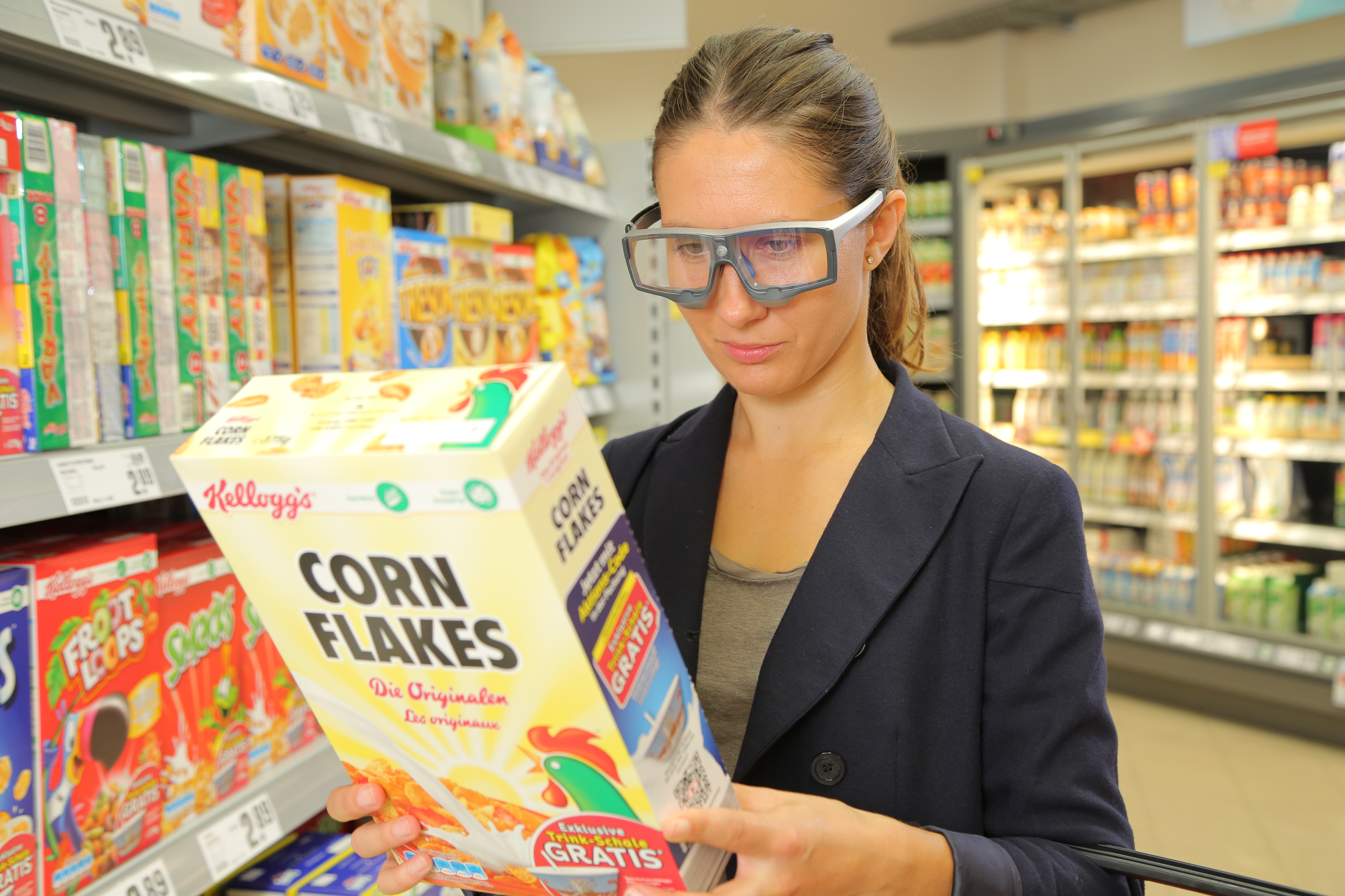
On “Black Friday” in the US, shoppers will come out in search of great deals, and researchers can measure the many physiological factors that motivate shoppers other than deals and price cuts.

Black Friday is one of the most exciting days for consumers in the United States. With the holidays right around the corner, significant deals and price cuts incentivize people to do bulk gift shopping. But is price the only factor influencing shoppers to dedicate hours waiting in line for a product?

In a neurophysiology study by Alexander et al., researchers found that participants who received a coupon while shopping online had a significant increase in oxytocin and a decrease in cortisol. A BIOPAC research system was used to gather ECG, respiration, and skin conductance data while the participants browsed a commerce website.

Another article by Ha-Brookshire and Bhaduri measured ECG response to stimuli that tested consumers’ perceived trust of businesses. The BIOPAC MP36R was used to record participants’ heart rates while they read negative reviews of an apparel business. The results found lower heart rate and less trust were correlated with the less favorable comments.

Conversely, a consumer research study by Langner et al. found that consumers’ love of brands is similar to their love of friends. Signals measured included EDA, EMG, ECG, and BPV, and were recorded by BIOPAC MP160/MP150 hardware to measure arousal levels while participants viewed different brand and interpersonal stimuli.

BIOPAC offers a variety of equipment that can be used in studies similar to the ones presented. To find more information on solutions for recording and analyzing signals such as EEG, EMG, respiration, skin conductance using the MP160/MP150, MP36, and other platforms, you can visit the individual application pages on the BIOPAC website. If you are interested in reading other related studies, visit the publications page on the BIOPAC website.

BIOPAC Systems, Inc. provides life science researchers and educators with data acquisition and analysis systems that inspire people and enable greater discovery about life. Visit us at www.biopac.com.

Practical Tips for Recording Physiological Data in MRI

The MRI system incorporates a VERY strong magnetic field and both emits and records radio frequency (RF) energy. This has impacts on subject and experimenter safety and on data accuracy.

By Alan Macy, BIOPAC Systems, Inc.

When recording physiological signals from subjects in MRI systems, two issues are very important:

  1. The MRI system incorporates a VERY strong magnetic field. It is extremely important that no significant magnetically susceptible objects are taken into the room that houses the MRI System.  All materials have some degree of magnetic susceptibility. However, materials with mass magnetic susceptibility values, similar to or have magnitude less than that of water (-0.91E-8 m^3/kg), are typically permitted to be used in the vicinity of MRI Systems. No electrodes, electrode leads, transducers or cabling, that contain significant magnetically susceptible components, should be used when recording data from a subject in the MRI.
  2. The MRI System both emits and records radio frequency (RF) energy. This energy is typically in the range of 5 to 500 MHz. For a 3T MRI, the characteristic proton precession frequency will be 3T * 42.7 MHz or 128.1 MHz.  MRI generated RF energy will, at least, partially reflect from encountered conductors and induce current flow in those conductors. Signal reflections and induced currents can disrupt sensitive imaging operations by creating signal artifacts. Also, RF induced currents, if not properly controlled, can cause local heating.

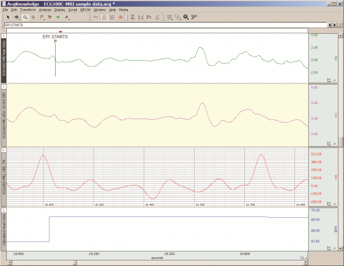
Care must be taken to limit the flow of RF energy both in and out of the room that houses the MRI. RF energy from the MRI superimposes with the very small biopotentials and transducer signals in the vicinity of the subject. Alternatively, RF energy from recording or other equipment in the MRI Control Room (e.g. digital clocks and power supply switching noise) can leak into the MRI chamber, via subject cabling, and cause degradation of the image created by the MRI. It can be complicated to record biopotentials, from a subject’s body, during an MRI scan. These signals are very tiny and the RF energy, and associated magnetic field shifting, generated by the MRI corrupts the recording. Accordingly, successful biopotential recording in the MRI involves the use of specialized amplifiers, optimized cabling, patch-panel filtering, advanced signal processing and/or synchronization with MRI scanning processes to perform clean measurements.

When RF energy is pulsed into the subject, this energy spreads throughout the subject because the subject is a volume conductor. As the MRI magnetic gradient shifts in synchrony with RF energy pulsing, the gradient shift induces a current flow in conductors that intersect with the field. From a practical standpoint, RF energy pulsing and gradient-induced current flows often manifest themselves as repetitive artifact signals directly in the band of the physiological generated biopotentials. This is because RF energy pulsing and magnetic field gradient switching are performed synchronously for imaging and the repetition rates for image slice generation usually fall within the range of 1-100 Hz. Depending upon the biopotential frequency range (from lowest to highest: EGG, EOG, ECG, EEG and EMG), different amplification and signal processing techniques can be used to isolate the signal of interest. Furthermore, it’s often helpful to control imaging processes so that the MRI artifact signal spectrum does not unduly impact the measured physiological signal spectrum.

For more information on BIOPAC’s MR Safe and MR Conditional solutions, visit BIOPAC’s MRI page or view BIOPAC’s full line of electrodes, amplifiers, and wearable, wireless transmitters and loggers.

Download a related and important application note that describes the relationship between MRI imaging sequences and associated physiological measurements, when subjects are monitored in the MRI. In particular, this note addresses newfound insights on the impact that imaging sequence types have upon simultaneously recorded electrocardiography (ECG) and impedance cardiography (ICG) subject measurements.

BIOPAC Systems, Inc. provides life science researchers and educators with data acquisition and analysis systems that inspire people and enable greater discovery about life.  Visit us at www.biopac.com.

Five Steps to a Better Physiology Lab – Preparation

Integrating new hardware and software into the Physiology lab curriculum…key points from instructors who have created successful labs that will greatly increase the chances of a rewarding lab experience for both students and instructor.

by William McMullen, BIOPAC Systems, Inc.

Integrating new hardware and software into the Physiology lab curriculum can be intimidating. Despite the best of early-summer intentions, in fall Instructors often find themselves apprehensively staring at a box full of unfamiliar equipment a few short days before the first lab. Luckily, the Biopac Student Lab excels in this situation, as it is easy to use and includes all materials necessary for students to prepare, record and analyze their data. This means that, in a crunch, if all one did was install the software and verify the hardware connections, most students should obtain meaningful data. Beyond the default setup, there are multiple customization options as described in the user support documentation. There are, however, some key points we have learned from instructors who have created successful labs that will greatly increase the chances of a rewarding lab experience for both students and instructor.

1. Organize the lab such that each student participates equally and each obtains recorded data from their bodies.
Students are more motivated when working with their own data. The Biopac Student Lab requires a minimum of three students for most lessons; a Recorder, a Director and a Subject. At the end of each lesson recordings, the students should rotate through these rolls and rerun the lesson until each student has recorded their own data. In order to provide enough recording time in a standard 2 hour lab period, the number of students per lab station should be four to five.

2. Setup basic Lesson Preferences before the first lab to minimize confusion.
Run each lesson* that will be used and setup the following:

  • If using lessons L02, L07-L09 or L12-L17, different models of transducers may be used. Check that the image and transducer descriptions in the lesson guide match the hardware used. If changes are needed, choose File > Lesson Preferences and select the desired transducer.
  • Disable undesired recording tasks by choosing File > Lesson Preferences > Lessons Recordings. If none of the recording tasks are desired (if using a custom procedure), disable all and only generic information will be displayed in the lesson guide.

*Note: If using the optional Curriculum Management System (available in BSL 4.1), only changes to the Host computer are required and all other computer/lab stations will be automatically updated. If not using this option, the changes must be made to each computer.

3. For Lessons L12 and L13 (Pulmonary Function), greatly reduce calibration time by matching the hardware with the software.
Label the lab station computer, the MP device and the Airflow Transducer (SS11LA/L) and make sure they are always matched. Run lesson 12 and check File > Preferences > Calibration Syringe Size to make sure it matches the syringe that will be used. Choose File > Preferences > Calibration Syringe Values and then select “Set once and use stored values”. Carefully run through the calibration using the syringe. From then on, when the students run the lesson, they will only need to calibrate for zero baseline (no syringe) which is easy and only takes a few seconds.

4. Students should have a basic understanding of the software before the first lab.
All Biopac Student Lab lessons use the same basic software tools and if students have a basic understanding of these tools in advance, they will experience greater success obtaining good data and having a positive lab experience. Students should be instructed to download the free Biopac Analysis software onto their personal computers.

This software not only provides the ability to analyze the data recorded in the lab but also allows students to prepare for a lab by studying the included tutorial videos and sample data. Some instructors give a test prior to the first lab to make sure students reviewed the BSL Tutorial Video.

5. Demonstrate proper electrode preparation and explain ways to minimize motion artifact. 
For lessons that use electrodes, good electrode contact and minimal EMG/motion artifact are keys to obtaining good data. Although this is explained in the procedures, it cannot be emphasized enough. Demonstrate the cleaning of the skin (with soap or alcohol) to remove dirt and oil and the abrading around the electrode site to remove the dead epidermal layers that impede electrical flow. Always place a drop of electrode gel on the electrode and wait at least 5 minutes after electrode placement before beginning a recording. To minimize EMG artifact, the student should be as relaxed and motionless as possible (unless procedure calls for movement). Talking, blinking (during EEG or EOG recording), laughing or deep breathing will add unwanted EMG artifact to the data.

These easily implemented steps will greatly assist students in obtaining good data from the very first lesson, leading to a positive lab experience and ultimately gaining a better understanding of Physiology.


BIOPAC Systems, Inc. provides life science researchers and educators with data acquisition and analysis systems that inspire people and enable greater discovery about life. BIOPAC lets you measure physiology anywhere, with innovative, compatible wireless, wearable solutions that can be used by anyone for meaningful discovery. We make high-quality scientific tools for physiology measurement and interpretation with superior compatibility and world-class customer service and support. Visit us at www.biopac.com.

error: Content is protected !!