ENS: Signals for Studying the Body’s “Second Brain”

We have all heard the saying “thinking with one’s stomach,” a cliché that suggests basing one’s critical thought processes on the whims of their digestive system. However, there may be more than a little truth to this saying. Often referred to as the body’s “second brain,” the enteric nervous system (ENS) comprises a complex network of neurons and ganglia embedded within the walls of the gastrointestinal tract. In recent years, researchers have increasingly recognized the significance of the ENS and its application in physiological research.

The ENS is a division of the autonomic nervous system (ANS) responsible for regulating the digestive process. It consists of millions of neurons spread throughout the esophagus, stomach, small intestine, and large intestine. These neurons form an intricate network that can function independently of the central nervous system (CNS) but also communicates with it. The ENS controls essential gastrointestinal functions, such as peristalsis, secretion of digestive enzymes, and blood flow regulation within the gut.

Several effective techniques are available to researchers to evaluate and measure ENS activity for their studies. Electrogastrography (EGG) provides a noninvasive approach to measuring the electrical activity of the stomach muscles. EEG can help researchers understand the rhythm and coordination of stomach contractions, aiding in the diagnosis of motility disorders.

In addition to EGG, a range of other physiological signals may be applied to ENS research. A recent study published in the June 2023 edition of Nature Communication investigated the gut-brain relationship by measuring physiological response to stimulation of the digestive system via different noninvasive techniques: EGG, electroencephalogram (EEG), skin conductance response (SCR), heart rate (HR), and heart rate variability (HRV). Research participants ingested a small vibrating probe that provided varying levels of stimulation as it passed through the lower digestive tract. EGG, EEG, and SCR signals were recorded along with hemodynamic signals via a BIOPAC data acquisition and analysis system. Researchers concluded that these techniques provided a viable alternative to more invasive approaches to measuring the body’s response to sensory stimulation of the digestive tract with, according to the published report, “implications for understanding gut feelings and gut-brain interactions in healthy and clinical populations.”

ENS ResearchMore invasive techniques for studying the ENS can still provide researchers with valuable data. A 2023 study by a team of South Korean researchers investigated how radial stretching affects intestinal contractions. The study also examined how drugs targeting the ENS affect such contractions to develop treatments for gastrointestinal disorders. Samples of intestinal tissue were taken from participants undergoing aggressive treatment for colorectal cancer. Samples were preserved in a tissue chamber and connected to a BIOPAC isometric force transducer to measure contractions, with data recorded and analyzed with a BIOPAC MP160 system running AcqKnowledge software. The study was able to detect regional differences in the stretch reflex of colon tissue via electrophysiological recordings of intestinal smooth muscles.

These are some of the ways in which researchers are currently using BIOPAC technologies to better understand the relationship between the ENS and other neurophysiological processes. For additional information on this topic, see our page on AcqKnowledge’s Gastric Myoelectric Activity feature and our EGG citations.


If you would like to know more about how BIOPAC can help you plan and equip your ENS study, reach out to your regional sales representative today.
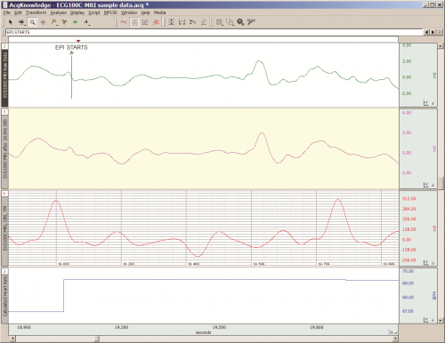
Practical Tips for Recording Physiological Data in MRI

The MRI system incorporates a VERY strong magnetic field and both emits and records radio frequency (RF) energy. This has impacts on subject and experimenter safety and on data accuracy.

By Alan Macy, BIOPAC Systems, Inc.

When recording physiological signals from subjects in MRI systems, two issues are very important:

  1. The MRI system incorporates a VERY strong magnetic field. It is extremely important that no significant magnetically susceptible objects are taken into the room that houses the MRI System.  All materials have some degree of magnetic susceptibility. However, materials with mass magnetic susceptibility values, similar to or have magnitude less than that of water (-0.91E-8 m^3/kg), are typically permitted to be used in the vicinity of MRI Systems. No electrodes, electrode leads, transducers or cabling, that contain significant magnetically susceptible components, should be used when recording data from a subject in the MRI.
  2. The MRI System both emits and records radio frequency (RF) energy. This energy is typically in the range of 5 to 500 MHz. For a 3T MRI, the characteristic proton precession frequency will be 3T * 42.7 MHz or 128.1 MHz.  MRI generated RF energy will, at least, partially reflect from encountered conductors and induce current flow in those conductors. Signal reflections and induced currents can disrupt sensitive imaging operations by creating signal artifacts. Also, RF induced currents, if not properly controlled, can cause local heating.

Care must be taken to limit the flow of RF energy both in and out of the room that houses the MRI. RF energy from the MRI superimposes with the very small biopotentials and transducer signals in the vicinity of the subject. Alternatively, RF energy from recording or other equipment in the MRI Control Room (e.g. digital clocks and power supply switching noise) can leak into the MRI chamber, via subject cabling, and cause degradation of the image created by the MRI. It can be complicated to record biopotentials, from a subject’s body, during an MRI scan. These signals are very tiny and the RF energy, and associated magnetic field shifting, generated by the MRI corrupts the recording. Accordingly, successful biopotential recording in the MRI involves the use of specialized amplifiers, optimized cabling, patch-panel filtering, advanced signal processing and/or synchronization with MRI scanning processes to perform clean measurements.

When RF energy is pulsed into the subject, this energy spreads throughout the subject because the subject is a volume conductor. As the MRI magnetic gradient shifts in synchrony with RF energy pulsing, the gradient shift induces a current flow in conductors that intersect with the field. From a practical standpoint, RF energy pulsing and gradient-induced current flows often manifest themselves as repetitive artifact signals directly in the band of the physiological generated biopotentials. This is because RF energy pulsing and magnetic field gradient switching are performed synchronously for imaging and the repetition rates for image slice generation usually fall within the range of 1-100 Hz. Depending upon the biopotential frequency range (from lowest to highest: EGG, EOG, ECG, EEG and EMG), different amplification and signal processing techniques can be used to isolate the signal of interest. Furthermore, it’s often helpful to control imaging processes so that the MRI artifact signal spectrum does not unduly impact the measured physiological signal spectrum.

For more information on BIOPAC’s MR Safe and MR Conditional solutions, visit BIOPAC’s MRI page or view BIOPAC’s full line of electrodes, amplifiers, and wearable, wireless transmitters and loggers.

Download a related and important application note that describes the relationship between MRI imaging sequences and associated physiological measurements, when subjects are monitored in the MRI. In particular, this note addresses newfound insights on the impact that imaging sequence types have upon simultaneously recorded electrocardiography (ECG) and impedance cardiography (ICG) subject measurements.

BIOPAC Systems, Inc. provides life science researchers and educators with data acquisition and analysis systems that inspire people and enable greater discovery about life.  Visit us at www.biopac.com.

error: Content is protected !!