Neuroergonomics: Measuring Cognitive Workload in Real-World Tasks

In some high-stakes jobs, the pressure can feel overwhelming. From air traffic controllers tracking dozens of aircraft simultaneously to surgeons guiding robotic instruments through delicate procedures, many modern occupations require people to process extraordinary amounts of information while maintaining accuracy and focus. As workplaces become more automated and technologically sophisticated, understanding how the brain and body respond to these demands has become a central challenge for ergonomics researchers. A growing area of study, known as neuroergonomics, combines neuroscience, physiology, and human factors research to better understand how people perform in complex real-world environments.

In human factors research, neuroergonomics conintues to gain traction by offering a more comprehensive picture of cognitive workload than traditional measures. Two individuals may perform a task equally well while experiencing dramatically different levels of mental effort. Excessive cognitive workload can contribute to fatigue, stress, errors, and reduced situational awareness, while insufficient workload can lead to disengagement and decreased vigilance. Researchers are therefore concentrating attention on measuring not only what people do but also how their brains and bodies respond as they do it.

Among the most valuable tools for studying cognitive workload are physiological signals that provide objective measurements of mental state. Electroencephalography (EEG) can reveal changes in neural activity associated with attention and cognitive effort, while functional near-infrared spectroscopy (fNIRS) measures changes in cortical blood oxygenation linked to mental workload. Electrocardiography (ECG) and heart rate variability (HRV) offer insight into autonomic nervous system activity, and eye-tracking measures such as pupil diameter and gaze behavior can indicate shifts in attention and cognitive demand. More often, researchers are combining multiple synchronized signals to create a more complete view of how people interact with demanding tasks and environments.

A team of researchers from Drexel University and Lockheed Martin investigated how multiple physiological signals can be combined to measure cognitive workload across a range of mental activities. Participants completed tasks involving working memory, vigilance, risk assessment, attention shifting, situation awareness, and inhibitory control while researchers simultaneously monitored EEG, fNIRS, ECG, photoplethysmography (PPG), electrooculography (EOG), and eye tracking. Brain activity was measured using an fNIR Devices system alongside other physiological monitoring technologies. The researchers found that different physiological measures varied in their sensitivity to specific task demands, with neuroimaging measures providing particularly valuable insight into changes in workload. The findings demonstrate the value of combining signals from the brain, heart, and eyes to create a broader perspective of cognitive effort in complex environments.

Researchers at Loughborough University in the United Kingdom examined how rising task complexity influences cognitive workload in modern manufacturing environments. Participants performed a series of assembly tasks while researchers recorded pupil diameter with Tobii eye-tracking and measured HRV with wearable ECG sensors. By comparing physiological responses across low-, medium-, and high-complexity tasks, the team identified significant changes in both pupillometry and cardiac indicators of cognitive load. Their findings suggest that physiological monitoring can provide an objective means of evaluating cognitive ergonomics in highly digitalized manufacturing environments and may help support the design of more sustainable, human-centered workplaces.
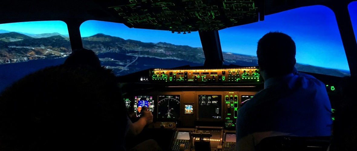
flight simAnother important challenge in neuroergonomics is determining which physiological measurements provide the most reliable indicators of mental workload. A collaborative team of researchers from France investigated how physiological measurements can be used to establish standardized levels of mental workload during aviation-inspired tasks. Using the Multi-Attribute Task Battery (MATB-II), participants performed simulated flight-related tasks under varying levels of demand while researchers measured EEG, eye-tracking, electrodermal activity, and cardiac activity. ECG and EDA signals were acquired using a BIOPAC data acquisition and analysis system running AcqKnowledge software. The study found that several cardiac, ocular, and electroencephalographic measures successfully distinguished high-workload conditions and correlated with subjective workload ratings. The researchers found that several physiological measures, particularly EEG, eye tracking, and cardiovascular metrics, reliably distinguished between different workload levels and closely tracked participants’ subjective assessments of task difficulty. Their findings suggest that multimodal physiological monitoring could support the development of adaptive systems that detect cognitive overload in real time, improving both performance and safety in demanding operational environments.

These studies highlight the increasingly multimodal nature of neuroergonomics research. Eye tracking provides insight into attention and visual processing, cardiovascular measures reveal how the autonomic nervous system responds to mental demands, and brain-imaging techniques such as EEG and fNIRS offer direct windows into neural workload. By combining these complementary perspectives, researchers are moving beyond simple measures of task performance toward a deeper understanding of how people think, adapt, and perform in complex environments. As workplaces continue to evolve alongside automation and intelligent technologies, neuroergonomics is helping researchers design systems that better support human capabilities, improve safety, and reduce the hidden costs of cognitive overload.


Are you planning a multimodal neuroergonomics study? Our team of experts can help you plan your research and identify the tools that will provide the best possible data.

2025: The Year in Review

As 2025 winds to a close, there is much to celebrate both here at BIOPAC and in the ever-expanding frontiers of research that our products engage with every day. Let’s take a quick look at where we have been over the past 12 months and what the future holds.

Popular Posts

Speaking of engagement, this is the time of year when we do a bit of self-assessment and revisit what topics our readers were most inspired by and most interested in. Apparently, many of our readers are closet gamers at heart, because our blog on research on game design and gaming impacts drove more eyes to our site than any other topic this year, followed by posts on cognitive neuroscience and aging and the human impact of isolation.

New Products and Updatesclimbing kid

On the heels of the 2024 release of our flagship MP200 data acquisition and analysis system and a significant upgrade to AcqKnowledge software, 2025 concludes with another milestone: the imminent release of AcqKnowledge 6.0.2. This latest version features a host of improvements and additions, including full integration of MedelOpt+, which blends whole-head functional near-infrared spectroscopy (fNIRS) with electroencephalography (EEG) in a single, highly flexible device.

BIOPAC in the Lab

Every year, BIOPAC products continue to drive the most exciting research in physiology, psychology, and neurology, and 2025 was no different. Each month, we highlight breakthrough studies in which BIOPAC played a role in gathering and analyzing key data. These are usually centered around a theme and feature research published in the last few years. To close out the year, we like to spotlight a couple of significant studies from 2025 that didn’t get mentioned.

A collaborative study by researchers from the University of Georgia and Beijing Normal University examined how mothers and children coordinate their physiological responses during stressful interactions. Using BIOPAC data acquisition systems coupled with wireless ECG transmitters, the team simultaneously recorded cardiac activity from each participant pair as they completed structured challenge tasks. This setup provided the temporal precision needed to track changes in heart rate and moment-to-moment autonomic synchrony, revealing how caregivers and children influence one another biologically during shared stress. The study found that greater synchrony was linked to both emotional dynamics and regulatory behaviors during the tasks, highlighting the deep coupling between social relationships and physiological responses. The wireless BIOPAC configuration enabled naturalistic interaction while capturing high-quality dyadic data, a persistent challenge in synchrony research.

In its first full year of availability, the MP200 is already making waves in the research community. At Guangdong University of Science and Technology in China, researchers examined how head-shoulder posture and cervical muscle activation differ between college students with chronic neck pain (CNP) and healthy individuals. The team employed surface electromyography (sEMG) recorded with a BIOPAC MP200 data acquisition system and AcqKnowledge software. Researchers placed surface electrodes over targeted neck muscles to record raw EMG signals during various strength tests and physical tasks. Students with CNP demonstrated significantly poorer head and shoulder alignment, reduced neck extensor strength, and altered EMG activity, indicating neuromuscular dysfunction associated with pain. Findings showed the connection between altered posture and impaired muscle function in chronic neck pain. The study demonstrates the effectiveness of surface EMG to support clinical diagnosis and rehabilitation.

For more inspiration as we head into 2026, explore our webinar library for tips, tricks, and methods directly from experts across a range of research fields.


If you are planning to launch a study for 2026, now is the time to reach out to your nearest BIOPAC customer service representative for help finding the right tools to get started.

Brain and Body: SPA-fNIRS Solves Physiological Signal Noise

Functional near-infrared spectroscopy (fNIRS) offers researchers a powerful noninvasive tool for studying brain activity by measuring changes in blood oxygen levels. These changes reflect how various brain regions respond to stimuli such as tasks that involve thinking, emotion, movement, and social interaction. These are often in natural, real-world settings that are not possible with other brain imaging methods. However, fNIRS is not without its own limitations. Systemic fluctuations in the body, such as breathing patterns, cardiovascular changes, and autonomic responses, can result in signal noise that is misinterpreted as brain activity by traditional fNIRS. The solution: a visit to the SPA.

Systemic Physiology Augmented fNIRS (SPA-fNIRS) is an advanced approach to fNIRS that goes beyond measuring local changes in cerebral oxygenation. While standard fNIRS measures cortical hemodynamics by tracking oxygenated (HbO) and deoxygenated (HbR) hemoglobin, noise originating from systemic physiological changes can corrupt those signals. Combining data from both neural and systemic physiological signals, SPA-fNIRS provides researchers with a more diverse and accurate view of how the brain and body interact in real time.

The method is being applied across various fields, including psychophysiology, cognitive neuroscience, exercise physiology, mental health, and human performance research. Studies have utilized SPA-fNIRS to investigate a range of topics, including meditation and emotional regulation, movement control, and social interaction. The broader goal is to move beyond studying the brain in isolation and to understand it as part of an integrated mind-body system. This holistic view is transforming how scientists interpret brain-function data, opening new paths for applied research in education, rehabilitation, and human-machine interface design.

soundResearchers in India investigated the impact of a traditional sound-based meditation technique, known as the Mind-Sound Resonance Technique (MSRT), on both brain and heart activity. Using fNIRS to track changes in oxygen levels in the prefrontal cortex, and a BIOPAC data acquisition and analysis system with AcqKnowledge software to record and analyze heart rate variability (HRV), the study employed a SPA-fNIRS design, integrating neural and physiological data in a single, synchronized dataset. HRV analysis allowed the researchers to quantify shifts in autonomic balance during and after meditation. The results showed increased oxygenation in the right prefrontal cortex and enhanced low-frequency HRV components, reflecting greater parasympathetic regulation. Together, these findings suggest that sound-based meditation not only improves brain oxygenation but also promotes calmer, more balanced heart-brain dynamics, illustrating how SPA-fNIRS methods can reveal the comprehensive physiological impact of mindfulness practices.

In her doctoral research at the Université de Lille in France, neuroscientist Aurore Guérin examined how the brain controls movement speed using a multimodal combination of fNIRS and physiological data recording systems. Participants performed repetitive arm movements at fast, natural, and slow tempos. At the same time, the study captured oxygenation in the prefrontal and motor cortices using fNIRS, alongside heart rate and respiration signals collected with a BIOPAC data acquisition system. A respiratory effort transducer measured breathing patterns, while a BioNomadix ECG module recorded cardiac activity for HRV analysis. All physiological signals were synchronized and analyzed in AcqKnowledge software, allowing the team to identify and remove systemic influences (e.g., heartbeat and breathing rhythms) from the fNIRS signals, following a SPA-fNIRS approach. The results revealed that faster movements primarily engaged motor regions, while slower, more deliberate pacing resulted in higher prefrontal activation, reflecting greater cognitive control. By integrating physiological monitoring with fNIRS, the study provided a clearer picture of how the brain and body coordinate timing and control during movement.

These studies illustrate how SPA-fNIRS is reshaping the way scientists investigate the interaction between brain and body. By combining neural and physiological data in a single synchronized system, researchers can separate brain activity from systemic noise and uncover the interplay between cognition, emotion, and autonomic regulation. For more information on this topic, watch our webinar on Multimodal SPA-fNIRS and check our our latest multimoldal fNIRS research products.


Are you planning a study that blends fNIRS with other physiological signals? Contact your local BIOPAC customer service representative to learn more about how SPA-fNIRS can benefit your research.

Boom! Pow! Clang! Exploring Stress Resilience with Startle Response and PPI

We have all felt that electrical jolt that comes with hearing a sudden, unexpected loud noise—the clang of a dropped cooking pan, the blast of a car horn, or the pop of a balloon. The reaction is instantaneous and nearly uncontrollable as muscles tense, eyes blink, pupils dilate, and one’s heart seems to leap from the chest. The startle response—a rapid, automatic reaction to sudden stimuli like loud noises—has a practical purpose, helping protect us from outside threats. It can also pose a danger if the response occurs while engaging in precision activities, such as driving a vehicle or operating machinery.

Within the psychophysiological field, the startle response can reveal how our brains and bodies process stress, attention, and emotional regulation. In pursuit of ways to mitigate its detrimental effects, researchers often study this reflex alongside prepulse inhibition (PPI). In this latter process, a weaker sound presented just before the startling noise reduces the strength of the response. To capture data when studying these phenomena, scientists use physiological signals such as electromyography (EMG) to measure the tiny muscle contractions around the eye during a blink, electrocardiography (ECG) for heart rate changes, electrodermal activity (EDA) for skin conductance, and respiration to track stress responses. In addition, researchers often pair these physiological measures with brain imaging techniques, like functional magnetic resonance imaging (fMRI) or electroencephalography (EEG), as well as cognitive tasks to build a complete picture of how startle responses connect with brain function and behavior.

Studies of startle response and PPI intersect with a wide range of fields, from neuroscience and psychiatry to aviation, defense, and occupational health. Clinical investigations of altered startle and PPI patterns are helping scientists uncover biomarkers of vulnerability or resilience. These studies are providing new insight into potential treatments for conditions like schizophrenia, PTSD, anxiety, and neurodegenerative disorders.

pilotIn applied contexts, like pilot training or high-stress professions, startle measures are used to understand how sudden stress impacts performance and decision-making. A study conducted by researchers at the University of Zagreb in Croatia with U.S. partners aimed to determine whether stress resilience markers, including startle response and PPI, could predict pilot performance under sudden startle events. Seventeen military pilot cadets completed baseline testing of physiological and cognitive features—including startle reflex habituation, PPI, heart rate variability (HRV), and multitasking ability—followed by flight simulator sessions with induced startle events. Physiological signals such as ECG, EMG (eye-blink startle reflex), respiration, and EDA were recorded via their respective BIOPAC amplifiers connected to a data acquisition and analysis system. Regression modeling showed that startle habituation, autonomic recovery, and cognitive task performance provided an accurate prediction of flight performance. The findings suggest that psychophysiological measures can help identify pilots who are more resistant to startle-induced performance degradation. Such insights could offer practical applications in pilot screening, training design, and aviation safety.

A team of Spanish researchers wanted to see how short-term (acute) stress affects both PPI and higher-level thinking (working memory) in healthy young adults. They used the Maastricht Acute Stress Test (MAST) to induce stress in one group, while another group did a non-stressful control task. After this, all participants heard loud noises that triggered the eye-blink startle response. In some trials, a prepulse immediately preceded the loud noise, which typically reduces the blink reflex. The startle (eye-blink) was measured from the orbicularis oculi muscle using BIOPAC Ag/AgCl electrodes connected to an Electromyography amplifier that fed data to a data acquisition system running AcqKnowledge software. This allowed the researchers to precisely record the intensity of a blink and the degree to which prepulse inhibited it. The team also tested working memory using tasks like digit span backwards and letter-number sequencing, in addition to other cognitive tests. They found that stress made startle responses stronger and reduced PPI, thereby degrading the participants’ ability to filter out the loud noise when it was preceded by a quieter one. Stress also impaired performance on working memory tasks compared to the non-stressed group. In practical terms, these results suggest that acute stress doesn’t just make one more reactive; it can interfere with basic sensory gating and cognitive control. This has implications for stressful jobs or situations (e.g., emergency responders or pilots) and possibly for understanding disorders where PPI or working memory is impaired.

These are just a few examples of how research into startle response and PPI can improve our understanding of the brain’s basic filtering and stress-response systems. The knowledge these studies provide may eventually translate into practical tools for diagnosis, prevention, and performance optimization. For additional information on research applications, check out our webinar on Startle and PPI.


Are you struggling to find the right tools to plan your startle response and PPI study? Our team of customer service experts will put you on the right path to gathering the best data possible for your research project.

Mind Over Matter: Cognitive Neuroscience and Aging

“Age is an issue of mind over matter. If you don’t mind, it doesn’t matter.”  —  Mark Twain.

While American author and humorist Mark Twain suggested that mental attitude could make aging less daunting, modern science shows that the changes our bodies and brains undergo over time are very real. Yet researchers are discovering that aging doesn’t have to mean inevitable cognitive decline. In fact, the growing field of cognitive neuroscience is revealing ways to track, understand, and even influence how our brains change with age, offering hope that we can preserve mental acuity, independence, and quality of life well into our later years.

When studying how aging affects the brain, cognitive neuroscience researchers use a variety of tools. In addition to standard behavioral tests like the Stroop test, n-back test, and verbal fluency exercises, which track reaction times, accuracy, and problem-solving strategies, they also rely on neuroimaging and physiological measurements to gather deeper insights. Neuroimaging techniques include electroencephalography (EEG), which monitors real-time electrical activity in the brain; functional near-infrared spectroscopy (fNIRS), which measures blood oxygen levels in specific cortical regions during cognitive tasks; and magnetic resonance imaging (MRI), which provides detailed structural and functional maps of the brain.

Other physiological signals play an essential role in this field, including heart rate variability (HRV), a key indicator of the autonomic nervous system’s ability to balance various physiological functions and handle stress. Electrodermal activity (EDA) allows researchers to track arousal among participants undergoing cognitive exercises. Electromyography (EMG) measures muscle activation during fine and gross motor tasks. Eye tracking and electrooculography (EOG) provide researchers with data on how aging impacts attention and the ability to process visual cues. Respiration and blood pressure provide vital information on vascular health, which is often linked to cognitive performance.

Taiwanese researchers from National Yang Ming Chiao Tung University in Taipei turned to HRV and fNIRS to uncover how aging and, more specifically, mild cognitive impairment (MCI), affect both brain and heart responses during cognitive tasks. Healthy young adults, cognitively healthy older adults, and older adults with MCI were asked to perform cognitive function and executive tests. Researchers recorded the participants’ prefrontal cortex (PFC) activation through changes in oxyhemoglobin using fNIRS and autonomic responses via HRV. The team gathered ECG data using a BIOPAC data acquisition module running AcqKnowledge data acquisition and analysis software, and equipped with an ECG amplifier. The study found that older adults who did not exhibit MCI showed lower global and executive cognitive performance, reduced HRV, but increased prefrontal activation. The research team interpreted this to be the result of compensatory neural effort. However, older adults with MCI showed even greater cognitive decline, but no further differences in HRV or prefrontal activation. The findings suggest that HRV and PFC activation patterns could serve as biomarkers for distinguishing healthy aging from early pathological decline.

To better understand how learning processes develop and change with age, researchers from Tilburg University in the Netherlands investigated how younger adults and older adults differ in their brain and body responses while learning a visuomotor sequence task. To capture these responses, the team measured EEG, skin conductance (EDA), heart rate, HRV, breathing rate, and eye movements during the task. Physiological data were collected using BIOPAC’s BioNomadix wireless dual ECG/respiration amplifier and PPG/EDA amplifier, which continuously recorded three-lead ECG, respiration, and EDA at 2,000 Hz, with signals processed and analyzed in AcqKnowledge software. This setup allowed the researchers to precisely synchronize physiological activity with learning performance. Both younger and older adults improved with practice, but older adults needed more trials to reach the same precision. Brainwave activity showed apparent age differences, while measures like HRV, respiration, and skin conductance did not. This suggests that EEG is a more sensitive marker of age-related learning changes and could help guide training programs for older adults.

readingA long-term study by researchers from Radboud University Medical Center in the Netherlands, together with UK collaborators, tracked a group of healthy older adults over nearly 11 years to see how their brain blood-flow regulation changed by their 80s. Using transcranial Doppler ultrasound to measure blood velocity in the middle cerebral arteries, the team also monitored control signals including heart rate, blood pressure, and breathing gases. Physiological data were recorded with a BIOPAC data acquisition and analysis system, with end-tidal CO₂ (EtCO₂) measured using capnography via a BIOPAC carbon dioxide measurement amplifier. The researchers found a slight decline in CO₂ reactivity but, surprisingly, an improvement in dynamic cerebral autoregulation (the brain’s ability to maintain steady blood flow despite blood pressure changes). These effects were most apparent in participants who later reported memory complaints, suggesting that while aging may reduce vascular responsiveness, the autoregulatory system may help compensate for early cognitive challenges. The findings show how vascular health in the brain relates to aging and memory, providing valuable insight for detecting early risks of dementia and shaping preventive strategies.

These studies highlight how combining brain imaging with physiological measurements is helping scientists reveal the complex relationship between aging, cognition, and overall health. Through cognitive neuroscience, researchers are identifying early signs of decline and gaining a better understanding of how the brain and body adapt over time. Research is paving the way to more effective screening, personalized interventions, and lifestyle strategies that may help people maintain mental sharpness and independence well into older age. For more information on the tools and techniques being applied to cognitive neuroscience research, see our Webinars and visit our page on Cognitive State Analysis.


Are you planning a study on how aging affects the brain or just looking to expand your cognitive neuroscience toolbox? Our customer service team is ready to help you find what you need to obtain the best possible data.

Aesthetically Pleasing: Exploring How the Brain Perceives the Arts

Beauty is in the eye of the beholder—a mostly subjective experience, so the saying implies. While we may share many common ideals about beauty and aesthetics, our reactions to art, music, and literature can diverge, converge, and overlap to a myriad of degrees. Neuroaesthetics explores how the brain perceives and responds to art, music, and other aesthetically evocative experiences. Combining neuroscience with psychology and the arts, researchers seek to understand why humans find certain images, sounds, or designs appealing and how these perceptions influence our emotions, cognition, and behavior. This interdisciplinary science has practical applications across a wide range of fields, including design, marketing, education, and mental health therapy, where understanding aesthetic experience can inform and enhance user engagement, learning, and emotional well-being.

Biometric signals provide insight into emotional and cognitive processing, allowing researchers to gain valuable data on how the brain creates and responds to aesthetically engaging stimuli. Commonly used techniques include electrodermal activity (EDA), which measures changes in skin conductance linked to emotional arousal; electroencephalography (EEG), which tracks brainwave patterns associated with attention and engagement; functional near-infrared spectroscopy (fNIRS), which monitors blood flow changes in the brain; and functional magnetic resonance imaging (fMRI), which provides high-resolution images of neural activity.

Additionally, the use of virtual reality (VR) and augmented reality (AR) is expanding in neuroaesthetics research, allowing scientists to create controlled, immersive environments that mimic real-world experiences. These technologies offer new ways to investigate how individuals engage with art and design in dynamic, interactive settings, pushing the boundaries of how we understand and harness aesthetic experiences.

Researchers in Shanghai, China, for example, investigated how different visual modes—2D and 3D VR—affect emotional arousal, demonstrating how immersive environments influence emotional experiences. The research team utilized EEG to monitor brain activity and EDA to measure skin conductance responses, both indicators of emotional arousal. EDA and EEG data were recorded while study participants wore VR headsets to view 2D and 3D environments. Data was collected and analyzed using a BIOPAC MP system with AcqKnowledge software. The findings revealed that 3D VR environments elicited more intense emotional stimulation than 2D settings, as evidenced by higher beta (21–30 Hz) EEG power and increased EDA responses. According to the published findings, results suggest that enhanced perception in 3D environments leads to greater emotional engagement, highlighting the potential of 3D VR in applications such as immersive therapy, virtual training, and interactive entertainment.

MusicA team of German researchers recently examined the intersection of neurology, psychology, music, and literature. Their findings, published in the journal NeuroImage, explored how melodic features influence the neural processing of spoken poems and songs, examining the interplay between language and music. Researchers employed fMRI to investigate the neural mechanisms underlying the processing of musical and nonmusical verse. The fMRI data revealed that both spoken poems and songs activated bilateral temporal auditory areas of the brain. One aspect of the study was the measurement of participants’ emotional responses to the stimulus, i.e., whether they liked the song or poem to which they were listening while the fMRI scan was conducted. To gather this data, participants interacted with a BIOPAC differential pressure transducer connected to the participants’ right and left index fingers to record the degree to which they enjoyed the music. The researchers stated the findings suggest that melodic properties are central to the aesthetic experience of both speech and music.

A study published by Britain’s Royal Society looked at the connection between aesthetic appreciation and emotional states, specifically how exposure to art or music can shift feelings of anxiety to curiosity. To understand how the brain perceives beauty, the researchers used EDA to assess participants’ physiological arousal during exposure to aesthetically pleasing stimuli, such as music. EDA data was gathered using a BIOPAC EDA amplifier connected to a data acquisition and analysis system while participants were administered anxiety-inducing electrical tactile stimulation during music listening sessions. The study demonstrated that aesthetic appreciation can reduce anxiety and promote curiosity by analyzing the EDA data alongside self-reported measures. The research team concluded that their results “might have potential applications across different human activities such as learning and therapy. The idea that aesthetic appreciation might affect anxious states and that expecting aesthetic rewards can transform anxiety into arousal and curiosity plays a central role in some psychotherapeutic models of human change.”

These examples provide a rough outline of a bigger picture, illustrating the many ways researchers use physiological signals to better understand the human mind’s response to art, music, and literature. For more information on the tools BIOPAC provides to assist in neurological and physiological research, see our online training and webinars.


Are you planning a research project to investigate the intersection of aesthetics and neuroscience? Contact a BIOPAC representative in your area to identify the right resources and equipment for a successful study.

A Remarkable 2024: Introducing Tools for a Better and Brighter Research Future

As 2024 winds down and we ready the confetti to ring in 2025, we at BIOPAC Systems Inc. want to toot our horn after wrapping up an exciting year of growth and innovation. This year brought some big wins for the research community, thanks to BIOPAC’s efforts to make physiological data acquisition and analysis more accessible and efficient than ever. Two major milestones were the launch of the MP200 system, the latest evolutionary step for our flagship data acquisition platform, and two feature-rich release updates to our AcqKnowledge data acquisition and analysis software.

Introducing the MP200: A New Achievement for Data Acquisition

MP200The MP system series has been the backbone of BIOPAC’s efforts to provide powerful and yet accessible data acquisition tools to researchers. Released in November, the newest incarnation of MP device, the MP200, ushers in a new era of precision, reliability, and flexibility for data acquisition. Designed to meet the evolving needs of researchers, the MP200 offers unparalleled scalability and compatibility with a wide range of physiological signals and measurements. Key enhancements include the following:

  • Enhanced Signal Quality: The MP200 uses advanced signal processing algorithms to ensure superior data integrity across diverse research applications, from neuroscience to exercise physiology.
  • Modular Design: The MP200’s modular architecture allows seamless integration with BIOPAC’s extensive sensors and amplifiers, creating near-limitless customization possibilities for complex experimental setups.
  • Future-Ready Connectivity: The MP200 facilitates real-time collaboration and analysis with high-speed data transfer protocols and cloud integration support.

Researchers will benefit from the MP200’s improved ability to streamline workflows and deliver insights with unmatched precision. This latest system exemplifies BIOPAC’s dedication to fostering groundbreaking discoveries in physiology and related fields.

AcqKnowledge Updates: Pioneering the Future of Data Analysis

AcqKnowledge data acquisition and analysis software has been a go-to tool for researchers for decades, and 2024 saw two major updates that brought exponential improvements: versions 5.0.10 and 6.0.0. These updates are packed with features that make data collection and analysis easier and more comprehensive.

One of the most impactful new licensed features is the integration of Lab Streaming Layer (LSL). This feature lets researchers sync and stream data from multiple sources in real time. Whether one is working on interdisciplinary projects or using several devices at once, LSL makes it simple to bring everything together for analysis. LSL also opens the door to a host of new products from our partners for which BIOPAC has added integration in AcqKnowledge. These include the Axon-R brain-computer interface (BCI) hub with augmented reality (AR) and artificial intelligence (AI) and the APEX wearable, wireless EEG system. Other new products have also been integrated into AcqKnowledge and are now available from BIOPAC, including new eye tracking products from Tobii and EyeLogic.

Another game-changing licensed addition, AcqKnowledge Cloud, allows users to securely store their data online, streamlining processes to access, share, and work on data from any location. Collaboration between teams is also easier, and users no longer need to worry about managing large files on their local system.

These updates are all about making work less complicated and more effective for researchers. With tools like these, diving into complex data, finding patterns, and pushing one’s research further has never been more straightforward.

Looking Ahead to 2025

More excitement is on the horizon for BIOPAC. Not only are several new product updates and releases in the works, but we will also be hosting our bi-annual 4T Human Physiology Conference on Tools, Trends, Techniques, and Technologies. Customers, educators, researchers, and other experts in Physiology and related fields will gather in Santa Barbara, California July 14-16. Participants will share knowledge, take part in hands-on workshops, and see what is shaping the future of research. Learn more and sign up today. Hopefully, we will be seeing you in 2025.

Everybody Hurts: Studying the Physiological and Neurological Components of Pain

A Buddhist saying states, “Pain is inevitable, but suffering is optional.” Pain’s dual nature has presented a conundrum for philosophy and the sciences. As a warning system, it protects us from harm, but it can also be the source of profound psychological and emotional damage. Pain is a complex and deeply subjective experience, yet it is one of the most critical areas of study in neuroscience.

Understanding pain involves not only exploring its physical origins but also decoding the neural circuits and physiological signals that contribute to its perception and regulation. Neuroscientific research into pain aims to answer fundamental questions about how pain is processed in the brain, how it varies from person to person, and how we can develop better treatments for chronic conditions.

This research uses various physiological signals to provide insights into pain mechanisms. Electroencephalography (EEG) captures brainwave activity, revealing how different regions of the brain respond to painful stimuli. Photoplethysmography (PPG) tracks blood flow changes, while electrocardiography (ECG) and skin conductance response (SCR) measure autonomic nervous system activity, offering clues about how the body reacts to stress and discomfort. Respiration patterns, too, are key indicators, as breathing can change in response to pain. Together, these signals help scientists map the body’s response to pain, shedding light on the underlying neural pathways and guiding the development of more effective pain management strategies. Understanding these physiological markers is a critical step toward personalized pain care and improving the quality of life for millions worldwide.

Pain research commonly focuses on the neurological mechanisms of pain to better understand its causes and how it can be treated. Researchers publishing their findings in the Journal of Biomedical and Health Informatics developed a device for studying pain perception biomarkers using thermal stimulation. Their device generates minutely controlled thermal stimuli using a computer-driven Peltier cell while simultaneously capturing EEG and PPG signals. Researchers identified the heat pain threshold (HPT) for each subject, defined as the maximum the subject can withstand when the Peltier cell gradually increases the temperature. The team used two-dimensional spectral entropy (SE) obtained from both the EEG and PPG signals to differentiate the condition of pain. EEG and PPG signals were fed to a BIOPAC MP36 data acquisition and analysis system running Biopac Student Lab (BSL) software. The study concluded that hemodynamics, brain dynamics, and their interactions can be used to discriminate thermal pain perception, which could have practical applications in monitoring pain perception. Possible applications include the study and treatment of “pathologies that affect the peripheral nervous system, such as small fiber neuropathies, fibromyalgia, or painful diabetic neuropathy.”

The pain mechanism associated specifically with fibromyalgia, one of the most common chronic pain disorders, was the focus of a study at the University of Jae´n in Spain. Despite its prevalence, much is still unknown about the underlying cause of this condition. Clinical and experimental studies of individuals diagnosed with fibromyalgia syndrome (FMS) have revealed autonomic nervous system (ANS) deficiencies. While both the parasympathetic and sympathetic components of the ANS have been studied, less is known about the sympathetic component as it relates to FMS. In this study, researchers used SCR as an indirect measure of the sweat response, which is controlled by the sympathetic mechanisms of the ANS. Researchers used a BIOPAC MP36 running AcqKnowledge to collect SCR data via an EDA lead set connected to participants’ fingers. The study found reduced skin conductance levels (SCL) and SCR in FMS patients compared to the non-FMS control group, indicating lower levels of sweating and reduced sympathetic activity in the skin. These factors provided the research team with a better understanding of the sympathetic nervous system’s role in FMS, with the potential for revealing new routes to better pain management and treatment.

For several decades, researchers and clinicians have studied the use of hypnosis to treat chronic pain. More recently, new technologies such as virtualpills reality have been incorporated into these techniques. A team of researchers from the University of Strasbourg in France studied the application of virtual reality hypnosis (VRH) to regulate HPT in healthy adult participants. Researchers used several physiological signals to measure autonomic function during the experiment, including ECG, respiration, SCR, and EEG. ECG signals were recorded using the BIOPAC ECG amplifier, while respiration rate was measured by a thermistor transducer that determined the difference in temperature between inhaled and exhaled air. SCRs were measured from the index and middle fingers of the dominant hand with a BIOPAC skin conductance transducer. Physiological signals were fed into a BIOPAC data acquisition system for analysis. A computer-generated immersive environment was created in VR and combined with a hypnotic auditory script. As with classical hypnosis treatments, the VRH scheme in this study began with an “induction period” followed by a “dissociation” period (separation between the mental state and the environment), followed by suggestions of pain reduction as analgesia. The study results supported a positive correlation between VRH and an increase in the participants’ pain threshold with promising implications for new non-pharmacological approaches to pain management and treatment.

These are just some of the ways that BIOPAC equipment is being used to explore the frontier of pain research and help ease the suffering of millions who deal with chronic pain and its physical and psychological effects. See our webinars page for additional information on how physiological signals can be applied to your next study.


Are you planning a study on pain or any related neurological or physiological area of research? Our team of experts is ready to help you find the tools to ensure success.

Mind and Machine: Exploring Brain-Computer Interfaces

In neuroscience, the frontier of brain-computer interfaces (BCI) holds the promise of bridging the gap between the human brain and external devices, offering revolutionary applications in fields ranging from robotics and prosthetics to communication and beyond. In the lab, scientists studying and developing new BCI technology leverage the human brain’s electrical and physiological signals.

Electroencephalography (EEG), functional near-infrared spectroscopy (fNIRS), and steady-state visually evoked potentials (SSVEP) are among the primary physiological and psychophysiological signals utilized in these investigations. Multimodal integration of two or more signal types allows researchers and engineers to take advantage of multiple data streams in their applications and studies.

EEG, which can measure electrical activity in the brain, is a cornerstone of BCI research. Researchers can decode various mental states and intentions by analyzing EEG patterns, enabling users to control external devices through mere thought. fNIRS complements EEG by measuring changes in blood oxygenation levels, offering insights into brain function with superior spatial resolution. SSVEP, on the other hand, capitalizes on the brain’s response to visual stimuli, providing a means for precise control in BCI applications.

The practical implications of BCI research extend far beyond the laboratory setting. In robotics, BCIs facilitate direct brain-to-machine communication, empowering users to manipulate robotic arms or navigate autonomous vehicles with their thoughts alone. A study published in the Journal of Information Science Engineering explored ways that an SSVEP-based BCI could be used to control assembly line robots via a headset measuring the operator’s EEG signals. The signals would communicate instructions to robots while operators identified defective parts, which the robots would pick out. During the experiment, EEG signals were obtained using a B-Alert X24 headset. The EEG-based BCI produced a 93.33% success rate in correctly identifying and removing defective parts from the assembly line process, allowing the operator to effectively “collaborate with the robot and communicate defective parts without manually operating the robot.”

Prosthetics benefit immensely from BCI advancements, enabling seamless integration with the user’s neural signals for intuitive control and enhanced functionality. A 2023 study by a team of researchers from the University of California Irvine explored the viability of creating low-cost cortical stimulators for bi-directional BCI (BD-BCI) research. BD-BCI takes brain-computer-interfaces to the next level with the brain not only providing motor commands to an artificial limb but also allowing it to receive sensory input from such a device. A BIOPAC data acquisition and analysis system was used to validate the charge balancing of subcortical electrocorticographic (ECoG) implants used in a stimulator. The charge balancing test was performed on phantom tissue designed to simulate real brain tissue. The resulting study successfully demonstrated “clinical validation of a fully-programmable electrical stimulator, integrated into an embedded BCI system.”

As researchers continue to refine BCI technologies, the possibilities for real-world applications are boundless. BIOPAC is among the companies on the cutting edge of BCI research, partnering with other innovators to provide the latest in multimodal BCI technologies such as Axon-R. For additional information on Axon-R, check out our recent webinar on the device.


Are you planning to explore the frontiers of BCI in the lab? Our regional customer reps have all the knowledge you need to integrate BIOPAC products into your BCI study.

Imaging is Everything: Data Acquisition in the MRI Environment

The marriage of physiological data recording and magnetic resonance imaging (MRI) has allowed scientists to unravel intricate connections between the mind and body. However, this synergy comes with its unique set of challenges that researchers must be aware of to ensure the integrity of both data and the well-being of study participants.

One of the foremost challenges in recording physiological data within the MRI lab is the strong magnetic field. Traditional monitoring equipment can be rendered useless or, worse, pose extreme safety risks due to their ferromagnetic components. Another critical consideration is the impact of radiofrequency (RF) pulses used in the imaging process. RF pulses can introduce artifacts in physiological recordings, affecting the quality of data collected. In previous posts we have discussed the basics of integrating data collection in the functional MRI (fMRI) environment as well as techniques for avoiding artifacts and signal noise while recording data in the MRI.

Researchers must also contend with challenges related to the confined space and noise levels within an MRI environment. A 2021 study conducted by the Lab for Autonomic Neuroscience, Imaging and Cognition (LANIC) at Jena University Hospital in Jena, Germany, focused on how the functional MRI (fMRI) environment itself affected outcomes in autonomic research. Specifically, the study investigated the influence of a loud and cramped environment during MRI on resting heart rate variability (HRV) measures and whether a familiarization session with MRI equipment might lower anxiety levels among participants. The research team recorded an electrocardiogram (ECG) and a photoplethysmogram (PPG) over 15 minutes from participants using a BIOPAC data acquisition and analysis system connected to finger-attached PPG sensors during non-MRI recording. For recording in the fMRI, researchers used a separate BIOPAC data acquisition system equipped with an MR-Conditional PPG module. While the study did establish a link between participant anxiety and HRV levels in the lab and fMRI environment, it did not find that the familiarization sessions had a significant impact on HRV outcomes.

gas maskDespite the challenges, the recording of physiological signals in the MRI environment provides unique opportunities for gathering critical data. A study published in the journal Military Medicine in 2020 tested the effectiveness of F MRI (not to be confused with fMRI) scans in identifying cases of War Lung Injury (WLI) in Iraq and Afghanistan war veterans. WRI refers to reductions in lung function resulting from exposure to environmental factors such as sandstorms and burn pits. F MRI employs magnetic resonance imaging with perfluorinated gases as inhaled contrast agents to evaluate various pulmonary diseases. If successful, the approach would provide an alternative to surgical biopsy for diagnosing suspected WLI. The researchers measured respiratory waveforms throughout the imaging sessions, during both normal room air breathing and PFP-oxygen gas mixture breathing using a BIOPAC MR-Conditional pneumotachometer. The study found that “F MRI provides better insight regarding lung function abnormalities” than other standard methods and that “F MRI could provide a functional tool to assist in identifying subjects with the disorder and potentially could aid in the evaluation of novel therapeutic approaches.”

BIOPAC provides a wealth of tools and information resources for gathering data in the MRI environment. This includes multiple webinars on recording data in both MRI and fMRI settings. As the field continues to evolve, BIOPAC’s innovative solutions pave the way for groundbreaking discoveries at the intersection of physiological signal recording and medical imaging.


If you are interested in recording and analyzing physiological signals in your next MRI-based research study, BIOPAC sales representatives are standing by to help you choose the right tools and resources for the job.

error: Content is protected !!