No Pain, All Gain: How EMG and Other Physiological Signals Aid Injury Avoidance

It can happen anywhere, anytime: on the pickleball court, during a pristine nature hike, or even while stepping off the front porch on the way to work. An ankle turns, a knee buckles, and suddenly you’re on the ground wondering what just happened. While such injuries often seem random, researchers who study human movement know they are rarely without warning.

Kinesiologists, scientists who study the mechanics and physiology of movement, have found that subtle changes in muscle activation, fatigue, coordination, balance, and neuromuscular control often emerge long before a strain, sprain, or overuse injury becomes apparent. To better understand these hidden danger signs, researchers increasingly rely on physiological measurements that reveal how the body’s muscular, nervous, cardiovascular, and biomechanical systems respond to activity. Such insights help identify injury risk factors and guide strategies designed to keep people moving safely and effectively.

Among the many tools used in kinesiology research, electromyography (EMG) is one of the most common because it reveals when and how muscles activate during movement. However, EMG is only one part of the story. Researchers also study force production, joint motion, movement acceleration, cardiovascular activity, respiration, muscle oxygenation, and neural activity to better understand how the body adapts to physical demands. Together, these measures provide a more complete picture of movement and help uncover patterns that observation alone may miss.

The applications of this work extend far beyond sports. Physiological monitoring helps clinicians evaluate rehabilitation progress, guide return-to-activity decisions, support fall-prevention efforts among older adults, and help design safer workplaces and training programs. Increasingly, researchers are shifting from simply treating injuries to identifying and addressing the factors that contribute to them before they occur.

Researchers in India conducted a cross-sectional study comparing elite and recreational university badminton athletes to explore whether physiological and neuromuscular characteristics could serve as injury-risk markers. The investigators collected a range of performance and neuromuscular measurements, including surface EMG recordings, to examine differences between competitive levels and establish preliminary benchmarks for injury-risk stratification. Surface muscle activity was recorded using a BIOPAC two-channel data acquisition system as part of the neuromuscular assessment protocol. The study found that elite athletes exhibited distinct physiological and neuromuscular profiles compared to recreational players, suggesting that objective physiological markers may help identify movement patterns and performance characteristics associated with increased injury vulnerability. These conclusions support the growing use of physiological profiling to identify athletes who may benefit from targeted injury-prevention strategies before injuries occur.

A team of researchers in Tunisia investigated how fatigue of the ankle evertor muscles affects force perception and neuromuscular function in healthy adults. Ankle injuries are among the most common musculoskeletal injuries in both athletic and recreational populations, making ankle stability an important area of study. Using surface EMG to measure muscle activity before and after a fatigue protocol, the researchers examined how localized fatigue affected participants’ ability to accurately reproduce force levels. Researchers used a BIOPAC EMG amplifier and data acquisition system to record electrical activity from muscles involved in ankle stabilization. The researchers found that fatigue impaired force perception and altered neuromuscular responses, suggesting that fatigue may reduce the body’s ability to control ankle movements accurately. The study results add to growing evidence that fatigue-related physiological changes can increase injury risk, particularly during prolonged exercise or competition when neuromuscular control begins to decline.

A multi-institutional team of researchers from the United States examined neuromuscular function in individuals who had undergone anterior cruciate ligament (ACL) reconstruction, comparing them with healthy controls. The researchers wanted to better understand whether the relationships between muscle performance, muscle activation, and nervous system function change after ACL surgery. Participants completed a series of strength and neuromuscular assessments, with quadriceps muscle activity recorded using a BIOPAC data acquisition system with AcqKnowledge software and an EMG amplifier. BIOPAC isolated stimulators were also used during testing to help determine how fully participants could activate their quadriceps muscles. The study found that several relationships observed in healthy knees were no longer present after ACL reconstruction, suggesting that changes in neuromuscular control can persist long after surgery and rehabilitation are complete. These findings highlight how physiological monitoring can uncover hidden deficits that may not be apparent during routine recovery assessments, helping clinicians make more informed return-to-sport decisions and potentially reducing the risk of future injury.

As these examples demonstrate, physiological measurements give researchers powerful tools to identify injury risk before injuries occur, understand how fatigue alters movement control, and evaluate recovery following musculoskeletal injury. By revealing the physiological mechanisms that influence human movement, signals such as EMG and related neuromuscular measures help bridge the gap between laboratory research and practical injury-prevention strategies. As kinesiology research continues to evolve, physiological monitoring will likely play an increasingly important role in supporting safer movement, more effective rehabilitation, and improved long-term physical performance.


Are you planning a kinesiology-focused study? BIOPAC local sales representatives have the information you need to design and equip your research project.

Muscles in Motion: Electromyography in Physiological Research

When we think about the human muscular system, movement often comes to mind. Activities such as running, lifting, and maintaining posture all rely on coordinated skeletal muscle activity, while cardiac muscle drives circulation and smooth muscle supports essential functions like digestion. Yet muscles do more than move us from point A to B; they also reflect what is happening inside the body. Patterns of muscle activation can reveal information about physical effort, fatigue, coordination, and recovery, as well as emotional expression, stress, and cognitive engagement. Electromyography (EMG) is a widely used technique for measuring the electrical signals generated by muscles during contraction, providing researchers with a direct and objective means to study both physiological function and the links among the nervous system, behavior, and mental state. By capturing muscle activity that may be too subtle to observe visually, EMG enables precise investigation of processes ranging from motor control and performance to emotional and stress-related responses.

There are several types of EMG, each suited to different research needs. Surface EMG (sEMG) uses small sensors placed on the skin to detect activity from nearby muscles and is the most common approach in behavioral and human performance studies. Intramuscular EMG involves inserting a fine-needle electrode directly into a muscle to record activity from specific muscle fibers, often used in clinical or specialized research settings. Facial EMG (fEMG), a form of surface EMG, focuses on facial muscles. By measuring muscle activity associated with smiling or frowning, fEMG allows researchers to study emotional responses, even when those responses are subtle or not consciously expressed.

One of the key strengths of EMG is its ease of integration with other physiological measurements. In many studies, EMG is recorded alongside electrocardiography (ECG), which measures heart activity; electroencephalography (EEG), which records electrical activity in the brain; functional near-infrared spectroscopy (fNIRS), which tracks changes in brain oxygenation; and eye tracking, which monitors visual attention and gaze patterns. By synchronizing these signals in time, researchers can better understand how different systems in the body work together. For example, combining EMG with EEG can reveal how brain activity leads to muscle movement, while pairing EMG with ECG can show how physical effort relates to heart responses during stress or exercise. This multimodal approach is central to advancing research in areas such as emotion, cognition, and human performance.

EMG also has important real-world applications. In rehabilitation, it helps clinicians monitor muscle recovery after injury. In ergonomics, it provides objective data about muscle strain in the workplace. In sports science, EMG helps optimize training and performance by identifying how and when muscles are activated. Increasingly, EMG is also used in human–machine interfaces, such as systems that allow people to control prosthetic limbs using their muscle signals. Its ability to detect fine-grained changes in muscle activity makes EMG a valuable tool across both research and applied settings.

Best practices for EMG data quality begin with careful electrode placement and attention to potential sources of noise. For sEMG and fEMG, electrodes should be placed over the “belly” of the target muscle, aligned with the muscle fibers, and spaced consistently to capture clean, interpretable signals. Gently cleaning the skin and, when appropriate, lightly abrading the surface helps reduce impedance and improve signal quality. In fEMG studies, precise and repeatable placement over well-defined facial muscles is critical, as these muscles are small and closely spaced. To minimize noise and artifacts, researchers should secure electrodes and cables to prevent movement, ask participants to limit unnecessary body motion, and ensure proper grounding of the recording system. Electrical interference from nearby equipment, power lines, or poor cable management can also contaminate EMG signals, so shielded cables and a controlled recording environment are recommended. Finally, synchronizing EMG with other physiological signals using a common acquisition system helps prevent timing errors and reduces the need for later corrections, resulting in cleaner data and more reliable conclusions.

hand mechanicsRecent studies demonstrate how EMG continues to expand into new and innovative areas. In a study published in Results in Engineering, a team of researchers from China developed a low-computational method for recognizing hand gestures from EMG signals to improve prosthetic device control. The researchers extracted key features from EMG recordings and used a lightweight machine-learning model, a multilayer perceptron (MLP), to classify gestures in real time. EMG data were collected using a four-channel BIOPAC data acquisition system running Biopac Student Lab (BSL) software. Surface Ag/AgCl electrodes were placed on four forearm muscles to capture relevant muscular activity. The study showed that accurate gesture recognition can be achieved without heavy computational requirements, making the system more practical for everyday use. This work highlights how EMG can support the development of more responsive, accessible assistive technologies.
Another study, conducted by a team of researchers from Mahidol University in Thailand and presented in the Institute of Electrical and Electronics Engineers (IEEE) proceedings, explored how combining EMG with motion sensors can improve ergonomic assessments. Focusing on occupational health, the study integrated surface EMG with data from an inertial measurement unit (IMU), a sensor that tracks movement and orientation. The researchers used the IMU to calculate Rapid Upper Limb Assessment (RULA) scores—a common method for evaluating postural risk—while EMG data collected via a BIOPAC four-channel research system provided insight into muscle activity. By combining these measures, the team found that posture alone did not always reflect actual muscle strain. The inclusion of EMG revealed additional muscle stress that might otherwise go unnoticed, suggesting that multimodal approaches can lead to more accurate workplace assessments and better injury-prevention strategies.

Researchers from Louisiana State University and the Pennington Biomedical Research Center in Baton Rouge, Louisiana, examined how training experience influences muscle activation and fatigue during resistance exercise performed with and without blood flow restriction (BFR) in young men. The study, published in Physiological Reports, compared trained and untrained participants while they completed knee extension exercises under low-, medium-, and high-load conditions. The team used sEMG to measure muscle excitation and total muscle activation in the vastus lateralis, with participants fitted with a portable BIOPAC BioNomadix Wireless EMG system. EMG signals were sampled at 2000 Hz and analyzed using a data acquisition system with AcqKnowledge software, allowing the researchers to quantify both peak muscle excitation and overall activation across repetitions. The results showed that low-load exercise with blood flow restriction produced lower peak muscle excitation but substantially greater total muscle activation compared to traditional high-load exercise, regardless of training status. These findings suggest that low-load BFR training may provide an effective alternative for stimulating muscle adaptations while reducing mechanical stress, with potential applications in rehabilitation and strength training programs.

Taken together, these examples illustrate the versatility and effectiveness of EMG in neuroscience, psychophysiological research, and beyond. Whether used to study emotion, improve physical training and workplace safety, or develop assistive technologies, EMG provides a direct link between muscle activity and human behavior. When combined with other physiological measures, it offers a more comprehensive understanding of how the brain and body interact. As research tools and techniques continue to advance, EMG will remain an essential method for exploring brain-body connections. For additional information on how to design studies around EMG, best practices for gathering EMG data, and how to integrate EMG with other signals, check out our webinar on EMG acquisition and analysis.


If EMG will be part of your next physiological or psychophysiological research project, reach out to our knowledgeable staff, who are standing by to help you with each stage of the planning and design process.

Virtual Phobia: How VR is Transforming Anxiety and Fear Research

“Do the thing you fear and the death of fear is certain.”

These words by American poet and philosopher Ralph Waldo Emerson assert a head-on approach to overcoming life’s anxieties and phobias. It’s a strategy backed by the latest research methodology, which, thanks to technology, can leverage the relative safety of virtual environments to study how participants react to and manage anxiety.

Anxiety disorders are among the most prevalent mental health conditions and are often maintained through avoidance of fear‑provoking situations. A cornerstone of evidence‑based treatment is exposure therapy, in which individuals gradually and safely confront feared experiences to reduce anxiety over time. In recent decades, virtual reality (VR) has emerged as an effective way to deliver exposure therapy and to study anxiety under controlled conditions. Virtual reality exposure therapy (VRET) uses immersive, computer‑generated environments to recreate anxiety‑triggering situations, such as public speaking, social interaction, or medical procedures, while allowing researchers and clinicians to control intensity, pacing, and repetition.

Research shows that VRET can be as effective as traditional, real‑world exposure for many anxiety‑related conditions, including specific phobias, social anxiety, panic disorder, and symptoms of post‑traumatic stress disorder (PTSD). One of VR’s key advantages is its balance between realism and experimental control. Environments can be standardized across participants, exposure levels can be adjusted in small steps, and scenarios can be repeated across sessions without the logistical challenges of invivo exposure. At the same time, well‑designed VR experiences reliably evoke emotional and stress responses, making them valuable for both treatment and research.

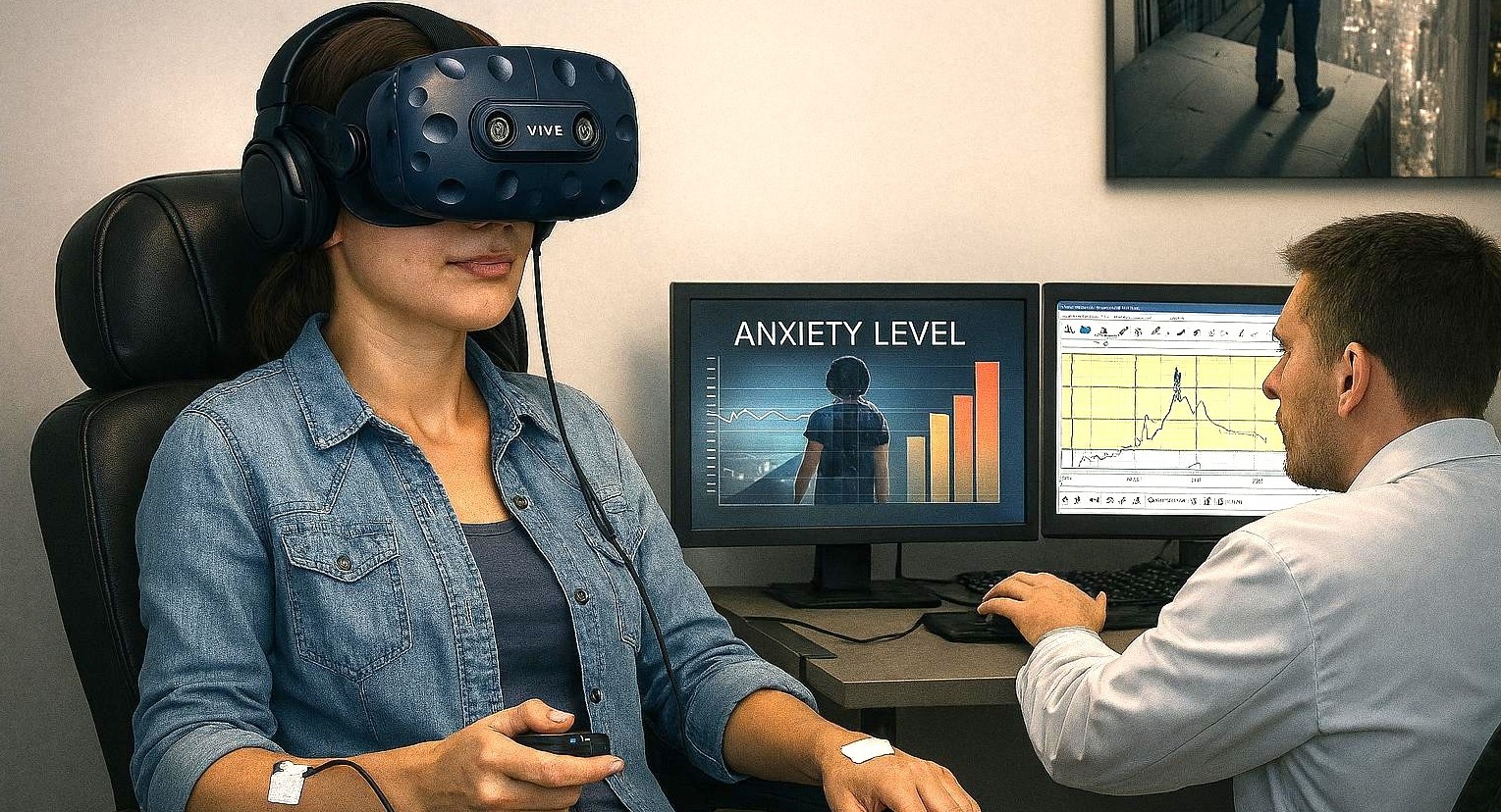
The scientific impact of VR is amplified when immersive environments are combined with multimodal psychophysiological measurements—such as electrocardiography (ECG), electroencephalography (EEG), functional near‑infrared spectroscopy (fNIRS), respiration, electrodermal activity (EDA), and eye tracking. In multimodal studies, researchers gain a deeper, objective understanding of how the brain and body respond to emotional challenges. By synchronizing VR events with physiological data, researchers can observe how the body responds moment by moment as fear is triggered, maintained, and eventually reduced.

Researchers at the University of Cyprus investigated whether a single session of VR exposure could meaningfully reduce public speaking anxiety in university students. Participants were placed in a virtual auditorium and asked to give a speech in front of realistic virtual audiences. The researchers could adjust audience size and behavior, allowing them to gradually increase the social challenge while maintaining full experimental control. To capture objective stress responses, heart activity was recorded using a Lead I electrocardiogram configuration, with two disposable silver/silver‑chloride electrodes placed on the forearms. Signals were amplified with a BIOPAC ECG100C bio‑amplifier, acquired using a MP data acquisition system with AcqKnowledge software for recording and analysis. This setup enabled precise measurement of heart rate and heart rate variability (HRV) throughout the task. The virtual environment was created using the Vizard Virtual Reality Toolkit, which was integrated with the BIOPAC system so that physiological data could be synchronized with key moments in the VR experience, such as the start of the speech. Results showed significant reductions in both self‑reported anxiety and physiological arousal following a single VR exposure session, demonstrating how VR and physiological monitoring can be combined to assess rapid emotional change.

Researchers at the University of Macau examined how social rewards and emotional cues influence anxiety during interpersonal interaction. Participants engaged in a virtual reality social interaction game designed to mimic real social exchanges while allowing researchers to control feedback and outcomes. During the task, heart activity was recorded using ECG with an MP data acquisition system. By synchronizing physiological data with events in the virtual environment, the research team could examine how anxiety‑related responses evolved as social interactions unfolded. The findings suggested that anxiety responses were more strongly related to sensitivity to others’ emotional cues than to reward outcomes themselves. This study highlights how immersive VR combined with synchronized physiological recording can provide insight into the subtle emotional dynamics of social anxiety, processes that are difficult to capture using self‑report measures alone.

dentalanxietyIn a multi‑institutional effort led by researchers at the University of Toledo, researchers developed a study protocol to better understand how dental fear develops and how early exposure might help prevent it. The study uses a virtual reality dental clinic to expose participants to dental‑related sights and sounds across multiple sessions and contexts. The virtual environment was programmed using Vizard software, and participants interacted with it using an HTC Vive Pro headset and handheld controllers, allowing natural movement and exploration. Skin conductance responses (SCR) were recorded using a BIOPAC MP system paired with a wireless BioNomadix EDA transmitter, enabling participants to move freely while skin conductance and other signals are collected. This wireless, immersive setup is especially well suited for fear‑learning research, where comfort and natural movement are critical. Although the publication focuses on study design rather than completed outcomes, it demonstrates how VR combined with physiological monitoring can support translational research aimed at understanding and preventing the development of fears and anxiety.

These studies show how VR and synchronized physiological measurement are expanding the study of anxiety, stress, and fear. VR provides immersive, repeatable experiences that reflect real‑world challenges, while physiological signals reveal how the body responds beneath conscious awareness. For additional information on how to integrate VR into multimodal research, visit our Webinar page for instructive trainings on this topic and others.


If you are planning to integrate VR into your next multimodal study, reach out to our skilled sales staff who can assist you through each step of the process.

Mind-Body Connection: Biofeedback in Research

The connection between the mind and body has long intrigued scientists, driving research into how our thoughts, emotions, and mental states can influence physical health and well-being. Biofeedback has become a recognized approach for helping people manage stress, reduce pain, and even enhance cognitive and physical performance. Biofeedback involves using sensors and real-time data to monitor physiological functions that are typically automatic, such as heart rate, breathing, muscle tension, and skin conductance. With this information, individuals can learn to gain greater awareness and, in some cases, voluntarily control these bodily processes.

Common physiological signals used in biofeedback include electrodermal activity (EDA), which reflects sweat gland activity and emotional arousal; electroencephalogram (EEG), which measures electrical activity in the brain; electrocardiogram (ECG), which measures heart electrical activity; heart rate variability (HRV), an index of autonomic nervous system flexibility; and respiration rate, among others. These signals offer researchers a window into the body’s stress responses, regulatory mechanisms, and even aspects of cognition and emotional control.

To complement physiological monitoring, researchers are increasingly integrating tools like stimulus presentation systems, virtual reality (VR) environments, and eye tracking technology into biofeedback studies. These tools allow scientists to study how people react to controlled stimuli, immerse participants in realistic scenarios, and track where and how long someone focuses visually, all while collecting detailed data on their physiological responses. This combination of immersive experiences and real-time monitoring helps researchers investigate how the body responds to stress or cognitive challenges and whether individuals can be trained to influence those responses.

Swiss researchers at ETH Zurich investigated whether individuals could voluntarily control their arousal states using real-time feedback on their pupil size. Pupil size is a physiological marker closely linked to the brain’s arousal systems, particularly the locus coeruleus. Study participants employed mental strategies to adjust their pupil diameter, receiving visual feedback through a dynamically changing circle that reflected their pupil size. The participants’ cardiovascular data, including ECG and respiration, were recorded to provide insights into the physiological effects of pupil self-regulation. The research team employed a BIOPAC data acquisition and analysis system running AcqKnowledge software to record ECG data continuously at a sampling rate of 1,000 Hz. Additionally, a BIOPAC respiratory effort transducer was used to measure respiration, although these respiratory data were not reported in the publication. The study’s findings demonstrated that pupil-based biofeedback enabled participants to modulate not only their pupil size but also associated neural activity and cardiovascular responses, such as heart rate. The results suggest a non-invasive method for individuals to control their autonomic functions, with potential applications in managing stress and anxiety disorders.

mind-bodyVagally mediated heart rate variability (vmHRV) refers to the natural changes in the time between heartbeats that are controlled by the vagus nerve, which is closely tied to how well a person can adapt to stress and regulate their body. Researchers from Université Paris Cité explored whether a brief session of vmHRV biofeedback could help people better control their memory, specifically whether it could help them tell the difference between real and false memories. In the study, 71 healthy young adults completed a VR memory task after receiving either vmHRV biofeedback or a simple breathing exercise. To track their physiological responses, the team used a BIOPAC data acquisition and analysis system and recorded heart activity via a three-lead ECG configured according to Einthoven’s triangle using Lead II. The results showed that this short biofeedback session had no immediate effect on memory accuracy or on vmHRV levels during the task. This suggests that a single session of HRV biofeedback may not be enough to influence memory-related brain functions, underlining the complex relationship between the body’s stress responses and cognitive control.

​A study published in the Journal of Human Kinetics looked at whether just one 10-minute session of HRV biofeedback, using a special breathing technique called resonance frequency breathing, could affect the level of activity of certain nerve cells. Researchers measured this by looking at a reflex in the lower leg muscle (the soleus) called the Hoffman reflex (H-reflex). Twelve healthy men took part in both the HRV biofeedback session and a regular breathing session in a random order. To measure muscle activity, researchers used a BIOPAC surface electrode on the leg and a reference electrode on the hand. The signals were recorded with a BIOPAC data acquisition system and analyzed with AcqKnowledge software, which helped line up the timing of muscle activity with electrical stimulation. The results showed that HRV biofeedback lowered the strength of the reflex and a related ratio (Hmax/Mmax), suggesting the spinal nerve cells were less excitable. These findings point to the potential of short HRV biofeedback sessions to affect the nervous system in ways that could be useful for physical therapy or improving movement control.

To learn more about how to integrate biofeedback techniques into your next research project, check out BIOPAC’s webinar, Biofeedback: Theory, Techniques, and Tools for Research.


Are you planning a research project that will utilize biofeedback data? Our expert customer service representatives can help you find the right tools for all your biofeedback needs.


A Remarkable 2024: Introducing Tools for a Better and Brighter Research Future

As 2024 winds down and we ready the confetti to ring in 2025, we at BIOPAC Systems Inc. want to toot our horn after wrapping up an exciting year of growth and innovation. This year brought some big wins for the research community, thanks to BIOPAC’s efforts to make physiological data acquisition and analysis more accessible and efficient than ever. Two major milestones were the launch of the MP200 system, the latest evolutionary step for our flagship data acquisition platform, and two feature-rich release updates to our AcqKnowledge data acquisition and analysis software.

Introducing the MP200: A New Achievement for Data Acquisition

MP200The MP system series has been the backbone of BIOPAC’s efforts to provide powerful and yet accessible data acquisition tools to researchers. Released in November, the newest incarnation of MP device, the MP200, ushers in a new era of precision, reliability, and flexibility for data acquisition. Designed to meet the evolving needs of researchers, the MP200 offers unparalleled scalability and compatibility with a wide range of physiological signals and measurements. Key enhancements include the following:

  • Enhanced Signal Quality: The MP200 uses advanced signal processing algorithms to ensure superior data integrity across diverse research applications, from neuroscience to exercise physiology.
  • Modular Design: The MP200’s modular architecture allows seamless integration with BIOPAC’s extensive sensors and amplifiers, creating near-limitless customization possibilities for complex experimental setups.
  • Future-Ready Connectivity: The MP200 facilitates real-time collaboration and analysis with high-speed data transfer protocols and cloud integration support.

Researchers will benefit from the MP200’s improved ability to streamline workflows and deliver insights with unmatched precision. This latest system exemplifies BIOPAC’s dedication to fostering groundbreaking discoveries in physiology and related fields.

AcqKnowledge Updates: Pioneering the Future of Data Analysis

AcqKnowledge data acquisition and analysis software has been a go-to tool for researchers for decades, and 2024 saw two major updates that brought exponential improvements: versions 5.0.10 and 6.0.0. These updates are packed with features that make data collection and analysis easier and more comprehensive.

One of the most impactful new licensed features is the integration of Lab Streaming Layer (LSL). This feature lets researchers sync and stream data from multiple sources in real time. Whether one is working on interdisciplinary projects or using several devices at once, LSL makes it simple to bring everything together for analysis. LSL also opens the door to a host of new products from our partners for which BIOPAC has added integration in AcqKnowledge. These include the Axon-R brain-computer interface (BCI) hub with augmented reality (AR) and artificial intelligence (AI) and the APEX wearable, wireless EEG system. Other new products have also been integrated into AcqKnowledge and are now available from BIOPAC, including new eye tracking products from Tobii and EyeLogic.

Another game-changing licensed addition, AcqKnowledge Cloud, allows users to securely store their data online, streamlining processes to access, share, and work on data from any location. Collaboration between teams is also easier, and users no longer need to worry about managing large files on their local system.

These updates are all about making work less complicated and more effective for researchers. With tools like these, diving into complex data, finding patterns, and pushing one’s research further has never been more straightforward.

Looking Ahead to 2025

More excitement is on the horizon for BIOPAC. Not only are several new product updates and releases in the works, but we will also be hosting our bi-annual 4T Human Physiology Conference on Tools, Trends, Techniques, and Technologies. Customers, educators, researchers, and other experts in Physiology and related fields will gather in Santa Barbara, California July 14-16. Participants will share knowledge, take part in hands-on workshops, and see what is shaping the future of research. Learn more and sign up today. Hopefully, we will be seeing you in 2025.

Measuring Mindfulness: Ancient Awareness for Modern Research

The concept of mindfulness has garnered significant attention in recent years, transcending its roots in ancient meditation practices to become a focal point in modern scientific research, particularly in the fields of physiology and psychophysiology. Understanding mindfulness involves delving into its essence: a state of heightened awareness and acceptance of the present moment, free from judgment or attachment to external stimuli. But how exactly does this concept intersect with the realms of physiology and psychophysiology, and how do researchers measure its impact on the human body and mind?

At its core, mindfulness entails being fully present and engaged in the current experience, whether it be a sensation, thought, or emotion. This mental state has been associated with numerous health benefits, including stress reduction, improved cognitive function, and enhanced emotional well-being. In the realm of physiology and psychophysiology, researchers are keen to explore how mindfulness practices influence the body’s physiological processes and how these changes can be quantified and understood.

One of the primary methods used to measure mindfulness in laboratory settings is through the analysis of physiological signals, such as electrodermal activity (EDA) and heart rate variability (HRV). During mindfulness practices, individuals often experience a shift in these physiological parameters. For example, studies have shown that mindfulness meditation can lead to decreased EDA, indicating a reduction in sympathetic nervous system activity and arousal levels. Similarly, practitioners may exhibit increased HRV, reflecting greater parasympathetic nervous system dominance and improved adaptability to stressors.

A collaborative study by a team of researchers from Wake Forest University, the University of Utah, and San Diego State University examined the role of HRV in mindfulness-based pain relief practices. Specifically, the study looked at the role of high-frequency heart rate variability (HF HRV), a marker of parasympathetic nervous system (PNS) activity, during mindfulness-based pain relief versus a sham-breathing exercise that served as a control. Respiration and electrocardiogram (ECG) data was fed into a BIOPAC data acquisition and analysis system. While the study results were inconclusive, researchers determined that further study was needed to explore “parasympathetic activation in mindfulness-based pain relief as compared to other meditative techniques.”

lotusA study published in the 2023 edition of the Journal of Psychiatric Research compared how rest and breath-focused mindfulness could be used to regulate and control various symptoms of post-traumatic stress disorder (PTSD) and dissociative symptoms related to past trauma in female study participants. Researchers used skin conductance response (SCR), one of the components of EDA, to measure autonomic function as it relates to PTSD symptoms. SCR data were collected during alternating resting control and breath-focused mindfulness conditions via BIOPAC EDA electrodes connected to a BIOPAC data acquisition and analysis system. The study found that rest and breathing exercises had produced positive results for individuals with low to moderate levels of trauma but were less effective with higher levels.

One notable feature of BIOPAC’s technology is its ability to integrate multiple physiological signals simultaneously, allowing researchers to better understand how mindfulness practices impact various bodily systems. An example is a study by a group of researchers in Israel that examined the effectiveness of mindfulness-based interventions (MBI) in improving mental well-being and resilience. MBIs are non-religious adaptations of contemplative Buddhist practices that are used to reduce suffering and increase well-being. Researchers used a BIOPAC BioNomadix system to wirelessly record a range of physiological signals, including ECG, electromyography (EMG), and EDA. Their goal was to determine whether mindfulness could positively influence how interoceptive signals from the autonomic nervous system regulate one’s sense of well-being. The study concluded that “mindfulness seems to increase the sensitivity of regulatory selections to interoceptive signals, and this is associated with subjective well-being.”

Mindfulness represents a fascinating intersection between ancient contemplative traditions and modern scientific inquiry. To learn more about how BIOPAC is helping researchers uncover new insights into the physiological aspects of mindfulness and its potential implications for health and well-being, check out our webinar on The Science of Mindfulness.


Are you looking for techniques and tools to measure mindfulness in your study? Contact your regional BIOPAC sales representative to integrate everything you will need to gather and analyze data.

VR Integration: Enhancing Research with Virtual Environments

Measuring the human body and brain’s response to real-world conditions and stimuli has long challenged researchers. This problem has usually been dealt with in one of two ways. The first approach is to take research participants into the field—not impossible with today’s mobile data recording technology but not always safe or convenient. The second option is to imitate real-world conditions in the lab using screens, audio equipment, and even scent delivery, but in the end, it is difficult to completely remove the lab setting from the participant’s experience—that is, until the introduction of Virtual Reality (VR) technology in research.

Just as it has revolutionized everything from entertainment to training, VR has provided researchers with the opportunity to create immersive experiences in the lab that mimic the real world. Advances in technology have not only made these simulated environments more realistic, but they have also made it easier to integrate a wide range of signals and stimulus delivery to enhance the recording of physiological and psychophysiological data.

BIOPAC, through cooperation with several partner companies, offers a range of data recording options that integrate seamlessly with third-party VR devices. fNIR Devices, for example, designs solutions for recording functional near-infrared spectroscopy (fNIRS) signals that can easily be incorporated with standard VR head-mounted displays (HMD) to measure neural activity and hemodynamic response in the prefrontal cortex while participants experience VR environments. BIOPAC partner Seenel has created MedelOpt, a line of whole-head wearable devices that combine electroencephalography (EEG) and fNIRS, including a VR option that integrates an HMD. Finally, partner company WorldViz offers a full spectrum of VR solutions from complete VR kits to full-room Projection VR technology to software that can be combined with both wired and wireless data recording technologies to provide a complete array of bio-signal data, from eye tracking to electrodermal activity.

A Canadian study published in 2023 used a combination of BIOPAC and WorldViz products to investigate how sensory feedback and arousal influence neuromusVR Shadowcular control and posture. Researchers measured arousal via electrodermal activity (EDA) and psychosocial questionnaires, rating perceived fear and anxiety in both real-world situations in VR environments that simulated standing both at ground level and on a raised platform. Participants wore a commercial HMD running WorldViz’s Vizard VR toolkit software. Electrical nerve stimulation was provided via a BIOPAC linear isolated stimulator. The study found the “physiological and perceptual indicators of fear and anxiety increased” in the VR platform simulation, providing “novel evidence that VR can be used to modulate upright postural control.”

A similar study published in the 2022 issue of the Journal of Sport Rehabilitation examined the effect non-physical mental training has on balance skills. The study used fNIRS technology to measure hemodynamic response in the prefrontal cortex of participants experiencing virtual environments through VR HMDs. Participants were instructed to imagine themselves imitating balance exercises viewed through an Oculus VR HMD while also fitted with an fNIR Devices Imager headband. Data was gathered using Cobi Studio software and analyzed via fNIRSoft software. The study identified several advantages of virtual reality mental training (VRMT) over conventional mental training (CMT).

These are just a few examples of how VR technology can be integrated with a variety of methodologies for gathering research data. For additional information on the subject, check out our Virtual Reality Resources, which include an array of online training on integrating VR into research.


Are you planning a research product and need help integrating VR with other data acquisition and analysis equipment? Contact your local BIOPAC sales rep to find the right solutions for you.

Put a Ring on It: Wearables Offer New Research Options

Rings, watches, bracelets, and belts—we think of them as indispensable fashion accessories. A bit of bling can put the finishing touch on an outfit for a night on the town, but what about a day in the lab? Wearable technology is transforming the landscape of physiology research by enabling real-time, noninvasive data collection in a variety of settings.

For many, their first introduction to the potential of wearable physiology technology came through smartwatches and fitness bracelets. These consumer electronics offer a convenient and affordable way for anyone from professional athletes to fitness hobbyists to monitor their health data, which often includes pulse, blood oxygen levels, steps, and other basic physiological signals.

As the underlying technology has become more sophisticated, researchers have embraced the convenience offered by such devices. Portable, unobtrusive, and wireless wearables have entered the research device market and now allow a range of signals to be monitored under a variety of circumstances and environments.

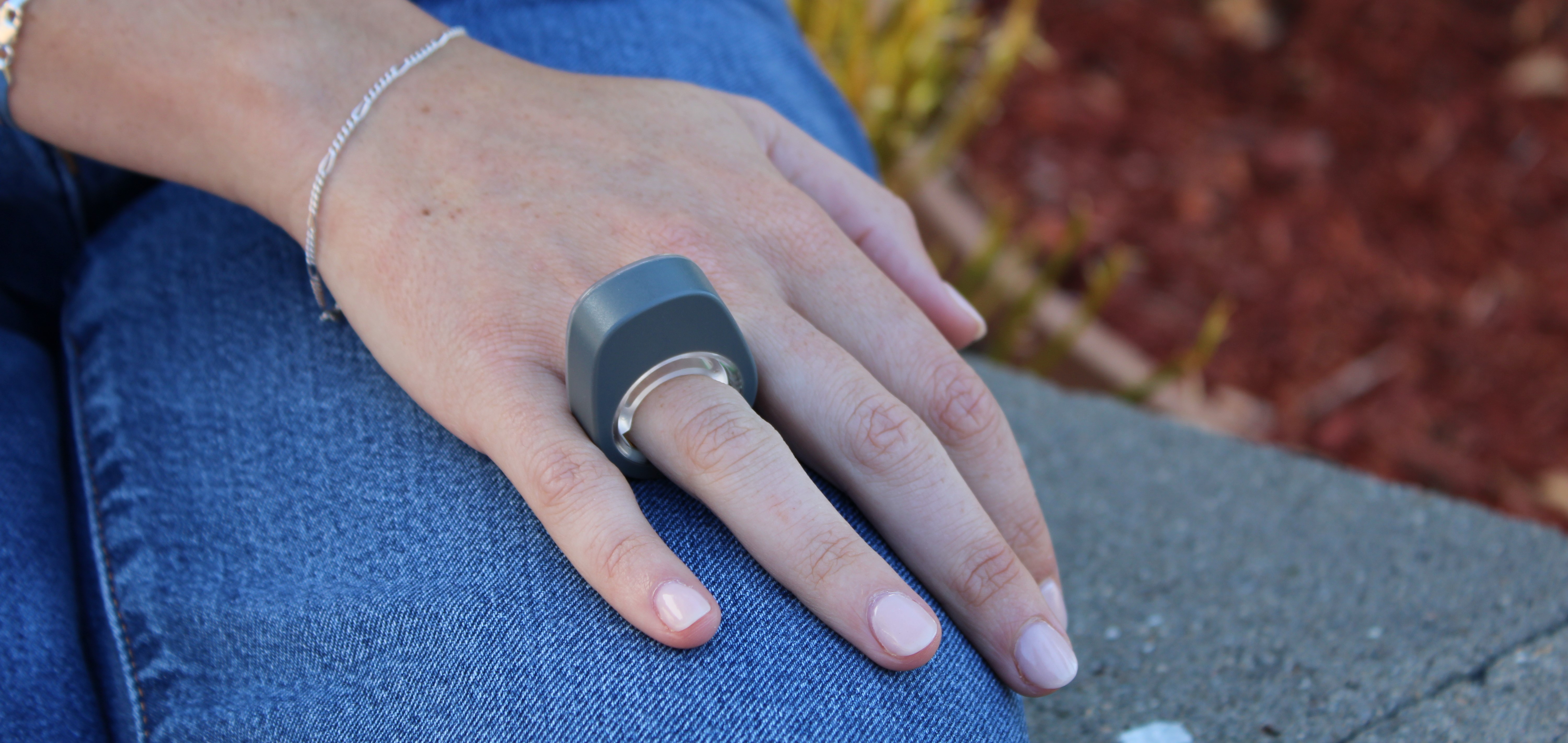
smartphoneBIOPAC recently expanded its established line of wireless devices by introducing the Research Ring, a wearable device with a specific focus on physiological signals such as Photoplethysmography (PPG), Electrodermal Activity (EDA), Electrocardiography (ECG), and body temperature. Products like the Research Ring allow the capture of high-quality data via an unintrusive and accessible wearable device, making them ideal for a wide range of potential applications. These include psychophysiology research, sports and exercise studies, sleep and stress research, and aging and wellness studies, to name just a few.

BIOPAC is no newcomer to providing the research and education communities with wireless and portable solutions designed for gathering physiological data. The Bionomadix line of wearable remote monitoring devices set the standard for such products and have been used in thousands of cited studies. For example, researchers conducting a study of on-the-job stress among healthcare professionals collected EDA and PPG signals using a BIOPAC wireless PPG and EDA amplifier and a wireless respiration with ECG amplifier. Wireless technology proved to be a key component of the study’s success as it allowed the data to be gathered without interfering with the immersive quality of the simulated work environment.

A team of researchers from the University of Porto in Portugal utilized a BIOPAC wearable physiological signal logger to collect EDA and ECG data while studying the correlation between physiological signals and expressive writing. In the study, published in the journal Experimental Psychology, participants were able to engage in writing exercises without the distraction of more cumbersome and intrusive wired solutions.

BIOPAC’s latest entry into its line of wearable wireless data collection devices promises to add greater convenience and versatility to this burgeoning sector. For more information on the new Research Ring, check out the recent webinar BIOPAC hosted with ring developer and partner, Senstream.


If you are interested in adding wearable devices to your next physiology study, reach out to your regional sales representative today to learn which BIOPAC wireless solutions are right for you.

ENS: Signals for Studying the Body’s “Second Brain”

We have all heard the saying “thinking with one’s stomach,” a cliché that suggests basing one’s critical thought processes on the whims of their digestive system. However, there may be more than a little truth to this saying. Often referred to as the body’s “second brain,” the enteric nervous system (ENS) comprises a complex network of neurons and ganglia embedded within the walls of the gastrointestinal tract. In recent years, researchers have increasingly recognized the significance of the ENS and its application in physiological research.

The ENS is a division of the autonomic nervous system (ANS) responsible for regulating the digestive process. It consists of millions of neurons spread throughout the esophagus, stomach, small intestine, and large intestine. These neurons form an intricate network that can function independently of the central nervous system (CNS) but also communicates with it. The ENS controls essential gastrointestinal functions, such as peristalsis, secretion of digestive enzymes, and blood flow regulation within the gut.

Several effective techniques are available to researchers to evaluate and measure ENS activity for their studies. Electrogastrography (EGG) provides a noninvasive approach to measuring the electrical activity of the stomach muscles. EEG can help researchers understand the rhythm and coordination of stomach contractions, aiding in the diagnosis of motility disorders.

In addition to EGG, a range of other physiological signals may be applied to ENS research. A recent study published in the June 2023 edition of Nature Communication investigated the gut-brain relationship by measuring physiological response to stimulation of the digestive system via different noninvasive techniques: EGG, electroencephalogram (EEG), skin conductance response (SCR), heart rate (HR), and heart rate variability (HRV). Research participants ingested a small vibrating probe that provided varying levels of stimulation as it passed through the lower digestive tract. EGG, EEG, and SCR signals were recorded along with hemodynamic signals via a BIOPAC data acquisition and analysis system. Researchers concluded that these techniques provided a viable alternative to more invasive approaches to measuring the body’s response to sensory stimulation of the digestive tract with, according to the published report, “implications for understanding gut feelings and gut-brain interactions in healthy and clinical populations.”

ENS ResearchMore invasive techniques for studying the ENS can still provide researchers with valuable data. A 2023 study by a team of South Korean researchers investigated how radial stretching affects intestinal contractions. The study also examined how drugs targeting the ENS affect such contractions to develop treatments for gastrointestinal disorders. Samples of intestinal tissue were taken from participants undergoing aggressive treatment for colorectal cancer. Samples were preserved in a tissue chamber and connected to a BIOPAC isometric force transducer to measure contractions, with data recorded and analyzed with a BIOPAC MP160 system running AcqKnowledge software. The study was able to detect regional differences in the stretch reflex of colon tissue via electrophysiological recordings of intestinal smooth muscles.

These are some of the ways in which researchers are currently using BIOPAC technologies to better understand the relationship between the ENS and other neurophysiological processes. For additional information on this topic, see our page on AcqKnowledge’s Gastric Myoelectric Activity feature and our EGG citations.


If you would like to know more about how BIOPAC can help you plan and equip your ENS study, reach out to your regional sales representative today.

4T|Phys 2023: BIOPAC Shares Tools, Trends, Techniques & Technology at UCSB

They hailed from across the globe, from Arkansas to Zanzibar and all points between. Students, teachers, researchers, and innovators gathered on the campus of UC Santa Barbara, overlooking the Pacific Ocean. They came to learn, share, and explore breakthroughs and best practices in physiology and related fields. 4T|Phys kicked off July 17 under clear California summer skies, an idyllic setting for discovering the Tools, Trends, Techniques, and Technology driving physiology education and exploration.

4T|Phys 2023 was the third installment in BIOPAC’s highly successful series of biennial industry conferences that began in 2017. After a two-year hiatus during the global Covid-19 pandemic, 4T came roaring back this summer with more workshops, more tracks, more sponsors, more presenters, and more countries represented than ever before. Over the course of three days, nearly 100 attendees participated in hands-on sessions with some of the top minds in physiology research, experiencing the latest tools for gathering and analyzing data.

BIOPAC’s in-house experts were joined by an array of authorities from the fields of physiology, psychology, imaging (fNIRS and fMRI), neuroscience, cardiovascular research, eye tracking, virtual reality (VR), biology, and engineering. Participants took part in workshop schedules drawn from five separate tracks that included fundamentals, which focused on the basics of data acquisition and analysis. The research track took attendees on a deeper dive into techniques and technologies, including recording evoked potentials, screen-based eye tracking, impedance cardiography using trans-radial bioimpedance velocimetry (TREV), stimulus presentation, exercise physiology, and many others. Participants were also able to tour UC Santa Barbara’s state-of-the-art Brain Imaging Lab to see up close how BIOPAC products are being used in both functional MRI (fMRI) and TREV studies.

“It was an incredible experience that left a lasting impact on me by helping me to understand more about physiological data collection and analysis,” said Dr. Parth Trivedi, a 4T|Phys attendee. “The choice of presenters and the topics covered were truly inspiring, offering valuable knowledge and fresh perspectives that enriched my understanding.”

4T|Phys 2023 Attendee MapThis year’s conference featured several new tracks such as a dedicated VR track led by partner company WorldViz. The WorldViz team showcased virtual reality solutions in a series of workshops that introduced how VR is being integrated in physiology and psychology research, explained how to create effective and immersive VR experiments, and demonstrated their latest PRISM technology for creating fully immersive projected 3D virtual environments.

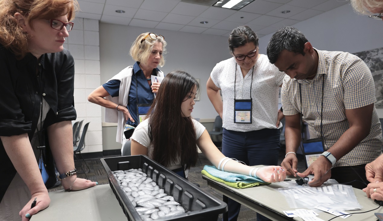
Several other partner companies introduced their technologies and products both in the 4T exhibition hall and in workshop sessions. Argus Science presented their ETVision eye-tracking glasses that synch seamlessly with AcqKnowledge. Cognixion demonstrated the Cognixion One Axon-R—Research Edition wearable BCI-AR computing platform, which is being used to help individuals with neurodegenerative conditions and severe motor impairments communicate and interact with their environments assisted by augmented reality. Seenel Imaging showed off their MedelOpt Systems for whole-head EEG with fNIRS, including new VR integration. Participants also got a chance to try on the Research Ring by Senstream, a new user-friendly wearable research ring, which allows easy recording of EDA, PPG, Temperature, and ECG signals. CNSystems showed off the company’s NIBP continuous noninvasive blood pressure (NIBP) and hemodynamics devices.

If you missed the educational and networking opportunities at 4T|Phys 2023, don’t despair. We will be back again in a few years, bigger and better than ever. Watch this space for updates on our next installment. In the meantime, see our Video Tutorials and Webinars On Demand for trainings and seminars on our products and partners.


Make sure you never miss a BIOPAC event and keep up with the latest Tools, Trends, Techniques, and Technologies by contacting your local BIOPAC sales representative.

error: Content is protected !!