
Under the Skin: Digging Deeper into the Physiology of Fear

From the thrill of cinematic jump scares to debilitating phobias that can turn daily life for some into a waking nightmare, our sense of fear can feel like both a blessing and a curse. Fear is one of the most fundamental human emotions, serving as a necessary safeguard for recognizing and responding to danger. Despite its primal origins, the mechanisms of fear remain complex. For decades, psychophysiologists have studied fear using measures such as the startle reflex and skin conductance, building a deep understanding of how the body reacts to threats. Today, however, researchers are beginning to look beyond these traditional markers. Technologies such as virtual reality (VR) expand how researchers can stimulate the fear response. At the same time, a broad range of signals and techniques can measure not only the body’s autonomic responses, but also how people allocate attention to threats and how the brain processes fear-related information.

One reason fear remains such a compelling topic is that it sits at the intersection of cognition, emotion, memory, and physiology. Fear responses can be observed through changes in sweating, heart activity, breathing patterns, pupil size, neural activity, and even social behavior. As researchers seek to gain a better understanding of anxiety disorders, phobias, post-traumatic stress disorder (PTSD), and related conditions, they increasingly rely on multiple synchronized signals to capture a more complete picture of how fear is learned, remembered, and generalized.

Among the most widely used measures is electrodermal activity (EDA), often recorded as skin conductance response (SCR), which reflects changes in sympathetic nervous system arousal. More recently, eye tracking and pupillometry have emerged as valuable tools for measuring vigilance and attention, while functional near-infrared spectroscopy (fNIRS) has enabled monitoring of changes in cortical activity during fear-related tasks in more naturalistic settings. Together, these approaches help researchers move beyond viewing fear as a single physiological reaction and toward understanding it as a coordinated brain-body process.

A team of researchers at South China Normal University in China explored how the duration of memory retrieval influences the reconsolidation and extinction of fear memories. Using a four-day fear-conditioning paradigm, participants were conditioned to associate visual cues with mild electric shocks. Simultaneously, skin conductance responses were recorded using an MP36 running Biopac Student Lab System. The experiment was designed and implemented with E-Prime stimulus presentation software. The team found that briefly reactivating a fear memory before training participants to view the stimulus as safe was most effective at preventing the fear from returning. Longer periods of memory reactivation produced weaker effects. The study highlights how electrodermal activity can reveal subtle changes in fear-memory processing and may ultimately contribute to improved behavioral interventions for anxiety-related disorders.

fingers and faceA study conducted at the University of Würzburg in Germany examined how anxiety sensitivity influences attention and memory during threat and safety learning. Participants learned associations between neutral faces and aversive outcomes while researchers recorded both electroencephalography (EEG) and pupil diameter. Pupillometry data were acquired using a Tobii eye tracker, allowing researchers to measure moment-to-moment changes in attention. Individuals with higher anxiety sensitivity showed larger pupil responses and enhanced neural markers of attention when viewing both threatening and safe cues, reflecting a generalized pattern of hypervigilance. Interestingly, this heightened attentional engagement did not translate into better memory performance. The findings suggest that pupillometry provides a valuable window into how anxious individuals process potential threats and may help researchers better understand mechanisms underlying pathological anxiety.

Another perspective on fear comes from research at Sichuan Normal University in China, where investigators look at how trait anxiety influences observational fear learning. Unlike traditional conditioning studies, participants learned about danger by watching another person experience threat-related events. Researchers measured skin conductance using a BioNomadix wireless EDA plus data acquisition and analysis system and monitored prefrontal brain activity using fNIRS. The study found that individuals with high trait anxiety exhibited stronger physiological fear responses and increased medial prefrontal cortex activation, even when observing threats indirectly. The work demonstrates that fear can be acquired socially and that neural measures such as fNIRS can reveal aspects of fear processing that are not readily captured by autonomic measures alone. These findings may have important implications for understanding how fear and anxiety spread through social environments and how vulnerability to anxiety disorders develops.

These studies illustrate how fear research leverages multimodal approaches to incorporate multiple physiological perspectives. Skin conductance continues to provide valuable information about autonomic arousal, while pupillometry reveals how attention is divided between threat and safety cues, and fNIRS offers insight into the neural processes involved in fear learning and regulation. As multimodal research platforms become increasingly accessible, researchers are gaining unprecedented insight into how fear is acquired, maintained, and modified. By combining signals from the body, the eyes, and the brain, modern psychophysiology is shining a light on the shadowy labyrinth that is one of humanity’s most powerful and important emotions. To learn more about how to construct a fear stimulation lab for your next fear-conditioning research project, check out our webinar on the subject.


If you are planning a multimodal fear conditioning study, our staff can help identify the right equipment and software for stimulus presentation and data recording and analysis.
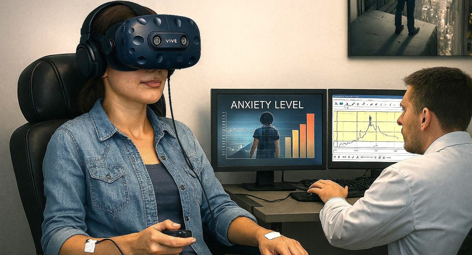

 

Virtual Phobia: How VR is Transforming Anxiety and Fear Research

“Do the thing you fear and the death of fear is certain.”

These words by American poet and philosopher Ralph Waldo Emerson assert a head-on approach to overcoming life’s anxieties and phobias. It’s a strategy backed by the latest research methodology, which, thanks to technology, can leverage the relative safety of virtual environments to study how participants react to and manage anxiety.

Anxiety disorders are among the most prevalent mental health conditions and are often maintained through avoidance of fear‑provoking situations. A cornerstone of evidence‑based treatment is exposure therapy, in which individuals gradually and safely confront feared experiences to reduce anxiety over time. In recent decades, virtual reality (VR) has emerged as an effective way to deliver exposure therapy and to study anxiety under controlled conditions. Virtual reality exposure therapy (VRET) uses immersive, computer‑generated environments to recreate anxiety‑triggering situations, such as public speaking, social interaction, or medical procedures, while allowing researchers and clinicians to control intensity, pacing, and repetition.

Research shows that VRET can be as effective as traditional, real‑world exposure for many anxiety‑related conditions, including specific phobias, social anxiety, panic disorder, and symptoms of post‑traumatic stress disorder (PTSD). One of VR’s key advantages is its balance between realism and experimental control. Environments can be standardized across participants, exposure levels can be adjusted in small steps, and scenarios can be repeated across sessions without the logistical challenges of invivo exposure. At the same time, well‑designed VR experiences reliably evoke emotional and stress responses, making them valuable for both treatment and research.

The scientific impact of VR is amplified when immersive environments are combined with multimodal psychophysiological measurements—such as electrocardiography (ECG), electroencephalography (EEG), functional near‑infrared spectroscopy (fNIRS), respiration, electrodermal activity (EDA), and eye tracking. In multimodal studies, researchers gain a deeper, objective understanding of how the brain and body respond to emotional challenges. By synchronizing VR events with physiological data, researchers can observe how the body responds moment by moment as fear is triggered, maintained, and eventually reduced.

Researchers at the University of Cyprus investigated whether a single session of VR exposure could meaningfully reduce public speaking anxiety in university students. Participants were placed in a virtual auditorium and asked to give a speech in front of realistic virtual audiences. The researchers could adjust audience size and behavior, allowing them to gradually increase the social challenge while maintaining full experimental control. To capture objective stress responses, heart activity was recorded using a Lead I electrocardiogram configuration, with two disposable silver/silver‑chloride electrodes placed on the forearms. Signals were amplified with a BIOPAC ECG100C bio‑amplifier, acquired using a MP data acquisition system with AcqKnowledge software for recording and analysis. This setup enabled precise measurement of heart rate and heart rate variability (HRV) throughout the task. The virtual environment was created using the Vizard Virtual Reality Toolkit, which was integrated with the BIOPAC system so that physiological data could be synchronized with key moments in the VR experience, such as the start of the speech. Results showed significant reductions in both self‑reported anxiety and physiological arousal following a single VR exposure session, demonstrating how VR and physiological monitoring can be combined to assess rapid emotional change.

Researchers at the University of Macau examined how social rewards and emotional cues influence anxiety during interpersonal interaction. Participants engaged in a virtual reality social interaction game designed to mimic real social exchanges while allowing researchers to control feedback and outcomes. During the task, heart activity was recorded using ECG with an MP data acquisition system. By synchronizing physiological data with events in the virtual environment, the research team could examine how anxiety‑related responses evolved as social interactions unfolded. The findings suggested that anxiety responses were more strongly related to sensitivity to others’ emotional cues than to reward outcomes themselves. This study highlights how immersive VR combined with synchronized physiological recording can provide insight into the subtle emotional dynamics of social anxiety, processes that are difficult to capture using self‑report measures alone.

dentalanxietyIn a multi‑institutional effort led by researchers at the University of Toledo, researchers developed a study protocol to better understand how dental fear develops and how early exposure might help prevent it. The study uses a virtual reality dental clinic to expose participants to dental‑related sights and sounds across multiple sessions and contexts. The virtual environment was programmed using Vizard software, and participants interacted with it using an HTC Vive Pro headset and handheld controllers, allowing natural movement and exploration. Skin conductance responses (SCR) were recorded using a BIOPAC MP system paired with a wireless BioNomadix EDA transmitter, enabling participants to move freely while skin conductance and other signals are collected. This wireless, immersive setup is especially well suited for fear‑learning research, where comfort and natural movement are critical. Although the publication focuses on study design rather than completed outcomes, it demonstrates how VR combined with physiological monitoring can support translational research aimed at understanding and preventing the development of fears and anxiety.

These studies show how VR and synchronized physiological measurement are expanding the study of anxiety, stress, and fear. VR provides immersive, repeatable experiences that reflect real‑world challenges, while physiological signals reveal how the body responds beneath conscious awareness. For additional information on how to integrate VR into multimodal research, visit our Webinar page for instructive trainings on this topic and others.


If you are planning to integrate VR into your next multimodal study, reach out to our skilled sales staff who can assist you through each step of the process.

Back to the Wild: Measuring the Effects of Human Interaction with Nature

“I think that I cannot preserve my health and spirits, unless I spend four hours a day at least—and it is commonly more than that—sauntering through the woods and over the hills and fields, absolutely free from all worldly engagements.” —Henry David Thoreau

In his 1861 essay “Walking,” transcendentalist writer and philosopher Henry David Thoreau extolled the virtues, both physical and mental, of communing with nature. While the psychological and physiological benefits of human connection with the natural environment are well established, modern science has taken a closer look at these interactions. Today’s researchers often rely on interdisciplinary approaches that blend psychology, physiology, neuroscience, and environmental science to investigate the mechanisms behind nature’s restorative effects on mind and body. With continued urban growth and shrinking access to wild rural spaces, as well as the digitization of our daily lives, this field has become more relevant than ever.

Researchers study human interactions with nature to better understand the tangible health and psychological benefits associated with exposure to natural environments, such as reduced stress, improved mood, and enhanced cognitive function. Theories such as the Biophilia Hypothesis and Attention Restoration Theory (ART) suggest that humans have a built-in affinity for nature, which can support mental and emotional well-being. Psychophysiological research seeks to uncover the processes underlying these effects and to compare how different environments—whether urban, natural, or virtual—impact our bodies and minds.

The study of human-nature interaction encompasses a wide range of practical applications, including healthcare and mental wellness. Beyond health applications, this field informs decisions and policies regarding urban planning and architecture, such as the design of biophilic buildings and green spaces in cities. Research also aims to help individuals focus their attention, enhance creativity, and promote emotional well-being in both work and educational settings.

A study published in the Journal of Environmental Psychology examined how immersion in nature and exposure to nature-related images affect attention and could be used in attention restoration therapy and related applications. Researchers measured participants’ neural activity before, during, and after a four-day group camping trip in Utah using electroencephalography (EEG) to capture reward positivity (RewP), a brain signal associated with reward processing. During these periods, researchers measured how participants responded to extrinsic rewards, including monetary incentives. To ensure data integrity and minimize motion artifacts in the outdoor setting, the team utilized BIOPAC’s wireless BioNomadix EEG system and verified electrode contact using the EL-CHECK impedance checker. The study’s findings showed a significant decrease in neural responsiveness to extrinsic (monetary) rewards following immersion in nature. This suggests that increased time spent immersed in natural environments may recalibrate the brain’s reward sensitivity and promote intrinsic motivation. These findings have implications for a variety of behaviors related to extrinsic stimuli, such as the treatment of addiction.mountaintop

Not all research into human-nature interactions focuses on immersion in real-world environments. With the evolution of virtual reality (VR) technology, new research leverages artificial environments to elicit biophilic responses. A study by researchers at the Southwest Jiaotong University School of Architecture in China, published in Frontiers in Public Health, investigated how exposure to biophilic design elements affects stress recovery in older adults. A group of 25 participants over the age of 60 were exposed to stressors and then underwent sessions with randomized VR environments that included green walls and outdoor views. The researchers measured two key physiological signals: heart rate variability (HRV), which assesses parasympathetic activity, and skin conductance level (SCL), a marker of sympathetic arousal. Electrocardiogram (ECG) data were collected using a BIOPAC ECG amplifier, while electrodermal activity was gathered via an EDA amplifier. Both signals were recorded using a BIOPAC data acquisition and analysis system. Results found that participants exposed to environments with green features demonstrated significantly higher HRV and lower SCL, indicating enhanced autonomic recovery and reduced stress. These findings suggest that the biophilic benefits of nature exposure can be made available to individuals virtually when real-world immersion is not possible or available.

As these studies demonstrate, there exists a wide and varied frontier to explore how human interaction with nature shapes our physiological, neurological, and psychological well-being. For more information on the tools to assist with such research, visit our Webinars library for detailed video trainings on how to use these technologies in your nature-related study.


Whether your study participants are hitting the trail, climbing a mountain, or just relaxing in the garden, BIOPAC customer service reps are ready with the right solutions to take your next research project back to nature.

Game On: Applying Physiological Data in Video Game Design and Research

Ever since players first started feeding quarters into arcade games and began plugging in home systems in the late twentieth century, researchers have looked for practical tools for both shaping video game design and measuring their effects on players. Physiological signals filled this niche area of research. Researchers began investigating the connection between physiological science and gaming in the 1980s and 1990s, when early studies explored the effects of video games on heart rate and arousal. These first studies focused on issues such as aggression and overstimulation, but research has since expanded to encompass the cognitive and emotional aspects of gameplay. An early research project by NASA, for example, examined how biofeedback could be applied to game design to enhance traditional game control methods while also improving players’ cognitive abilities.

Today, physiological monitoring is not only a method for psychological inquiry but also a vital asset in improving user experience (UX) research, game accessibility, and even driving therapeutic game development. As wearable sensors and game engines grow more sophisticated, the integration of physiological signals into game design is opening new frontiers for immersive, responsive, and health-conscious gaming. By analyzing real-time bodily signals, such as heart rate (HR), electroencephalography (EEG), electromyography (EMG), electrodermal activity (EDA), respiration, photoplethysmography (PPG), and electrooculography (EOG), researchers and developers can gain a deeper understanding of how players emotionally and physically respond to game elements.

A study published in the International Journal of Human-Computer Studies investigated whether players’ self-reported preferences for first-person shooter (FPS) game dynamics match their emotional reactions during both gameplay and viewing. Researchers recruited 24 active gamers and divided them into two groups: those who enjoyed the fast-paced action of FPS games and those who didn’t. While participants played an FPS game and later watched a recording of it, the team measured electrodermal activity (EDA), heart rate, and facial muscle responses, specifically expressions associated with frowning (corrugator supercilii) and smiling (zygomaticus major), to capture emotional arousal and valence. Physiological signal data were recorded using BIOPAC EDA, PPG, and EMG amplifiers, which were connected to a data acquisition and analysis system running AcqKnowledge software. Although both groups reported more positive feelings and higher excitement after actively playing rather than watching, the data revealed a deeper story: players who disliked the game showed more substantial rises in arousal during play. Those who favored the dynamics, however, showed steadier emotional responses across both conditions. Muscle activity patterns also differed. Those who enjoyed FPS dynamics exhibited sharper increases in frowning-related muscle activity during play than watchers, indicating nuanced emotional engagement. Overall, the study found that what people say they prefer does reflect in their bodies’ emotional reactions. Still, those preferences also shape whether playing or spectating feels more intense, highlighting how combining self-reports with physiological signals can deepen an understanding of game design and player experience.

controllerA team of researchers from Bournemouth University’s Centre for Digital Entertainment investigated how players’ bodies react during intense engagement, or flow, in virtual reality (VR) games. The goal was to capture objective physiological signals, such as heart rate and eye-blink patterns, alongside subjective self-reports to see how well a game’s design induced flow versus boredom or anxiety. The researchers had participants play a custom VR “tower defense” game while monitored with BIOPAC equipment, including a two-channel Biopac Student Lab (BSL) data acquisition system and physiological modules to record HRV via ECG, as well as EOG and EEG signals. The team then utilized clustering and machine-learning classifiers to decode flow states with high accuracy. The research team also successfully identified that high flow correlated with fewer blinks and longer blink intervals, indicating a state of deep concentration. The study not only demonstrates the value of combining physiological and self-report data but also shows how wearable sensors can inform design decisions in VR games by providing real-time feedback on player experience.

Beyond game design, a primary area of research focus has been on the long-term psychological, neurological, and physiological impact that games have on players. A study published in the Journal of Media Psychology explored how experiences with violent video games might reduce prosocial behavior, such as the motivation to help others, through physiological changes. Researchers recruited 155 Chinese college students who reported their exposure to violent games (in terms of violence, bloodiness, and duration) and completed simple self-reports measuring their willingness to help and the time they took to help others in controlled scenarios. Researchers used a BIOPAC data acquisition unit running AcqKnowledge software to record relevant physiological data. A three-lead ECG amplifier, respiration module, and respiration belt were used to record ECG and breathing signals across baseline, violent-film task, and during recovery periods. Researchers calculated the reduction in respiratory sinus arrhythmia (RSA), a marker of parasympathetic nervous system engagement. They found that higher levels of violent-game experience predicted less RSA reduction during stress, which in turn was linked to reduced prosocial behavior. The study’s findings suggest that playing violent video games may impair physiological readiness to respond in an empathetic manner to others, showing RSA reduction to be a key influence. The study’s findings demonstrate how physiological and psychophysiological signals provide a deeper understanding of the impacts of video games, while also guiding design decisions.

Physiological signals help researchers identify moments of excitement, stress, immersion, or frustration. Such insights are increasingly being used to refine gameplay mechanics, balance difficulty, and enhance player engagement through adaptive or personalized game experiences. They also reveal the short- and long-term effects gameplay has on both individuals and society. For more ideas on how to apply these signals in your research, visit our Webinars page for information on wearable devices, mobile data acquisition, and other helpful topics.


Are you planning a research project related to video games? Reach out to our customer service specialists to get all the help you need in mapping out and equipping your study team.

Mind-Body Connection: Biofeedback in Research

The connection between the mind and body has long intrigued scientists, driving research into how our thoughts, emotions, and mental states can influence physical health and well-being. Biofeedback has become a recognized approach for helping people manage stress, reduce pain, and even enhance cognitive and physical performance. Biofeedback involves using sensors and real-time data to monitor physiological functions that are typically automatic, such as heart rate, breathing, muscle tension, and skin conductance. With this information, individuals can learn to gain greater awareness and, in some cases, voluntarily control these bodily processes.

Common physiological signals used in biofeedback include electrodermal activity (EDA), which reflects sweat gland activity and emotional arousal; electroencephalogram (EEG), which measures electrical activity in the brain; electrocardiogram (ECG), which measures heart electrical activity; heart rate variability (HRV), an index of autonomic nervous system flexibility; and respiration rate, among others. These signals offer researchers a window into the body’s stress responses, regulatory mechanisms, and even aspects of cognition and emotional control.

To complement physiological monitoring, researchers are increasingly integrating tools like stimulus presentation systems, virtual reality (VR) environments, and eye tracking technology into biofeedback studies. These tools allow scientists to study how people react to controlled stimuli, immerse participants in realistic scenarios, and track where and how long someone focuses visually, all while collecting detailed data on their physiological responses. This combination of immersive experiences and real-time monitoring helps researchers investigate how the body responds to stress or cognitive challenges and whether individuals can be trained to influence those responses.

Swiss researchers at ETH Zurich investigated whether individuals could voluntarily control their arousal states using real-time feedback on their pupil size. Pupil size is a physiological marker closely linked to the brain’s arousal systems, particularly the locus coeruleus. Study participants employed mental strategies to adjust their pupil diameter, receiving visual feedback through a dynamically changing circle that reflected their pupil size. The participants’ cardiovascular data, including ECG and respiration, were recorded to provide insights into the physiological effects of pupil self-regulation. The research team employed a BIOPAC data acquisition and analysis system running AcqKnowledge software to record ECG data continuously at a sampling rate of 1,000 Hz. Additionally, a BIOPAC respiratory effort transducer was used to measure respiration, although these respiratory data were not reported in the publication. The study’s findings demonstrated that pupil-based biofeedback enabled participants to modulate not only their pupil size but also associated neural activity and cardiovascular responses, such as heart rate. The results suggest a non-invasive method for individuals to control their autonomic functions, with potential applications in managing stress and anxiety disorders.

mind-bodyVagally mediated heart rate variability (vmHRV) refers to the natural changes in the time between heartbeats that are controlled by the vagus nerve, which is closely tied to how well a person can adapt to stress and regulate their body. Researchers from Université Paris Cité explored whether a brief session of vmHRV biofeedback could help people better control their memory, specifically whether it could help them tell the difference between real and false memories. In the study, 71 healthy young adults completed a VR memory task after receiving either vmHRV biofeedback or a simple breathing exercise. To track their physiological responses, the team used a BIOPAC data acquisition and analysis system and recorded heart activity via a three-lead ECG configured according to Einthoven’s triangle using Lead II. The results showed that this short biofeedback session had no immediate effect on memory accuracy or on vmHRV levels during the task. This suggests that a single session of HRV biofeedback may not be enough to influence memory-related brain functions, underlining the complex relationship between the body’s stress responses and cognitive control.

​A study published in the Journal of Human Kinetics looked at whether just one 10-minute session of HRV biofeedback, using a special breathing technique called resonance frequency breathing, could affect the level of activity of certain nerve cells. Researchers measured this by looking at a reflex in the lower leg muscle (the soleus) called the Hoffman reflex (H-reflex). Twelve healthy men took part in both the HRV biofeedback session and a regular breathing session in a random order. To measure muscle activity, researchers used a BIOPAC surface electrode on the leg and a reference electrode on the hand. The signals were recorded with a BIOPAC data acquisition system and analyzed with AcqKnowledge software, which helped line up the timing of muscle activity with electrical stimulation. The results showed that HRV biofeedback lowered the strength of the reflex and a related ratio (Hmax/Mmax), suggesting the spinal nerve cells were less excitable. These findings point to the potential of short HRV biofeedback sessions to affect the nervous system in ways that could be useful for physical therapy or improving movement control.

To learn more about how to integrate biofeedback techniques into your next research project, check out BIOPAC’s webinar, Biofeedback: Theory, Techniques, and Tools for Research.


Are you planning a research project that will utilize biofeedback data? Our expert customer service representatives can help you find the right tools for all your biofeedback needs.


Aesthetically Pleasing: Exploring How the Brain Perceives the Arts

Beauty is in the eye of the beholder—a mostly subjective experience, so the saying implies. While we may share many common ideals about beauty and aesthetics, our reactions to art, music, and literature can diverge, converge, and overlap to a myriad of degrees. Neuroaesthetics explores how the brain perceives and responds to art, music, and other aesthetically evocative experiences. Combining neuroscience with psychology and the arts, researchers seek to understand why humans find certain images, sounds, or designs appealing and how these perceptions influence our emotions, cognition, and behavior. This interdisciplinary science has practical applications across a wide range of fields, including design, marketing, education, and mental health therapy, where understanding aesthetic experience can inform and enhance user engagement, learning, and emotional well-being.

Biometric signals provide insight into emotional and cognitive processing, allowing researchers to gain valuable data on how the brain creates and responds to aesthetically engaging stimuli. Commonly used techniques include electrodermal activity (EDA), which measures changes in skin conductance linked to emotional arousal; electroencephalography (EEG), which tracks brainwave patterns associated with attention and engagement; functional near-infrared spectroscopy (fNIRS), which monitors blood flow changes in the brain; and functional magnetic resonance imaging (fMRI), which provides high-resolution images of neural activity.

Additionally, the use of virtual reality (VR) and augmented reality (AR) is expanding in neuroaesthetics research, allowing scientists to create controlled, immersive environments that mimic real-world experiences. These technologies offer new ways to investigate how individuals engage with art and design in dynamic, interactive settings, pushing the boundaries of how we understand and harness aesthetic experiences.

Researchers in Shanghai, China, for example, investigated how different visual modes—2D and 3D VR—affect emotional arousal, demonstrating how immersive environments influence emotional experiences. The research team utilized EEG to monitor brain activity and EDA to measure skin conductance responses, both indicators of emotional arousal. EDA and EEG data were recorded while study participants wore VR headsets to view 2D and 3D environments. Data was collected and analyzed using a BIOPAC MP system with AcqKnowledge software. The findings revealed that 3D VR environments elicited more intense emotional stimulation than 2D settings, as evidenced by higher beta (21–30 Hz) EEG power and increased EDA responses. According to the published findings, results suggest that enhanced perception in 3D environments leads to greater emotional engagement, highlighting the potential of 3D VR in applications such as immersive therapy, virtual training, and interactive entertainment.

MusicA team of German researchers recently examined the intersection of neurology, psychology, music, and literature. Their findings, published in the journal NeuroImage, explored how melodic features influence the neural processing of spoken poems and songs, examining the interplay between language and music. Researchers employed fMRI to investigate the neural mechanisms underlying the processing of musical and nonmusical verse. The fMRI data revealed that both spoken poems and songs activated bilateral temporal auditory areas of the brain. One aspect of the study was the measurement of participants’ emotional responses to the stimulus, i.e., whether they liked the song or poem to which they were listening while the fMRI scan was conducted. To gather this data, participants interacted with a BIOPAC differential pressure transducer connected to the participants’ right and left index fingers to record the degree to which they enjoyed the music. The researchers stated the findings suggest that melodic properties are central to the aesthetic experience of both speech and music.

A study published by Britain’s Royal Society looked at the connection between aesthetic appreciation and emotional states, specifically how exposure to art or music can shift feelings of anxiety to curiosity. To understand how the brain perceives beauty, the researchers used EDA to assess participants’ physiological arousal during exposure to aesthetically pleasing stimuli, such as music. EDA data was gathered using a BIOPAC EDA amplifier connected to a data acquisition and analysis system while participants were administered anxiety-inducing electrical tactile stimulation during music listening sessions. The study demonstrated that aesthetic appreciation can reduce anxiety and promote curiosity by analyzing the EDA data alongside self-reported measures. The research team concluded that their results “might have potential applications across different human activities such as learning and therapy. The idea that aesthetic appreciation might affect anxious states and that expecting aesthetic rewards can transform anxiety into arousal and curiosity plays a central role in some psychotherapeutic models of human change.”

These examples provide a rough outline of a bigger picture, illustrating the many ways researchers use physiological signals to better understand the human mind’s response to art, music, and literature. For more information on the tools BIOPAC provides to assist in neurological and physiological research, see our online training and webinars.


Are you planning a research project to investigate the intersection of aesthetics and neuroscience? Contact a BIOPAC representative in your area to identify the right resources and equipment for a successful study.

Pour One Out: The Physiology of Alcohol Consumption

“Dry January,” the cutting out of alcohol consumption during the first month of the calendar year, entered the public vernacular a little over a decade ago. The idea was first promoted in 2013 by a British charity to encourage alcohol abstinence, or at the very least, healthy moderation. The idea took off as a counterbalance to the excesses of indulgence that often characterize the end-of-year holiday season. As if on cue, the U.S. surgeon general released a report earlier this month linking increased cancer risk to even moderate alcohol use and called for alcohol products to carry a warning label like those found on tobacco products.

Since the early 1990s, the prevailing belief among some in the medical community held that moderate amounts of alcohol may not necessarily be detrimental to overall health and may even have beneficial effects. Despite the surgeon general’s report, the consensus in the benefits versus risks debate remains elusive, a fact summarized well in a recent article in The Atlantic and an accompanying podcast by journalist Derek Thompson. Without wading further into the controversy, it is worth pointing out that there is a rich and growing body of research into the effects of alcohol on human physiology, cognitive and psychological well-being, and longevity, all of which rely on some form of life science metrics to measure these effects.

Physiological signals offer a powerful window into the biological and neurological effects of alcohol consumption, providing objective, real-time insights into the body’s and brain’s response. By measuring electrical, chemical, and physical signals generated by the body, researchers can examine changes in heart rate, brain activity, muscle tone, and more. Commonly used signals in alcohol studies include electroencephalography (EEG) to monitor brain activity, electromyography (EMG) for muscle function, electrocardiography (ECG) for heart rhythms, and electrodermal activity (EDA) and galvanic skin response (GSR) for stress and arousal levels. Each tool captures aspects of how alcohol influences the human system, from cognitive processing impairments to autonomic regulation shifts. In research settings, these signals are applied to explore both short- and long-term effects of alcohol. For example, EEG studies can track alterations in brainwave patterns, shedding light on reduced attention, memory disruption, or impaired decision-making during intoxication. ECG and GSR data, meanwhile, reveal how alcohol modulates cardiovascular activity and stress responses, highlighting its impact on the autonomic nervous system (ANS).

A 2021 study by researchers at Wake Forest University and John Hopkins University School of Medicine used heart rate variability (HRV) to examine the relationship between alcohol abstinence, ANS function, and whole brain functional brain networks among everyday drinkers. Whole brain functional brain networks demonstrate how different parts of the brain work together as a connected system. This study combined functional brain wave mapping generated with resting-state MRI scans during three-day periods of alcohol consumption and abstinence. The effects of abstinence were measured using HRV. Alcohol craving in patients with an alcohol use disorder (AUD) has been associated with high-frequency heart rate variability (HF-HRV) with acute consumption being related to decreasing HF-HRV. During the study, ECG data was collected via a BIOPAC data collection and analysis system. The study concluded that “while brain networks do differ across drinking states in risky drinkers, the change is primarily driven by HRV.”woman drinking

A current study by a team of German researchers explores the effectiveness of Virtual Reality (VR) in Cue Exposure Therapy (CET), a strategy in Cognitive Behavioral Therapy (CBT) used for treating AUD. Research participants are exposed to VR scenarios with alcohol-related cues. VR with integrated eye tracking offers advantages over real-life scenarios when developing new treatment options, according to the study authors. Participants wore head-mounted displays (HMD) to immerse them in three different scenarios while heart rate, HRV, pupillometry, and EDA were measured. Respiration and ECG data are gathered via a wireless BioNomadix BN-RSPEC while PPG and EDA are gathered with a BN-PPGED. Physiological signal data is then fed to a BIOPAC data acquisition system running AcqKnowledge software for analysis. While the study is ongoing, researchers believe it will lead to a better understanding of the induction of craving in VR cue exposure…” and that it could offer “new diagnostic and therapeutic perspectives…providing a basis for future biofeedback training in VR exposure therapy.”

These are a few of the ways physiological signals are being applied in alcohol-related research. For more information on integrating HRV analysis into your next research study, see our webinar on the topic. Better yet, get hands-on training on a full range of research equipment and techniques at our biannual conference on Tools, Trends, Techniques, and Technologies in Human Physiology, 4T|Phys.


Are you planning an alcohol or addiction-related study? BIOPAC has the tools and our regional sales staff is ready to help you with everything you need.

Everybody Hurts: Studying the Physiological and Neurological Components of Pain

A Buddhist saying states, “Pain is inevitable, but suffering is optional.” Pain’s dual nature has presented a conundrum for philosophy and the sciences. As a warning system, it protects us from harm, but it can also be the source of profound psychological and emotional damage. Pain is a complex and deeply subjective experience, yet it is one of the most critical areas of study in neuroscience.

Understanding pain involves not only exploring its physical origins but also decoding the neural circuits and physiological signals that contribute to its perception and regulation. Neuroscientific research into pain aims to answer fundamental questions about how pain is processed in the brain, how it varies from person to person, and how we can develop better treatments for chronic conditions.

This research uses various physiological signals to provide insights into pain mechanisms. Electroencephalography (EEG) captures brainwave activity, revealing how different regions of the brain respond to painful stimuli. Photoplethysmography (PPG) tracks blood flow changes, while electrocardiography (ECG) and skin conductance response (SCR) measure autonomic nervous system activity, offering clues about how the body reacts to stress and discomfort. Respiration patterns, too, are key indicators, as breathing can change in response to pain. Together, these signals help scientists map the body’s response to pain, shedding light on the underlying neural pathways and guiding the development of more effective pain management strategies. Understanding these physiological markers is a critical step toward personalized pain care and improving the quality of life for millions worldwide.

Pain research commonly focuses on the neurological mechanisms of pain to better understand its causes and how it can be treated. Researchers publishing their findings in the Journal of Biomedical and Health Informatics developed a device for studying pain perception biomarkers using thermal stimulation. Their device generates minutely controlled thermal stimuli using a computer-driven Peltier cell while simultaneously capturing EEG and PPG signals. Researchers identified the heat pain threshold (HPT) for each subject, defined as the maximum the subject can withstand when the Peltier cell gradually increases the temperature. The team used two-dimensional spectral entropy (SE) obtained from both the EEG and PPG signals to differentiate the condition of pain. EEG and PPG signals were fed to a BIOPAC MP36 data acquisition and analysis system running Biopac Student Lab (BSL) software. The study concluded that hemodynamics, brain dynamics, and their interactions can be used to discriminate thermal pain perception, which could have practical applications in monitoring pain perception. Possible applications include the study and treatment of “pathologies that affect the peripheral nervous system, such as small fiber neuropathies, fibromyalgia, or painful diabetic neuropathy.”

The pain mechanism associated specifically with fibromyalgia, one of the most common chronic pain disorders, was the focus of a study at the University of Jae´n in Spain. Despite its prevalence, much is still unknown about the underlying cause of this condition. Clinical and experimental studies of individuals diagnosed with fibromyalgia syndrome (FMS) have revealed autonomic nervous system (ANS) deficiencies. While both the parasympathetic and sympathetic components of the ANS have been studied, less is known about the sympathetic component as it relates to FMS. In this study, researchers used SCR as an indirect measure of the sweat response, which is controlled by the sympathetic mechanisms of the ANS. Researchers used a BIOPAC MP36 running AcqKnowledge to collect SCR data via an EDA lead set connected to participants’ fingers. The study found reduced skin conductance levels (SCL) and SCR in FMS patients compared to the non-FMS control group, indicating lower levels of sweating and reduced sympathetic activity in the skin. These factors provided the research team with a better understanding of the sympathetic nervous system’s role in FMS, with the potential for revealing new routes to better pain management and treatment.

For several decades, researchers and clinicians have studied the use of hypnosis to treat chronic pain. More recently, new technologies such as virtualpills reality have been incorporated into these techniques. A team of researchers from the University of Strasbourg in France studied the application of virtual reality hypnosis (VRH) to regulate HPT in healthy adult participants. Researchers used several physiological signals to measure autonomic function during the experiment, including ECG, respiration, SCR, and EEG. ECG signals were recorded using the BIOPAC ECG amplifier, while respiration rate was measured by a thermistor transducer that determined the difference in temperature between inhaled and exhaled air. SCRs were measured from the index and middle fingers of the dominant hand with a BIOPAC skin conductance transducer. Physiological signals were fed into a BIOPAC data acquisition system for analysis. A computer-generated immersive environment was created in VR and combined with a hypnotic auditory script. As with classical hypnosis treatments, the VRH scheme in this study began with an “induction period” followed by a “dissociation” period (separation between the mental state and the environment), followed by suggestions of pain reduction as analgesia. The study results supported a positive correlation between VRH and an increase in the participants’ pain threshold with promising implications for new non-pharmacological approaches to pain management and treatment.

These are just some of the ways that BIOPAC equipment is being used to explore the frontier of pain research and help ease the suffering of millions who deal with chronic pain and its physical and psychological effects. See our webinars page for additional information on how physiological signals can be applied to your next study.


Are you planning a study on pain or any related neurological or physiological area of research? Our team of experts is ready to help you find the tools to ensure success.

Back to School: Studying the New Frontiers in Digital Learning

As students return to the classroom and the academic year kicks off, it’s worth acknowledging that the campus is not just a setting for learning. The classroom and the academic processes within it are ripe for study. The application of physiological and psychological signals in education research has become increasingly vital to understanding academic performance and optimizing teaching methods.

With the proliferation of online teaching, particularly during the COVID-19 pandemic, researchers have gained unprecedented access to diverse digital learning environments. By analyzing data like eye-tracking, heart rate, and brain activity, alongside cognitive and emotional responses, educators and scientists can better assess the effectiveness of these tools, offering insights that could revolutionize how we approach learning in the digital age.

A study by researchers in Montreal, Canada examined the impact various video formats had on student engagement and performance during online learning sessions. The research team employed a combination of electrodermal activity (EDA) and facial movement to measure cognitive response to video stimuli. Two groups of study participants were each shown different formats of online learning courses. The first group watched an infographic-based video comprising animated graphics, images, and text. The second viewed a video capture of a professor lecturing in a classroom setting. Cognitive and emotional engagement data were gathered from both groups via BIOPAC wireless EDA amplifiers connected to a BIOPAC data acquisition and analysis system. Further measurement of emotional engagement was conducted using a facial recognition system with facial emotion analysis software. Results of the study showed that while the recorded lecture provided greater emotional engagement over a shorter period, the infographic video results showed a higher level of emotional and cognitive engagement over longer periods resulting in heightened levels of student performance overall.

Increased reliance on computer-focused teaching methods has sparked interest in the psychological and physiological impacts of this approach. A study by a pair of researchers at the Technical University of Munich looked at the physical and emotional responses students experienced in computer-based learning environments (CBLE) to determine if these metrics provided insight into academic performance. The research team gathered a combination of EDA, heart rate (HR), and self-reported data from 32 participants in a CBLE lab. EDA was gathered using a BIOPAC lead set connected to an MP36 data acquisition system running Biopac Student Lab software. Raw electrocardiogram (ECG) data was also collected via the MP36, while heart rate variability (HRV) was calculated using third-party analysis software. Researchers compared physiological data to participant responses. Negative academic affective responses to CBLEs were reflected in increased EDA and HR levels. EDA also showed a positive correlation with learning performance, while increased student boredom was reflected in decreased heart rate. Researchers concluded that EDA could be used as an accurate predictor of academic success in CBLEs.

virtual classroomWhile the addition of virtual learning environments has created new opportunities in digital education, it has also drawn considerable interest as well as sparked some debate among educators regarding their effectiveness and impact on students. Researchers from the University of California, Santa Barbara and the University of Wisconsin published the results of a collaborative study into how immersive virtual reality (IVR) systems are being used in education and their effect on both cognitive and affective processing. Study participants were divided into two student groups. One viewed a biology lesson on the human bloodstream as an interactive animated journey in IVR while the other viewed a slideshow on a desktop monitor. EEG and ECG data were gathered using a B-Alert wireless EEG system while PPG and EDA signals were collected via a BioNomadix Wireless Amplifier. All data was fed into an MP160 data acquisition system running AcqKnowledge. The study concluded that students using the IVR environments not only performed at lower levels than their counterparts using non-IVR methods, but data also suggested higher levels of distraction and reduced engagement in the IVR group based on EEG measures than those who viewed the slideshow. According to the published study, “results support the notion that immersive environments create high affective and cognitive distraction, which leads to poorer learning outcomes than desktop environments.”

The frontier of digital learning is just beginning to be explored. BIOPAC has the tools for gathering a wide range of physiological and psychological data on digital and virtual learning environments. For additional information on integrating digital solutions into your study, see our Webinars page.


Are you planning an education study using physiological signals and data, VR, facial recognition, or eye tracking? Our regional sales staff is ready to help you find the right tools for the job.

VR Integration: Enhancing Research with Virtual Environments

Measuring the human body and brain’s response to real-world conditions and stimuli has long challenged researchers. This problem has usually been dealt with in one of two ways. The first approach is to take research participants into the field—not impossible with today’s mobile data recording technology but not always safe or convenient. The second option is to imitate real-world conditions in the lab using screens, audio equipment, and even scent delivery, but in the end, it is difficult to completely remove the lab setting from the participant’s experience—that is, until the introduction of Virtual Reality (VR) technology in research.

Just as it has revolutionized everything from entertainment to training, VR has provided researchers with the opportunity to create immersive experiences in the lab that mimic the real world. Advances in technology have not only made these simulated environments more realistic, but they have also made it easier to integrate a wide range of signals and stimulus delivery to enhance the recording of physiological and psychophysiological data.

BIOPAC, through cooperation with several partner companies, offers a range of data recording options that integrate seamlessly with third-party VR devices. fNIR Devices, for example, designs solutions for recording functional near-infrared spectroscopy (fNIRS) signals that can easily be incorporated with standard VR head-mounted displays (HMD) to measure neural activity and hemodynamic response in the prefrontal cortex while participants experience VR environments. BIOPAC partner Seenel has created MedelOpt, a line of whole-head wearable devices that combine electroencephalography (EEG) and fNIRS, including a VR option that integrates an HMD. Finally, partner company WorldViz offers a full spectrum of VR solutions from complete VR kits to full-room Projection VR technology to software that can be combined with both wired and wireless data recording technologies to provide a complete array of bio-signal data, from eye tracking to electrodermal activity.

A Canadian study published in 2023 used a combination of BIOPAC and WorldViz products to investigate how sensory feedback and arousal influence neuromusVR Shadowcular control and posture. Researchers measured arousal via electrodermal activity (EDA) and psychosocial questionnaires, rating perceived fear and anxiety in both real-world situations in VR environments that simulated standing both at ground level and on a raised platform. Participants wore a commercial HMD running WorldViz’s Vizard VR toolkit software. Electrical nerve stimulation was provided via a BIOPAC linear isolated stimulator. The study found the “physiological and perceptual indicators of fear and anxiety increased” in the VR platform simulation, providing “novel evidence that VR can be used to modulate upright postural control.”

A similar study published in the 2022 issue of the Journal of Sport Rehabilitation examined the effect non-physical mental training has on balance skills. The study used fNIRS technology to measure hemodynamic response in the prefrontal cortex of participants experiencing virtual environments through VR HMDs. Participants were instructed to imagine themselves imitating balance exercises viewed through an Oculus VR HMD while also fitted with an fNIR Devices Imager headband. Data was gathered using Cobi Studio software and analyzed via fNIRSoft software. The study identified several advantages of virtual reality mental training (VRMT) over conventional mental training (CMT).

These are just a few examples of how VR technology can be integrated with a variety of methodologies for gathering research data. For additional information on the subject, check out our Virtual Reality Resources, which include an array of online training on integrating VR into research.


Are you planning a research product and need help integrating VR with other data acquisition and analysis equipment? Contact your local BIOPAC sales rep to find the right solutions for you.

error: Content is protected !!