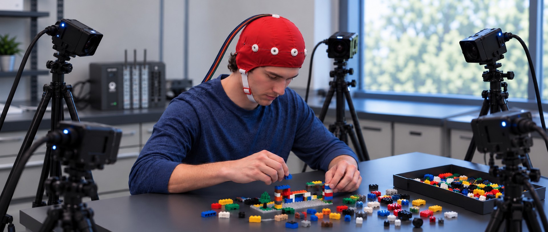
Capturing More Than Physiology: Multi-Camera and Multimodal Research

In the early 20th century, Cubist painters like Pablo Picasso and Georges Braque argued that traditional linear perspective on a subject was an illusion. Something was always hidden from the viewer, distorted, or lost altogether. Just as a Cubist painter might fracture their subject to reveal its true shape, a physiology lab uses multiple synchronized camera angles to bypass a single lens’ limitations.

The same principle applies to modern physiological and psychophysiological research. Physiological signals such as electrocardiogram (ECG), electroencephalogram (EEG), electrodermal activity (EDA), respiration, and blood flow provide valuable insight into how the body responds to stimuli, environments, and social interactions. Yet physiological data alone often leaves important questions unanswered. What was the participant doing? What was happening in the environment? Did social interaction, movement, or behavioral events coincide with a change in the recorded signal?

To answer these questions, many studies now combine physiological recordings with multiple video streams, eye tracking, audio, environmental monitoring, and other forms of media capture. This multimodal approach lets investigators examine physiological measurements alongside the events occurring around them, revealing a richer, more complete picture of human and animal behavior. As these data streams multiply, so does the need to capture, synchronize, and review them together.

Researchers at Macquarie University in Australia and collaborating institutions recently described a multimodal laboratory designed to study face-to-face communication and social interaction. The platform centered on a BIOPAC data acquisition system, amplifiers, and amplifier input module that acquired ECG, EDA, photoplethysmography (PPG), respiration, and skin temperature. The researchers combined physiological measurements with a Tobii eye tracker, wearable sensors, audio recording, motion analysis systems, and six video cameras positioned throughout the laboratory. The project showed that adding more recording sources often complicates data collection coordination, and that integrating multiple acquisition systems requires careful synchronization and workflow design. The camera system played a particularly important role. They used multiple viewing angles for behavioral coding, gesture analysis, participant observation, and synchronization verification. Rather than serving only as a record of the experiment, the video data became an important part of the analysis itself. For researchers studying communication, teamwork, education, or human-computer interaction, the work illustrates how behavioral observations can enhance interpretation of physiological measurements.

A team from the Swedish University of Agricultural Sciences (SLU) explored similar ideas outside the traditional laboratory by developing the SLU Multisensory Outdoor Laboratory (SENSOLA). The researchers used BIOPAC data acquisition and analysis systems with AcqKnowledge software and BioNomadix wireless devices to collect respiration and PPG measurements as participants moved through natural and built environments. Their platform combined those recordings with eye tracking, stimulation, video, audio, GPS data, participant feedback, and environmental measurements. One key observation was that physiological measurements become much more insightful when paired with information about a participant’s surroundings and experiences. Video proved especially valuable because it documented environmental conditions, participant behavior, and unexpected events that might otherwise be difficult to identify during later analysis. As physiological research moves beyond controlled laboratory settings, projects such as SENSOLA demonstrate the value of capturing physiological and observational data together.
ratPreclinical research is increasingly applying the same principles. In a recent toxicology study from Japan, investigators developed the ToxQb platform around a BIOPAC data acquisition system and AcqKnowledge software with NDT to collect EEG and ECG data from laboratory rats. The physiological measurements were integrated with overhead, front, and side-view cameras, along with thermal imaging systems. The goal was to monitor behavioral and physiological changes simultaneously over extended periods. By reviewing camera footage alongside EEG and ECG recordings, researchers could relate changes in movement, posture, and activity to physiological events. The authors concluded that combining these diverse data streams provided deeper insight than any single measurement could. The study demonstrates that multimodal acquisition extends beyond human research, across a wide range of life-science applications.

Although these studies span communication research, environmental psychology, and toxicology, they point to a common trend: researchers are capturing much more than physiology. Like the artists who challenged the limitations of a single viewpoint, today’s researchers increasingly rely on multiple synchronized perspectives to reveal a more complete picture of physiology, behavior, and environment. Multiple cameras, video recordings, eye tracking, environmental sensors, and behavioral observations can provide valuable context for interpreting physiological measurements. Learn how using BIOPAC’s MultiCapture feature in AcqKnowledge 6 to integrates multi-camera video and other media into research via our webinar on the topic.


Are you planning to integrate multiple camera perspectives in your next study? Our sales team can help find the right equipment and show you how to leverage AcqKnowledge‘s MultiCapture feature to achieve the best possible results.
Brain and Body: SPA-fNIRS Solves Physiological Signal Noise

Functional near-infrared spectroscopy (fNIRS) offers researchers a powerful noninvasive tool for studying brain activity by measuring changes in blood oxygen levels. These changes reflect how various brain regions respond to stimuli such as tasks that involve thinking, emotion, movement, and social interaction. These are often in natural, real-world settings that are not possible with other brain imaging methods. However, fNIRS is not without its own limitations. Systemic fluctuations in the body, such as breathing patterns, cardiovascular changes, and autonomic responses, can result in signal noise that is misinterpreted as brain activity by traditional fNIRS. The solution: a visit to the SPA.

Systemic Physiology Augmented fNIRS (SPA-fNIRS) is an advanced approach to fNIRS that goes beyond measuring local changes in cerebral oxygenation. While standard fNIRS measures cortical hemodynamics by tracking oxygenated (HbO) and deoxygenated (HbR) hemoglobin, noise originating from systemic physiological changes can corrupt those signals. Combining data from both neural and systemic physiological signals, SPA-fNIRS provides researchers with a more diverse and accurate view of how the brain and body interact in real time.

The method is being applied across various fields, including psychophysiology, cognitive neuroscience, exercise physiology, mental health, and human performance research. Studies have utilized SPA-fNIRS to investigate a range of topics, including meditation and emotional regulation, movement control, and social interaction. The broader goal is to move beyond studying the brain in isolation and to understand it as part of an integrated mind-body system. This holistic view is transforming how scientists interpret brain-function data, opening new paths for applied research in education, rehabilitation, and human-machine interface design.

soundResearchers in India investigated the impact of a traditional sound-based meditation technique, known as the Mind-Sound Resonance Technique (MSRT), on both brain and heart activity. Using fNIRS to track changes in oxygen levels in the prefrontal cortex, and a BIOPAC data acquisition and analysis system with AcqKnowledge software to record and analyze heart rate variability (HRV), the study employed a SPA-fNIRS design, integrating neural and physiological data in a single, synchronized dataset. HRV analysis allowed the researchers to quantify shifts in autonomic balance during and after meditation. The results showed increased oxygenation in the right prefrontal cortex and enhanced low-frequency HRV components, reflecting greater parasympathetic regulation. Together, these findings suggest that sound-based meditation not only improves brain oxygenation but also promotes calmer, more balanced heart-brain dynamics, illustrating how SPA-fNIRS methods can reveal the comprehensive physiological impact of mindfulness practices.
In her doctoral research at the Université de Lille in France, neuroscientist Aurore Guérin examined how the brain controls movement speed using a multimodal combination of fNIRS and physiological data recording systems. Participants performed repetitive arm movements at fast, natural, and slow tempos. At the same time, the study captured oxygenation in the prefrontal and motor cortices using fNIRS, alongside heart rate and respiration signals collected with a BIOPAC data acquisition system. A respiratory effort transducer measured breathing patterns, while a BioNomadix ECG module recorded cardiac activity for HRV analysis. All physiological signals were synchronized and analyzed in AcqKnowledge software, allowing the team to identify and remove systemic influences (e.g., heartbeat and breathing rhythms) from the fNIRS signals, following a SPA-fNIRS approach. The results revealed that faster movements primarily engaged motor regions, while slower, more deliberate pacing resulted in higher prefrontal activation, reflecting greater cognitive control. By integrating physiological monitoring with fNIRS, the study provided a clearer picture of how the brain and body coordinate timing and control during movement.

These studies illustrate how SPA-fNIRS is reshaping the way scientists investigate the interaction between brain and body. By combining neural and physiological data in a single synchronized system, researchers can separate brain activity from systemic noise and uncover the interplay between cognition, emotion, and autonomic regulation. For more information on this topic, watch our webinar on Multimodal SPA-fNIRS and check our our latest multimoldal fNIRS research products.


Are you planning a study that blends fNIRS with other physiological signals? Contact your local BIOPAC customer service representative to learn more about how SPA-fNIRS can benefit your research.

Measuring Mindfulness: Ancient Awareness for Modern Research

The concept of mindfulness has garnered significant attention in recent years, transcending its roots in ancient meditation practices to become a focal point in modern scientific research, particularly in the fields of physiology and psychophysiology. Understanding mindfulness involves delving into its essence: a state of heightened awareness and acceptance of the present moment, free from judgment or attachment to external stimuli. But how exactly does this concept intersect with the realms of physiology and psychophysiology, and how do researchers measure its impact on the human body and mind?

At its core, mindfulness entails being fully present and engaged in the current experience, whether it be a sensation, thought, or emotion. This mental state has been associated with numerous health benefits, including stress reduction, improved cognitive function, and enhanced emotional well-being. In the realm of physiology and psychophysiology, researchers are keen to explore how mindfulness practices influence the body’s physiological processes and how these changes can be quantified and understood.

One of the primary methods used to measure mindfulness in laboratory settings is through the analysis of physiological signals, such as electrodermal activity (EDA) and heart rate variability (HRV). During mindfulness practices, individuals often experience a shift in these physiological parameters. For example, studies have shown that mindfulness meditation can lead to decreased EDA, indicating a reduction in sympathetic nervous system activity and arousal levels. Similarly, practitioners may exhibit increased HRV, reflecting greater parasympathetic nervous system dominance and improved adaptability to stressors.

A collaborative study by a team of researchers from Wake Forest University, the University of Utah, and San Diego State University examined the role of HRV in mindfulness-based pain relief practices. Specifically, the study looked at the role of high-frequency heart rate variability (HF HRV), a marker of parasympathetic nervous system (PNS) activity, during mindfulness-based pain relief versus a sham-breathing exercise that served as a control. Respiration and electrocardiogram (ECG) data was fed into a BIOPAC data acquisition and analysis system. While the study results were inconclusive, researchers determined that further study was needed to explore “parasympathetic activation in mindfulness-based pain relief as compared to other meditative techniques.”

lotusA study published in the 2023 edition of the Journal of Psychiatric Research compared how rest and breath-focused mindfulness could be used to regulate and control various symptoms of post-traumatic stress disorder (PTSD) and dissociative symptoms related to past trauma in female study participants. Researchers used skin conductance response (SCR), one of the components of EDA, to measure autonomic function as it relates to PTSD symptoms. SCR data were collected during alternating resting control and breath-focused mindfulness conditions via BIOPAC EDA electrodes connected to a BIOPAC data acquisition and analysis system. The study found that rest and breathing exercises had produced positive results for individuals with low to moderate levels of trauma but were less effective with higher levels.

One notable feature of BIOPAC’s technology is its ability to integrate multiple physiological signals simultaneously, allowing researchers to better understand how mindfulness practices impact various bodily systems. An example is a study by a group of researchers in Israel that examined the effectiveness of mindfulness-based interventions (MBI) in improving mental well-being and resilience. MBIs are non-religious adaptations of contemplative Buddhist practices that are used to reduce suffering and increase well-being. Researchers used a BIOPAC BioNomadix system to wirelessly record a range of physiological signals, including ECG, electromyography (EMG), and EDA. Their goal was to determine whether mindfulness could positively influence how interoceptive signals from the autonomic nervous system regulate one’s sense of well-being. The study concluded that “mindfulness seems to increase the sensitivity of regulatory selections to interoceptive signals, and this is associated with subjective well-being.”

Mindfulness represents a fascinating intersection between ancient contemplative traditions and modern scientific inquiry. To learn more about how BIOPAC is helping researchers uncover new insights into the physiological aspects of mindfulness and its potential implications for health and well-being, check out our webinar on The Science of Mindfulness.


Are you looking for techniques and tools to measure mindfulness in your study? Contact your regional BIOPAC sales representative to integrate everything you will need to gather and analyze data.

4T|Phys 2023: BIOPAC Shares Tools, Trends, Techniques & Technology at UCSB

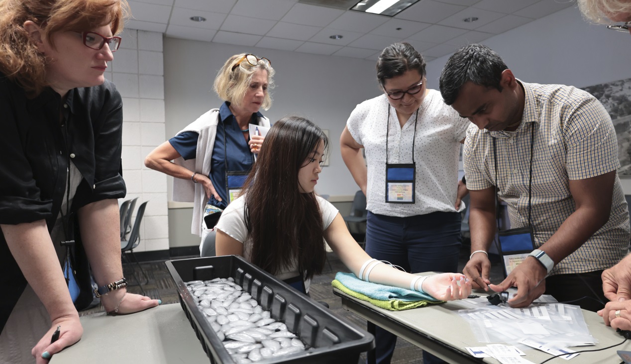
They hailed from across the globe, from Arkansas to Zanzibar and all points between. Students, teachers, researchers, and innovators gathered on the campus of UC Santa Barbara, overlooking the Pacific Ocean. They came to learn, share, and explore breakthroughs and best practices in physiology and related fields. 4T|Phys kicked off July 17 under clear California summer skies, an idyllic setting for discovering the Tools, Trends, Techniques, and Technology driving physiology education and exploration.

4T|Phys 2023 was the third installment in BIOPAC’s highly successful series of biennial industry conferences that began in 2017. After a two-year hiatus during the global Covid-19 pandemic, 4T came roaring back this summer with more workshops, more tracks, more sponsors, more presenters, and more countries represented than ever before. Over the course of three days, nearly 100 attendees participated in hands-on sessions with some of the top minds in physiology research, experiencing the latest tools for gathering and analyzing data.

BIOPAC’s in-house experts were joined by an array of authorities from the fields of physiology, psychology, imaging (fNIRS and fMRI), neuroscience, cardiovascular research, eye tracking, virtual reality (VR), biology, and engineering. Participants took part in workshop schedules drawn from five separate tracks that included fundamentals, which focused on the basics of data acquisition and analysis. The research track took attendees on a deeper dive into techniques and technologies, including recording evoked potentials, screen-based eye tracking, impedance cardiography using trans-radial bioimpedance velocimetry (TREV), stimulus presentation, exercise physiology, and many others. Participants were also able to tour UC Santa Barbara’s state-of-the-art Brain Imaging Lab to see up close how BIOPAC products are being used in both functional MRI (fMRI) and TREV studies.

“It was an incredible experience that left a lasting impact on me by helping me to understand more about physiological data collection and analysis,” said Dr. Parth Trivedi, a 4T|Phys attendee. “The choice of presenters and the topics covered were truly inspiring, offering valuable knowledge and fresh perspectives that enriched my understanding.”

4T|Phys 2023 Attendee MapThis year’s conference featured several new tracks such as a dedicated VR track led by partner company WorldViz. The WorldViz team showcased virtual reality solutions in a series of workshops that introduced how VR is being integrated in physiology and psychology research, explained how to create effective and immersive VR experiments, and demonstrated their latest PRISM technology for creating fully immersive projected 3D virtual environments.

Several other partner companies introduced their technologies and products both in the 4T exhibition hall and in workshop sessions. Argus Science presented their ETVision eye-tracking glasses that synch seamlessly with AcqKnowledge. Cognixion demonstrated the Cognixion One Axon-R—Research Edition wearable BCI-AR computing platform, which is being used to help individuals with neurodegenerative conditions and severe motor impairments communicate and interact with their environments assisted by augmented reality. Seenel Imaging showed off their MedelOpt Systems for whole-head EEG with fNIRS, including new VR integration. Participants also got a chance to try on the Research Ring by Senstream, a new user-friendly wearable research ring, which allows easy recording of EDA, PPG, Temperature, and ECG signals. CNSystems showed off the company’s NIBP continuous noninvasive blood pressure (NIBP) and hemodynamics devices.

If you missed the educational and networking opportunities at 4T|Phys 2023, don’t despair. We will be back again in a few years, bigger and better than ever. Watch this space for updates on our next installment. In the meantime, see our Video Tutorials and Webinars On Demand for trainings and seminars on our products and partners.


Make sure you never miss a BIOPAC event and keep up with the latest Tools, Trends, Techniques, and Technologies by contacting your local BIOPAC sales representative.

Eye-Catching: Mobile Tech Enhances Immersive Experiments

Freed from the limitations of stationary eye tracking technology, mobile eye tracking enables researchers to study their subjects while they perform a wide range of immersive tasks in natural settings.

Sixteenth-century pediatrician Thomas Phaer mused that “the eyes are the windows to the soul.” These sensory organs and their movements certainly provide a wealth of information to researchers studying the interaction of mind and body. Eye tracking has long been a key tool of physiological and psychophysiological research, and recent developments in mobile eye tracking take this valuable data resource out of the confines of the lab and into the real world.

Freed from the limitations of stationary eye tracking technology, mobile eye tracking enables researchers to study participants while they perform a wide range of immersive tasks in natural settings. These self-contained devices come in a variety of form factors including wearable glasses that provide untethered eye tracking data acquisition in real time that can be combined with other physiological data collected during live experiments.

Today’s lightweight mobile devices can record a broad spectrum of eye tracking data, including pupil size and position, gaze path, vergence angle, and areas of interest (AOI). The latter connects stimuli to eye-movement metrics, providing data on the participant’s psychophysiological response to stimuli within their field of vision. In previous blog posts and webinars, we have explored how mobile eye tracking technology gathers this data. Numerous studies have been published that demonstrate its potential in physiological and psychophysiological research. Settings and applications include user experience, sports and physical therapy, public safety and law enforcement, driving and equipment operation, and work environments from the office to the factory floor.

Mobile eye tracking technology allowed researchers at The University of Sheffield to take their experiments to the city streets for a 2020 study into how different lighting affects cyclists navigating urban environments at night. Mobile eye tracking provided data on the gaze behavior of nocturnal cyclists in parallel with skin conductance response (SCR) to better understand how obstacle detection is influenced by variations in type, location, and level of lighting. Bicycles and their riders were equipped with a BIOPAC data acquisition unit and audio stimulation units along with a BioNomadix EDA transmitter and electrodes to record the participants’ SCR. Eye tracking data, including AOI, were synchronized with SCR data and analyzed using AcqKnowledge software to determine how the riders reacted to a variety of lighting conditions. Researchers concluded that lighting plays a significant role in riders’ comfort levels at night and that improved lighting could increase urban bicycle use.

VGM playerLess intrusive by design, mobile eye tracking can provide indoor experiments with a heightened degree of immersion. A study at the University of British Columbia used mobile eye tracking glasses to create a more natural environment to investigate how the immersive effects of electronic gaming machines (EGM) impact gambling addiction. The study looked at how machine design triggered psychophysiological responses in participants by assigning various AOI to EGM screens. ECG data were recorded along with respiration effort via a BIOPAC respiration effort transducer and amplifier, which were fed to a data acquisition unit and synchronized with AOI data recorded via mobile eye tracking glasses. The study was able to successfully identify several key markers for game immersion using AOI that suggest behavioral links between immersion and gambling addiction.

Possible applications for mobile eye tracking in research continue to expand with the evolution of the technology. To learn more about recent developments in mobile eye tracking, check out our latest webinar on mobile eye tracking glasses with AcqKnowledge integration.


Learn more about how to integrate mobile eye tracking into your next research project by contacting your local BIOPAC sales representative.

Mind and Body: Signals to Enhance Your Psychophysiology Research

The human body and mind share an intrinsic link, both influencing and reflecting the health and general well-being of the other. Within the field of psychophysiology, researchers gain a wealth of insight into mental processes and disorders by studying physiological signals recorded from both the brain and body. Signals such as electrodermal activity (EDA), skin temperature (SKT), electroencephalogram (EEG), heart rate variability (HRV), and functional near-infrared spectroscopy (fNIRS) provide just a few of the tools helping us to better understand these mind-body interactions.

A wide range of research technologies has made these signals increasingly accessible in the lab as well as in everyday environments, allowing studies to place participants in increasingly naturalistic settings while gathering critical physiological data. In this way, researchers can provide a more accurate picture of how study participants react to stress and arousal, experience emotion, or interact within group environments.

One recent study used EDA and heart rate (HR) to examine the role of emotion in student success within computer-based learning environments (CBLE). As discussed in previous posts, a range of signals can be used to measure the body’s physiological response to emotion, otherwise known as “affect.” By gaining a better understanding of learners’ emotional responses to CBLE stimuli, researchers in this study hope to apply their data to improvements in CBLE design. EDA data was gathered from participants via SS57LA EDA leads while HR signals were collected with SS2LB shielded lead sets, both of which were connected to a BSL MP36 data acquisition and analysis system. Participants were shown imagery designed to evoke a negative emotional reaction before being asked to read a complex academic paper. The study was able to establish a link between the participants’ emotions and their comprehension of the reading material.

head with brain exposedIn another study, researchers used EDA and skin conductance response (SCR) to measure participants’ reactions to conditioned stimuli (CS) and unconditioned stimuli (US). The goal was to determine if fear-inducing memories could be modified either pharmacologically or through behavior modification to overcome fear. An MP36R System with AcqKnowledge software allowed analysis of SCR signals gathered via SS3LA finger transducers across a range of experiments designed to trigger fear-inducing memories. While the overall results were inconclusive, the SCR signals provided valuable feedback regarding the participants’ response to stimuli.

While EDA is a common thread through many of these psychophysiology studies, other signals provide researchers with a more complete and nuanced data set from which to draw their conclusions. Researchers exploring the factors influencing musical preferences combined EDA data with ECG and facial electromyography (fEMG) signals to assess how listeners reacted to familiar and unfamiliar musical stimuli. Data from all three signals were fed to an MP36 data acquisition and analysis system. The study found a strong correlation between musical preferences and familiarity with material as reflected by the subjects’ emotional responses.

These examples only scratch the surface of how psychophysiological research is being aided by an array of signals, all made accessible with available technology. To learn more about how to implement these signals in your next study, see BIOPAC’s Psychophysiology Applications webpage.


Contact your BIOPAC sales representative to learn more about the wide range of data-gathering tools available to help you construct your next psychophysiology study.

Skin Deep: Using EDA to Measure Affective Response

We say something “gets under our skin” when it elicits an unpleasant or irritating emotional response. This connection between our skin and emotion runs deeper than a simple idiomatic expression, however. Along with performing several critical functions—protective, regulatory, and sensory—our skin provides a window into our physiological and emotional states as we respond to a range of stimuli. Electrodermal Activity (EDA) is the mechanism providing these insights.

EDA, also known as galvanic skin response (GSR), measures eccrine activity resulting from the sympathetic nervous system’s response to environmental stimulation. As a function of the autonomic nervous system, it is an involuntary response to a range of sensory inputs, including touch, sight, sound, odor, and taste. EDA regulates the rate of sweat produced by sweat glands in the extremities, particularly in the fingertips, palms, and soles of the feet.

EDA increases in conjunction with the level of stress or nervousness experienced by an individual. For this reason, it provides researchers with a valuable means of measuring affective response to external stimuli. As we explored in a previous post, affect is the measurable physiological expression of emotion. While there are numerous physiological signals that researchers can employ to measure affect, EDA in particular functions as an indicator of arousal. For this reason, EDA has become one of the most relied upon signals employed by researchers exploring psychophysiological phenomena.

Violence StudyA recent Slovakian study published in Adaptive Human Behavior and Physiology illustrates how EDA is commonly employed in such research. Researchers studying the influence of gender differences on reaction to violent imagery used skin conductivity response (SCR), the mechanism behind EDA, to measure levels of arousal when participants were shown images of war and sports. The goal of the study was to test the “male warrior hypothesis” (MWH). The theory suggests that men have evolved psychological mechanisms to form aggressive alliances with the goal of acquiring or protecting reproductive resources in war and that team sports stimulate that same impulse. EDA was measured using BIOPAC electrodes and leads feeding data to an MP36 data acquisition unit. While the study found that visual perception of team sports and war imagery stimulated different levels of excitement as measured by EDA in men and women, it was inconclusive in showing that sports provide a substitute for war in the sample of participants.

A study conducted at the University of Ottawa and published in the Journal of Behavior Therapy and Experimental Psychiatry used EDA to examine whether response-focused emotion regulation (RF-ER) strategies provided an effective way to control evoked emotions—specifically, does smiling make one feel better in times of heightened stress? EDA data was gathered from participants using a BIOPAC skin conductance transducer and amplifier connected to a BIOPAC data acquisition unit while participants were shown images designed to evoke both arousal and negative responses. Participants were asked to either suppress their emotional response to images or employ expressive dissonance (e.g., smile when disturbed). The results of the study shed doubt on previously held assumptions that RF-ER strategies were effective in controlling emotion.

These are just a sampling of the ways in which EDA is being used in psychophysiological research. For additional information on the background, tools, and techniques for using EDA in your studies, visit our EDA Resource Directory, read our EDA Introductory Guide. and watch Electrodermal Activity: Intro to Recording and Analysis or any of our EDA webinars.


For additional information on how BIOPAC can assist you with your research using EDA and a range of other physiological signals, contact your local sales representative.

Guilt Trips: Taking Measure of Our Moral Compass

“So full of artless jealousy is guilt, It spills itself in fearing to be spilt”
—Gertrude, William Shakespeare’s “Hamlet”

In Act 4 of Shakespeare’s famous tragedy, Hamlet’s mother Gertrude points out that the harder one tries to conceal guilt, the easier it is to expose. Little does she realize that she may have hit upon how future researchers approach the study of guilt in the lab. For practical purposes, it is a primary motivator in moral action and impacts how an individual responds to certain stimuli.

Psychologists have developed a myriad of tests to gauge guilt in a clinical setting such as the Test of Self-Conscious Affect (TOSCA), a multiple-choice test that tracks responses to various moral scenarios. While such techniques have been useful in linking guilt with conditions such as depression and anxiety, they have also shown their limitations in providing a deeper understanding of guilt’s influence on behavior, development, and psychological health.

In a recent BIOPAC webinar, UCSB professor Dr. Hongbo Yu addressed these limitations. Scenario-based tasks, he points out, are non-committal in examining the causes of guilt and do a poor job of measuring behavioral tendencies linked to guilt. Interaction-based research methods, however, allow participants to experience emotions in real-time, allow direct control of the causes of guilt in the lab, and allow for direct measurement of responses to stimuli.

As discussed in prior posts on affect, the body’s physiological manifestation of emotion, there is a range of methods and physiological signals available to researchers for measuring affect and gaining a better understanding of emotional states like guilt. Dr. Yu utilized eye-tracking, electrodermal activity (EDA), and functional magnetic resonance imaging (fMRI) in his examples. These are just a few of the signals currently being used in the lab to measure guilt.

One of the practical applications of guilt research is detecting when a subject is concealing the truth, as one would with a polygraph test. A recent study conducted by researchers at the University of Wisconsin-Madison’s Department of Psychology sought to gauge deception among research participants taking the Concealed Information Test (CIT). The goal was to improve upon traditional polygraph test signals such as heart rate and respiratory rate. Respiration data was gathered using a respiration transducer belt while ECG signals were collected with BIOPAC electrodes. In addition, researchers gathered EDA data using a transducer and finger electrodes. The study found that thGuilty Girle EDA signal was significantly more effective in accurately measuring physiological responses during the test procedure.

A 2018 study by researchers at Liverpool Hope University looked at the link between guilt and aggression in children by examining activity in the autonomic nervous system during so-called “bad behavior.” The study measured respiratory sinus arrhythmia (RSA) and skin conductance in children 4–8 years old while they imagined engaging in antisocial activities. EDA and ECG signals were gathered via wireless amplifiers and fed to an MP150 data acquisition unit (superseded by the MP160) running AcqKnowledge software. The study found a linkage between physiological responses to aggression and guilt that may provide a better understanding of the mechanisms behind guilt and related behavior in early and middle childhood.

These are just a few examples of how physiological signals can be applied to measuring guilt in research. BIOPAC offers a wide range of data acquisition, stimulus presentation, and VR options suitable for creating custom guilt research environments in the lab.


For additional information, contact your BIOPAC representative to learn more about the range of signals that can be utilized to gather data on guilt and other emotional states in the lab.

VR—New Worlds to Explore

Use VR to Expand the Capabilities of Your Lab

The immersive technology of virtual reality (VR) literally offers whole new worlds to explore. Combining VR/Immersive Solutions with Physiology Data allows researchers to create virtual worlds that are impossible or prohibitively expensive in the real world. Additionally, researchers can synchronize VR events with physiological response data to record and analyze physiological, behavioral, and subjective response data. Multimodal stimulation can deliver visual (3D), auditory, olfactory, haptic, and/or electric stimulus to add an incredible degree of realism to the immersive world!

Interfacing a VR workstation with a data acquisition system can add a new dimension to your research by combining objective measures with a tightly controlled experimental protocol. Immersive environments can take the subject anywhere and allow you to unlock the boundaries of your physical lab space and budget.

  • Create rich 3D environments that are 100% personalized to match your protocol.
  • Send physiological data back to the VR world to make decisions based on the participant’s physiological responses

Participants can move around naturally within the virtual world while their physiological data is captured. Record any combination of signals: ECG, EDA, EEG, EOG, EMG, EGG, temperature, respiration, and pulse. Include eye tracking to track subjects’ gaze as they view the immersive environment during the experiment. Close synchronization between the stimuli and the physiological signals is achieved through digital markers.

Monitor data in real time and use detailed signal analysis for post-acquisition scoring of the data. Automated analysis routines can streamline the analysis process, eliminate subjectivity, and save time—see AcqKnowledge for some powerful options.

VR software provides you with an unlimited degree of flexibility to create a specific world and control people and items within the world. VR software is easy to use and adaptable, so researchers can make subtle changes to an experiment without having to redesign the entire protocol.

Immerse your subjects in a BIOPAC VR world and record physiological data as the virtual environment influences the subject. Applications are varied and include Psychology, Biomechanics & Kinesiology, Ergonomics, Marketing/Consumer Research, Healthcare, Computer Modeling/Simulation, Education, Training: hazard, medical, etc.,

Use a feedback loop for greater control and automation of the VR world. This allows the world to change in real time, based on the participant’s responses. Timing events are recorded for tight synchronization with physiological data for meaningful analysis.

See VR Resources here for more examples of how VR can expand the capabilities of your lab!

BIOPAC offers a wide array of wired and wireless equipment that can be used in your research. To find more information on solutions for recording and analyzing signals such as ECG, heart rate, respiration, and more using any platforms mentioned in this blog post, you can visit the individual application pages on the BIOPAC website.

Photo by stem.T4L on Unsplash

Understanding Stimulus Presentation Studies

Stimulus presentation studies are an important aspect of modern research that record and organize a participant’s reactions and physiological data to various inputs. These studies are commonly used in neuromarketing or psychophysiological experiments. First off, what is stimulus exactly? Put simply, a stimulus is a sensory input delivered to a participant with the purpose of eliciting a particular response. These stimuli can consist of images, words, lights, sounds, virtual reality, scent, or even mild electrical impulses.

The idea is to present varying types of stimulus to a participant at expected or unexpected intervals and record his/her response. For example, a collection of images might be presented (one at a time) ranging from pleasant (say an impressive vista) to neutral (say a street scene) to unpleasant (possibly a striking snake or other “fight or flight” image). Programmable stimulus systems (such as E-Prime or SuperLab) to present the stimuli, and can be used with a Chronos or M-Pod response keypad that the participant presses, each key representing a different response (e.g., pleasant image = press Key 1, unpleasant = press Key 2, etc.). A physiological signal commonly gathered from the participant is electrodermal activity (EDA), which measures the perspiration generated in a participant’s palms or fingertips. This “eccrine activity” has a strong correlation with emotional responses. For studies involving visual stimuli, a camera may be used to record the participant’s facial expressions. The expressions are simultaneously recorded and categorized using software such as FaceReader.

Stimulus experiments are generally set up using the hardware and software of the stimulus system and the resulting physiological data and stimulus markers recorded using a data acquisition system, such as BIOPAC’s MP160 with STP100C, MP36, or Smart Center Systems. These systems include AcqKnowledge or Biopac Student Lab life science software and the MP data acquisition units that interface with the stimulus hardware.

In practice, setting up stimulus studies is easier than it sounds on paper. The parameters and stimulus types are set up via drag-and-drop-interfaces in the E-Prime or SuperLab software, and the hardware interface set up using an I/O or USB connection between the stim hardware and the computer. The stimulus and BIOPAC data acquisition software can easily reside on the same computer, as can all stimulus and BIOPAC data acquisition hardware.

Stimulus studies can range from a simple setup, such as a participant responding to a sound for reaction time studies, to more randomized studies utilizing different types of stimulus delivered at varying pre-programmed intervals.

When the study is completed, data can be analyzed in AcqKnowledge software, and the various stimulus/responses organized automatically using AcqKnowledge’s automated Stim-Response Analysis routine.

For more information about conducting Stimulus studies using E-Prime, SuperLab, and AcqKnowledge software, we invite you to view the following free webinars and video tutorials:

Stimulus Presentation and Event Mark Up with E-Prime

Stimulus Presentation and Event Mark Up with SuperLab

AcqKnowledge Stim Synchronization and Analysis

Focus Areas – Automatically Create Using Define Between Events

BIOPAC offers a wide array of wired and wireless equipment that can be used in your research. To find more information on solutions for recording and analyzing signals such as ECG, heart rate, respiration and more using any platforms mentioned in this blog post, you can visit the individual application pages on the BIOPAC website.

error: Content is protected !!