Neuroergonomics: Measuring Cognitive Workload in Real-World Tasks

In some high-stakes jobs, the pressure can feel overwhelming. From air traffic controllers tracking dozens of aircraft simultaneously to surgeons guiding robotic instruments through delicate procedures, many modern occupations require people to process extraordinary amounts of information while maintaining accuracy and focus. As workplaces become more automated and technologically sophisticated, understanding how the brain and body respond to these demands has become a central challenge for ergonomics researchers. A growing area of study, known as neuroergonomics, combines neuroscience, physiology, and human factors research to better understand how people perform in complex real-world environments.

In human factors research, neuroergonomics conintues to gain traction by offering a more comprehensive picture of cognitive workload than traditional measures. Two individuals may perform a task equally well while experiencing dramatically different levels of mental effort. Excessive cognitive workload can contribute to fatigue, stress, errors, and reduced situational awareness, while insufficient workload can lead to disengagement and decreased vigilance. Researchers are therefore concentrating attention on measuring not only what people do but also how their brains and bodies respond as they do it.

Among the most valuable tools for studying cognitive workload are physiological signals that provide objective measurements of mental state. Electroencephalography (EEG) can reveal changes in neural activity associated with attention and cognitive effort, while functional near-infrared spectroscopy (fNIRS) measures changes in cortical blood oxygenation linked to mental workload. Electrocardiography (ECG) and heart rate variability (HRV) offer insight into autonomic nervous system activity, and eye-tracking measures such as pupil diameter and gaze behavior can indicate shifts in attention and cognitive demand. More often, researchers are combining multiple synchronized signals to create a more complete view of how people interact with demanding tasks and environments.

A team of researchers from Drexel University and Lockheed Martin investigated how multiple physiological signals can be combined to measure cognitive workload across a range of mental activities. Participants completed tasks involving working memory, vigilance, risk assessment, attention shifting, situation awareness, and inhibitory control while researchers simultaneously monitored EEG, fNIRS, ECG, photoplethysmography (PPG), electrooculography (EOG), and eye tracking. Brain activity was measured using an fNIR Devices system alongside other physiological monitoring technologies. The researchers found that different physiological measures varied in their sensitivity to specific task demands, with neuroimaging measures providing particularly valuable insight into changes in workload. The findings demonstrate the value of combining signals from the brain, heart, and eyes to create a broader perspective of cognitive effort in complex environments.

Researchers at Loughborough University in the United Kingdom examined how rising task complexity influences cognitive workload in modern manufacturing environments. Participants performed a series of assembly tasks while researchers recorded pupil diameter with Tobii eye-tracking and measured HRV with wearable ECG sensors. By comparing physiological responses across low-, medium-, and high-complexity tasks, the team identified significant changes in both pupillometry and cardiac indicators of cognitive load. Their findings suggest that physiological monitoring can provide an objective means of evaluating cognitive ergonomics in highly digitalized manufacturing environments and may help support the design of more sustainable, human-centered workplaces.

flight simAnother important challenge in neuroergonomics is determining which physiological measurements provide the most reliable indicators of mental workload. A collaborative team of researchers from France investigated how physiological measurements can be used to establish standardized levels of mental workload during aviation-inspired tasks. Using the Multi-Attribute Task Battery (MATB-II), participants performed simulated flight-related tasks under varying levels of demand while researchers measured EEG, eye-tracking, electrodermal activity, and cardiac activity. ECG and EDA signals were acquired using a BIOPAC data acquisition and analysis system running AcqKnowledge software. The study found that several cardiac, ocular, and electroencephalographic measures successfully distinguished high-workload conditions and correlated with subjective workload ratings. The researchers found that several physiological measures, particularly EEG, eye tracking, and cardiovascular metrics, reliably distinguished between different workload levels and closely tracked participants’ subjective assessments of task difficulty. Their findings suggest that multimodal physiological monitoring could support the development of adaptive systems that detect cognitive overload in real time, improving both performance and safety in demanding operational environments.

These studies highlight the increasingly multimodal nature of neuroergonomics research. Eye tracking provides insight into attention and visual processing, cardiovascular measures reveal how the autonomic nervous system responds to mental demands, and brain-imaging techniques such as EEG and fNIRS offer direct windows into neural workload. By combining these complementary perspectives, researchers are moving beyond simple measures of task performance toward a deeper understanding of how people think, adapt, and perform in complex environments. As workplaces continue to evolve alongside automation and intelligent technologies, neuroergonomics is helping researchers design systems that better support human capabilities, improve safety, and reduce the hidden costs of cognitive overload.


Are you planning a multimodal neuroergonomics study? Our team of experts can help you plan your research and identify the tools that will provide the best possible data.


The Nose Knows: Brain-Body Research and the Science of Olfactory Cognition

“Smell is a potent wizard that transports you across thousands of miles and all the years you have lived.” — Helen Keller

There is something uniquely intimate about the sense of smell. Unlike vision or hearing, olfactory signals travel a remarkably direct neural route from receptors in the nasal epithelium through the olfactory bulb, and straight into the brain’s limbic system, the seat of emotion and memory. A single scent can transport us instantly to a childhood kitchen, sharpen our alertness, or dissolve stress within seconds. Yet for all its power, olfaction has historically been one of the most understudied human senses. That landscape is changing. A new generation of multimodal brain-body research platforms, capable of simultaneously recording signals from the brain and the body, is giving scientists insight into how smell shapes thought, emotion, and cognition.

The olfactory system is the only sensory system that bypasses the thalamus, the brain’s central relay station, and connects directly to the amygdala and hippocampus, two structures critical to emotional processing (affect) and memory. This anatomical shortcut explains why odors are so potent at triggering emotional memories, often more vividly than visual or auditory cues. Beyond memory, researchers are increasingly interested in how odor exposure alters measurable physiological and neurological states in real time, ranging from modulating cognitive performance and stress to the early detection of neurodegenerative disease, in which olfactory dysfunction often appears years before other symptoms emerge.

What makes this research technically challenging is also what makes it scientifically rich: the olfactory experience simultaneously engages the central nervous system and the autonomic nervous system, meaning that studying it well requires capturing signals from both brain and body at once. Electroencephalography (EEG) provides direct data on the brain’s electrical activity with millisecond-level temporal resolution, making it well-suited to detecting rapid shifts in cognition and arousal following odor exposure. Functional near-infrared spectroscopy (fNIRS) offers a complementary picture, using near-infrared light to detect changes in cortical oxygenation, a measure of the brain’s metabolic response to stimulation. Wearable and tolerant of movement, fNIRS is particularly well-suited to naturalistic experimental designs. Electrodermal activity (EDA), or skin conductance, captures the autonomic nervous system’s arousal response through sweat gland activity, while electrocardiography (ECG) and heart rate variability (HRV) add a cardiovascular layer that can distinguish sympathetic from parasympathetic activity in fine detail. Today’s lab setups use a range of sophisticated scent delivery systems to integrate precise, controllable olfactory stimuli into experiments. The real power of modern multimodal platforms lies in their ability to synchronize all these channels within a single setup, time-locking each physiological response to a specific odor event and revealing the full brain-body picture that no single signal could show alone.

A research team at the All India Institute of Medical Sciences in Jodhpur investigated whether citrus odor could buffer the cognitive and physiological impact of working memory load. Thirty participants completed a demanding working memory test under both control and citrus-odor conditions, with the odor delivered via an aroma diffuser. ECG, photoplethysmography (PPG), and EDA were recorded using a BIOPAC data acquisition system with AcqKnowledge software for analysis, and dual-signal BioNomadix wireless EDA and PPG module. For EDA recording, disposable silver/silver chloride disc electrodes were applied to the hypothenar and thenar eminences, and the PPG sensor was placed on the index finger. Citrus odor significantly improved task accuracy while suppressing the rise in skin conductance typically associated with cognitive load. HRV measures, including pNN50, RMSSD, and HF power, shifted toward parasympathetic dominance, indicating a calmer, more regulated physiological state. The findings, published in the Annals of Neurosciences, suggest that citrus odor acts as a cognitive stress buffer and point to practical applications in educational, occupational, and clinical settings with high sustained cognitive demands.

A counterintuitive question motivated a study at Nagaoka University of Technology in Japan: could black pepper, widely known as a stimulant, actually calm the body’s stress response? A group of university students performed a 30-minute arithmetic calculation task under three aroma conditions: black pepper, ginger, and a scentless control. Scents were delivered at precise, reproducible intervals through an olfactometer. A BIOPAC data acquisition system recorded continuous ECG and skin conductance level (SCL) at 200 Hz with 16-bit resolution, with high-frequency HRV (HF-HRV) derived as a measure of cardiac parasympathetic activity. Electrodes were placed beneath the right clavicle and on the lower left abdomen following the Lead II placement protocol for ECG measurements using multi-lead ECG cables. SCL was recorded using skin conductance sensors placed on the palmar side of the middle phalanx of the index and ring fingers of the participant’s non-dominant hand. The results, published in the Journal of Physiological Anthropology, found that black pepper aroma reduced the stress-induced rise in heart rate by 38.9% compared to the scentless control, attenuated the decline in HRV by 32.9%, and blunted the increase in SCL compared to ginger. Crucially, subjective stress ratings did not differ across conditions, demonstrating that the body can respond to olfactory stimuli at a physiological level even when conscious perception does not register a difference. These findings have meaningful implications for workplace wellness and aromatherapy research.

lavenderA French consortium of researchers from the Université Bourgogne Franche-Comté and the Université Grenoble Alpes tested whether music, lavender odor, or their combination would accelerate autonomic recovery following acute cognitive stress. Ninety-nine participants completed a battery of cognitively demanding tasks. They were then randomly assigned to one of four 30-minute recovery conditions: silence, relaxing classical music, lavender essential oil, or music and lavender together. A data acquisition system running AcqKnowledge continuously recorded ECG and EDA using Ag-AgCl finger electrodes, with HF-HRV as an index of parasympathetic activity and EDA as a measure of sympathetic arousal. Published in Psychophysiology, the study found that both music and lavender independently facilitated autonomic recovery. The surprising finding, however, was the absence of any multisensory advantage: combining the two stimuli produced no greater benefit than either alone, suggesting that when multiple sensory channels converge on the same relaxation goal, they may share rather than combine their neural resources. The study underscores both the value of multimodal physiological recording in parsing the mechanisms of stress recovery and the importance of rigorously testing assumptions about how the senses work together.

These studies illustrate how much remains to be discovered at the intersection of smell, brain, and body, and how essential it is to measure all three at once. As synchronized multimodal platforms continue to evolve, the science of olfactory cognition continues to shape our fundamental understanding of how the brain works and the development of practical tools to support cognitive health and wellbeing. To learn more about the convergence of olfaction with brain-body research, be sure to check out our webinar on how scents influence cognition.


If you are planning a brain-body study that integrates multimodal data collection with stimulus presentation like scent delivery, our expert staff is ready to help find everything you need to get started.

2025: The Year in Review

As 2025 winds to a close, there is much to celebrate both here at BIOPAC and in the ever-expanding frontiers of research that our products engage with every day. Let’s take a quick look at where we have been over the past 12 months and what the future holds.

Popular Posts

Speaking of engagement, this is the time of year when we do a bit of self-assessment and revisit what topics our readers were most inspired by and most interested in. Apparently, many of our readers are closet gamers at heart, because our blog on research on game design and gaming impacts drove more eyes to our site than any other topic this year, followed by posts on cognitive neuroscience and aging and the human impact of isolation.

New Products and Updatesclimbing kid

On the heels of the 2024 release of our flagship MP200 data acquisition and analysis system and a significant upgrade to AcqKnowledge software, 2025 concludes with another milestone: the imminent release of AcqKnowledge 6.0.2. This latest version features a host of improvements and additions, including full integration of MedelOpt+, which blends whole-head functional near-infrared spectroscopy (fNIRS) with electroencephalography (EEG) in a single, highly flexible device.

BIOPAC in the Lab

Every year, BIOPAC products continue to drive the most exciting research in physiology, psychology, and neurology, and 2025 was no different. Each month, we highlight breakthrough studies in which BIOPAC played a role in gathering and analyzing key data. These are usually centered around a theme and feature research published in the last few years. To close out the year, we like to spotlight a couple of significant studies from 2025 that didn’t get mentioned.

A collaborative study by researchers from the University of Georgia and Beijing Normal University examined how mothers and children coordinate their physiological responses during stressful interactions. Using BIOPAC data acquisition systems coupled with wireless ECG transmitters, the team simultaneously recorded cardiac activity from each participant pair as they completed structured challenge tasks. This setup provided the temporal precision needed to track changes in heart rate and moment-to-moment autonomic synchrony, revealing how caregivers and children influence one another biologically during shared stress. The study found that greater synchrony was linked to both emotional dynamics and regulatory behaviors during the tasks, highlighting the deep coupling between social relationships and physiological responses. The wireless BIOPAC configuration enabled naturalistic interaction while capturing high-quality dyadic data, a persistent challenge in synchrony research.

In its first full year of availability, the MP200 is already making waves in the research community. At Guangdong University of Science and Technology in China, researchers examined how head-shoulder posture and cervical muscle activation differ between college students with chronic neck pain (CNP) and healthy individuals. The team employed surface electromyography (sEMG) recorded with a BIOPAC MP200 data acquisition system and AcqKnowledge software. Researchers placed surface electrodes over targeted neck muscles to record raw EMG signals during various strength tests and physical tasks. Students with CNP demonstrated significantly poorer head and shoulder alignment, reduced neck extensor strength, and altered EMG activity, indicating neuromuscular dysfunction associated with pain. Findings showed the connection between altered posture and impaired muscle function in chronic neck pain. The study demonstrates the effectiveness of surface EMG to support clinical diagnosis and rehabilitation.

For more inspiration as we head into 2026, explore our webinar library for tips, tricks, and methods directly from experts across a range of research fields.


If you are planning to launch a study for 2026, now is the time to reach out to your nearest BIOPAC customer service representative for help finding the right tools to get started.

Mind Over Matter: Cognitive Neuroscience and Aging

“Age is an issue of mind over matter. If you don’t mind, it doesn’t matter.”  —  Mark Twain.

While American author and humorist Mark Twain suggested that mental attitude could make aging less daunting, modern science shows that the changes our bodies and brains undergo over time are very real. Yet researchers are discovering that aging doesn’t have to mean inevitable cognitive decline. In fact, the growing field of cognitive neuroscience is revealing ways to track, understand, and even influence how our brains change with age, offering hope that we can preserve mental acuity, independence, and quality of life well into our later years.

When studying how aging affects the brain, cognitive neuroscience researchers use a variety of tools. In addition to standard behavioral tests like the Stroop test, n-back test, and verbal fluency exercises, which track reaction times, accuracy, and problem-solving strategies, they also rely on neuroimaging and physiological measurements to gather deeper insights. Neuroimaging techniques include electroencephalography (EEG), which monitors real-time electrical activity in the brain; functional near-infrared spectroscopy (fNIRS), which measures blood oxygen levels in specific cortical regions during cognitive tasks; and magnetic resonance imaging (MRI), which provides detailed structural and functional maps of the brain.

Other physiological signals play an essential role in this field, including heart rate variability (HRV), a key indicator of the autonomic nervous system’s ability to balance various physiological functions and handle stress. Electrodermal activity (EDA) allows researchers to track arousal among participants undergoing cognitive exercises. Electromyography (EMG) measures muscle activation during fine and gross motor tasks. Eye tracking and electrooculography (EOG) provide researchers with data on how aging impacts attention and the ability to process visual cues. Respiration and blood pressure provide vital information on vascular health, which is often linked to cognitive performance.

Taiwanese researchers from National Yang Ming Chiao Tung University in Taipei turned to HRV and fNIRS to uncover how aging and, more specifically, mild cognitive impairment (MCI), affect both brain and heart responses during cognitive tasks. Healthy young adults, cognitively healthy older adults, and older adults with MCI were asked to perform cognitive function and executive tests. Researchers recorded the participants’ prefrontal cortex (PFC) activation through changes in oxyhemoglobin using fNIRS and autonomic responses via HRV. The team gathered ECG data using a BIOPAC data acquisition module running AcqKnowledge data acquisition and analysis software, and equipped with an ECG amplifier. The study found that older adults who did not exhibit MCI showed lower global and executive cognitive performance, reduced HRV, but increased prefrontal activation. The research team interpreted this to be the result of compensatory neural effort. However, older adults with MCI showed even greater cognitive decline, but no further differences in HRV or prefrontal activation. The findings suggest that HRV and PFC activation patterns could serve as biomarkers for distinguishing healthy aging from early pathological decline.

To better understand how learning processes develop and change with age, researchers from Tilburg University in the Netherlands investigated how younger adults and older adults differ in their brain and body responses while learning a visuomotor sequence task. To capture these responses, the team measured EEG, skin conductance (EDA), heart rate, HRV, breathing rate, and eye movements during the task. Physiological data were collected using BIOPAC’s BioNomadix wireless dual ECG/respiration amplifier and PPG/EDA amplifier, which continuously recorded three-lead ECG, respiration, and EDA at 2,000 Hz, with signals processed and analyzed in AcqKnowledge software. This setup allowed the researchers to precisely synchronize physiological activity with learning performance. Both younger and older adults improved with practice, but older adults needed more trials to reach the same precision. Brainwave activity showed apparent age differences, while measures like HRV, respiration, and skin conductance did not. This suggests that EEG is a more sensitive marker of age-related learning changes and could help guide training programs for older adults.

readingA long-term study by researchers from Radboud University Medical Center in the Netherlands, together with UK collaborators, tracked a group of healthy older adults over nearly 11 years to see how their brain blood-flow regulation changed by their 80s. Using transcranial Doppler ultrasound to measure blood velocity in the middle cerebral arteries, the team also monitored control signals including heart rate, blood pressure, and breathing gases. Physiological data were recorded with a BIOPAC data acquisition and analysis system, with end-tidal CO₂ (EtCO₂) measured using capnography via a BIOPAC carbon dioxide measurement amplifier. The researchers found a slight decline in CO₂ reactivity but, surprisingly, an improvement in dynamic cerebral autoregulation (the brain’s ability to maintain steady blood flow despite blood pressure changes). These effects were most apparent in participants who later reported memory complaints, suggesting that while aging may reduce vascular responsiveness, the autoregulatory system may help compensate for early cognitive challenges. The findings show how vascular health in the brain relates to aging and memory, providing valuable insight for detecting early risks of dementia and shaping preventive strategies.

These studies highlight how combining brain imaging with physiological measurements is helping scientists reveal the complex relationship between aging, cognition, and overall health. Through cognitive neuroscience, researchers are identifying early signs of decline and gaining a better understanding of how the brain and body adapt over time. Research is paving the way to more effective screening, personalized interventions, and lifestyle strategies that may help people maintain mental sharpness and independence well into older age. For more information on the tools and techniques being applied to cognitive neuroscience research, see our Webinars and visit our page on Cognitive State Analysis.


Are you planning a study on how aging affects the brain or just looking to expand your cognitive neuroscience toolbox? Our customer service team is ready to help you find what you need to obtain the best possible data.

Aesthetically Pleasing: Exploring How the Brain Perceives the Arts

Beauty is in the eye of the beholder—a mostly subjective experience, so the saying implies. While we may share many common ideals about beauty and aesthetics, our reactions to art, music, and literature can diverge, converge, and overlap to a myriad of degrees. Neuroaesthetics explores how the brain perceives and responds to art, music, and other aesthetically evocative experiences. Combining neuroscience with psychology and the arts, researchers seek to understand why humans find certain images, sounds, or designs appealing and how these perceptions influence our emotions, cognition, and behavior. This interdisciplinary science has practical applications across a wide range of fields, including design, marketing, education, and mental health therapy, where understanding aesthetic experience can inform and enhance user engagement, learning, and emotional well-being.

Biometric signals provide insight into emotional and cognitive processing, allowing researchers to gain valuable data on how the brain creates and responds to aesthetically engaging stimuli. Commonly used techniques include electrodermal activity (EDA), which measures changes in skin conductance linked to emotional arousal; electroencephalography (EEG), which tracks brainwave patterns associated with attention and engagement; functional near-infrared spectroscopy (fNIRS), which monitors blood flow changes in the brain; and functional magnetic resonance imaging (fMRI), which provides high-resolution images of neural activity.

Additionally, the use of virtual reality (VR) and augmented reality (AR) is expanding in neuroaesthetics research, allowing scientists to create controlled, immersive environments that mimic real-world experiences. These technologies offer new ways to investigate how individuals engage with art and design in dynamic, interactive settings, pushing the boundaries of how we understand and harness aesthetic experiences.

Researchers in Shanghai, China, for example, investigated how different visual modes—2D and 3D VR—affect emotional arousal, demonstrating how immersive environments influence emotional experiences. The research team utilized EEG to monitor brain activity and EDA to measure skin conductance responses, both indicators of emotional arousal. EDA and EEG data were recorded while study participants wore VR headsets to view 2D and 3D environments. Data was collected and analyzed using a BIOPAC MP system with AcqKnowledge software. The findings revealed that 3D VR environments elicited more intense emotional stimulation than 2D settings, as evidenced by higher beta (21–30 Hz) EEG power and increased EDA responses. According to the published findings, results suggest that enhanced perception in 3D environments leads to greater emotional engagement, highlighting the potential of 3D VR in applications such as immersive therapy, virtual training, and interactive entertainment.

MusicA team of German researchers recently examined the intersection of neurology, psychology, music, and literature. Their findings, published in the journal NeuroImage, explored how melodic features influence the neural processing of spoken poems and songs, examining the interplay between language and music. Researchers employed fMRI to investigate the neural mechanisms underlying the processing of musical and nonmusical verse. The fMRI data revealed that both spoken poems and songs activated bilateral temporal auditory areas of the brain. One aspect of the study was the measurement of participants’ emotional responses to the stimulus, i.e., whether they liked the song or poem to which they were listening while the fMRI scan was conducted. To gather this data, participants interacted with a BIOPAC differential pressure transducer connected to the participants’ right and left index fingers to record the degree to which they enjoyed the music. The researchers stated the findings suggest that melodic properties are central to the aesthetic experience of both speech and music.

A study published by Britain’s Royal Society looked at the connection between aesthetic appreciation and emotional states, specifically how exposure to art or music can shift feelings of anxiety to curiosity. To understand how the brain perceives beauty, the researchers used EDA to assess participants’ physiological arousal during exposure to aesthetically pleasing stimuli, such as music. EDA data was gathered using a BIOPAC EDA amplifier connected to a data acquisition and analysis system while participants were administered anxiety-inducing electrical tactile stimulation during music listening sessions. The study demonstrated that aesthetic appreciation can reduce anxiety and promote curiosity by analyzing the EDA data alongside self-reported measures. The research team concluded that their results “might have potential applications across different human activities such as learning and therapy. The idea that aesthetic appreciation might affect anxious states and that expecting aesthetic rewards can transform anxiety into arousal and curiosity plays a central role in some psychotherapeutic models of human change.”

These examples provide a rough outline of a bigger picture, illustrating the many ways researchers use physiological signals to better understand the human mind’s response to art, music, and literature. For more information on the tools BIOPAC provides to assist in neurological and physiological research, see our online training and webinars.


Are you planning a research project to investigate the intersection of aesthetics and neuroscience? Contact a BIOPAC representative in your area to identify the right resources and equipment for a successful study.

Everybody Hurts: Studying the Physiological and Neurological Components of Pain

A Buddhist saying states, “Pain is inevitable, but suffering is optional.” Pain’s dual nature has presented a conundrum for philosophy and the sciences. As a warning system, it protects us from harm, but it can also be the source of profound psychological and emotional damage. Pain is a complex and deeply subjective experience, yet it is one of the most critical areas of study in neuroscience.

Understanding pain involves not only exploring its physical origins but also decoding the neural circuits and physiological signals that contribute to its perception and regulation. Neuroscientific research into pain aims to answer fundamental questions about how pain is processed in the brain, how it varies from person to person, and how we can develop better treatments for chronic conditions.

This research uses various physiological signals to provide insights into pain mechanisms. Electroencephalography (EEG) captures brainwave activity, revealing how different regions of the brain respond to painful stimuli. Photoplethysmography (PPG) tracks blood flow changes, while electrocardiography (ECG) and skin conductance response (SCR) measure autonomic nervous system activity, offering clues about how the body reacts to stress and discomfort. Respiration patterns, too, are key indicators, as breathing can change in response to pain. Together, these signals help scientists map the body’s response to pain, shedding light on the underlying neural pathways and guiding the development of more effective pain management strategies. Understanding these physiological markers is a critical step toward personalized pain care and improving the quality of life for millions worldwide.

Pain research commonly focuses on the neurological mechanisms of pain to better understand its causes and how it can be treated. Researchers publishing their findings in the Journal of Biomedical and Health Informatics developed a device for studying pain perception biomarkers using thermal stimulation. Their device generates minutely controlled thermal stimuli using a computer-driven Peltier cell while simultaneously capturing EEG and PPG signals. Researchers identified the heat pain threshold (HPT) for each subject, defined as the maximum the subject can withstand when the Peltier cell gradually increases the temperature. The team used two-dimensional spectral entropy (SE) obtained from both the EEG and PPG signals to differentiate the condition of pain. EEG and PPG signals were fed to a BIOPAC MP36 data acquisition and analysis system running Biopac Student Lab (BSL) software. The study concluded that hemodynamics, brain dynamics, and their interactions can be used to discriminate thermal pain perception, which could have practical applications in monitoring pain perception. Possible applications include the study and treatment of “pathologies that affect the peripheral nervous system, such as small fiber neuropathies, fibromyalgia, or painful diabetic neuropathy.”

The pain mechanism associated specifically with fibromyalgia, one of the most common chronic pain disorders, was the focus of a study at the University of Jae´n in Spain. Despite its prevalence, much is still unknown about the underlying cause of this condition. Clinical and experimental studies of individuals diagnosed with fibromyalgia syndrome (FMS) have revealed autonomic nervous system (ANS) deficiencies. While both the parasympathetic and sympathetic components of the ANS have been studied, less is known about the sympathetic component as it relates to FMS. In this study, researchers used SCR as an indirect measure of the sweat response, which is controlled by the sympathetic mechanisms of the ANS. Researchers used a BIOPAC MP36 running AcqKnowledge to collect SCR data via an EDA lead set connected to participants’ fingers. The study found reduced skin conductance levels (SCL) and SCR in FMS patients compared to the non-FMS control group, indicating lower levels of sweating and reduced sympathetic activity in the skin. These factors provided the research team with a better understanding of the sympathetic nervous system’s role in FMS, with the potential for revealing new routes to better pain management and treatment.

For several decades, researchers and clinicians have studied the use of hypnosis to treat chronic pain. More recently, new technologies such as virtualpills reality have been incorporated into these techniques. A team of researchers from the University of Strasbourg in France studied the application of virtual reality hypnosis (VRH) to regulate HPT in healthy adult participants. Researchers used several physiological signals to measure autonomic function during the experiment, including ECG, respiration, SCR, and EEG. ECG signals were recorded using the BIOPAC ECG amplifier, while respiration rate was measured by a thermistor transducer that determined the difference in temperature between inhaled and exhaled air. SCRs were measured from the index and middle fingers of the dominant hand with a BIOPAC skin conductance transducer. Physiological signals were fed into a BIOPAC data acquisition system for analysis. A computer-generated immersive environment was created in VR and combined with a hypnotic auditory script. As with classical hypnosis treatments, the VRH scheme in this study began with an “induction period” followed by a “dissociation” period (separation between the mental state and the environment), followed by suggestions of pain reduction as analgesia. The study results supported a positive correlation between VRH and an increase in the participants’ pain threshold with promising implications for new non-pharmacological approaches to pain management and treatment.
These are just some of the ways that BIOPAC equipment is being used to explore the frontier of pain research and help ease the suffering of millions who deal with chronic pain and its physical and psychological effects. See our webinars page for additional information on how physiological signals can be applied to your next study.


Are you planning a study on pain or any related neurological or physiological area of research? Our team of experts is ready to help you find the tools to ensure success.

Free Your EEG: Wireless and Wearable Systems for the Lab and Beyond

The evolution of wearable technology for recording physiological signals represents a transformative leap for research, particularly when it comes to electroencephalography (EEG). These advancements allow researchers to capture brain activity data in real-world settings, moving beyond the confines of laboratory-based experiments. Mobile and wearable EEG devices utilize wireless technology to transmit data, offering significant advantages over traditional methods.

Everyone aspires to more freedom—to speak, think, or move about as one pleases. Should researchers be any different? Why be shackled to the static wires and cables of the lab, when one can release study participants with the help of mobile and wearable technology?

The evolution of wearable technology for recording physiological signals represents a transformative leap for research, particularly when it comes to electroencephalography (EEG). These advancements allow researchers to capture brain activity data in real-world settings, moving beyond the confines of laboratory-based experiments. Mobile and wearable EEG devices utilize wireless technology to transmit data, offering significant advantages over traditional methods.

EEG is a cornerstone in the study of brain activity, providing insights into the electrical patterns generated by neuronal firing. EEG has traditionally been used in physiology and psychophysiology research to understand brain function, monitor cognitive states, and investigate neural responses to various stimuli. The conventional EEG setup typically involves a stationary system with numerous electrodes affixed to the scalp using a conductive gel, which can limit the subject’s movement and impact the naturalistic settings of studies.

One of the primary benefits of mobile and wearable EEG systems is their ability to record brain activity during dynamic, everyday activities. This capability provides a more comprehensive understanding of how the brain responds to real-world stimuli and stressors, as opposed to controlled, static environments. For instance, researchers can now study cognitive processes in natural settings like workplaces or homes, enhancing the ecological validity of their findings.

A study by the School of Architecture at the University of Liverpool provides an example in which researchers leveraged wearable EEG technology when comparing subjective and physiological responses to noise within wooden buildings. The research team conducted listening tests using EEG data recorded via the B-Alert X-24 wireless headset system, which allowed participants to “sit comfortably in a simulated living room” while listening to a variety of sounds that were both airborne and resonating through the floors. Participants rated their level of noise annoyance on an 11-point scale and results were compared to EEG data. In discussing the design of their study, the authors highlighted the importance of incorporating noninvasive, wearable technology, stating, “Among the several options available for measuring physiological reactions to auditory stimuli, contemporary technologies made possible the recording of EEG in a relatively easy way by means of portable devices. These offer the precious opportunity of acquiring data directly from the scalp, where brain activity is detectable almost instantaneously in synchrony with the stimulus presentation.”

Similarly, when studying occurrences of hyperventilation among a participant group with a range of anxiety-related disorders, researchers from Yale University turned to a BIOPAC BioNomadix wireless receiver to gather EEG signal data. By attaching individual electrodes to a third-party cap that connected wirelessly to a BIOPAC data acquisition and analysis system, they minimized the discomfort and restriction of movement that may have resulted from a wired solution, thereby influencing participants’ response to stimuli.

Researchers at the Transylvania University of Brașov in Romania faced similar demands that required a flexible noninvasive means to record surface EEG signals from participants as they responded to a variety of colors and patterns with the goal of better understanding the complexity of human color and texture perception. The research team compared participants’ subjective responses to EEG responses via Event-Related Potentials and computational measurements of color and texture.  EEG data was gathered with a cap-based BioNomadix wireless system and analyzed using AcqKnowledge software. The study’s authors explained their choice of wireless EEG recording devices as follows: “Since human users are the target, it is important to focus the technology development towards small-scale, wearable, wireless systems that provide flexibility, easy use and comfort to the user side, while at the same time maintaining high signal quality.”

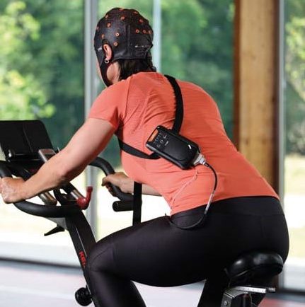
APEX cycleAdditional wearable EEG options available through BIOPAC include the MedelOpt Mobility EEG system, combining wearable EEG and functional near-infrared spectroscopy (fNIRS) technology with advanced signal processing and robust data transmission capabilities. APEX wireless EEG systems further enhance the landscape with their ability to record high-quality EEG data with flexibility for a range of applications from cognitive neuroscience to mental workload studies. Learn more about recording EEG data by watching our webinar: A Beginner’s Guide to EEG.


Are you interested in unlocking the benefits of mobile and wireless EEG for your next research project? Let our staff of experts find the right tools for you.

Mind and Machine: Exploring Brain-Computer Interfaces

In neuroscience, the frontier of brain-computer interfaces (BCI) holds the promise of bridging the gap between the human brain and external devices, offering revolutionary applications in fields ranging from robotics and prosthetics to communication and beyond. In the lab, scientists studying and developing new BCI technology leverage the human brain’s electrical and physiological signals.

Electroencephalography (EEG), functional near-infrared spectroscopy (fNIRS), and steady-state visually evoked potentials (SSVEP) are among the primary physiological and psychophysiological signals utilized in these investigations. Multimodal integration of two or more signal types allows researchers and engineers to take advantage of multiple data streams in their applications and studies.

EEG, which can measure electrical activity in the brain, is a cornerstone of BCI research. Researchers can decode various mental states and intentions by analyzing EEG patterns, enabling users to control external devices through mere thought. fNIRS complements EEG by measuring changes in blood oxygenation levels, offering insights into brain function with superior spatial resolution. SSVEP, on the other hand, capitalizes on the brain’s response to visual stimuli, providing a means for precise control in BCI applications.

The practical implications of BCI research extend far beyond the laboratory setting. In robotics, BCIs facilitate direct brain-to-machine communication, empowering users to manipulate robotic arms or navigate autonomous vehicles with their thoughts alone. A study published in the Journal of Information Science Engineering explored ways that an SSVEP-based BCI could be used to control assembly line robots via a headset measuring the operator’s EEG signals. The signals would communicate instructions to robots while operators identified defective parts, which the robots would pick out. During the experiment, EEG signals were obtained using a B-Alert X24 headset. The EEG-based BCI produced a 93.33% success rate in correctly identifying and removing defective parts from the assembly line process, allowing the operator to effectively “collaborate with the robot and communicate defective parts without manually operating the robot.”

Prosthetics benefit immensely from BCI advancements, enabling seamless integration with the user’s neural signals for intuitive control and enhanced functionality. A 2023 study by a team of researchers from the University of California Irvine explored the viability of creating low-cost cortical stimulators for bi-directional BCI (BD-BCI) research. BD-BCI takes brain-computer-interfaces to the next level with the brain not only providing motor commands to an artificial limb but also allowing it to receive sensory input from such a device. A BIOPAC data acquisition and analysis system was used to validate the charge balancing of subcortical electrocorticographic (ECoG) implants used in a stimulator. The charge balancing test was performed on phantom tissue designed to simulate real brain tissue. The resulting study successfully demonstrated “clinical validation of a fully-programmable electrical stimulator, integrated into an embedded BCI system.”

As researchers continue to refine BCI technologies, the possibilities for real-world applications are boundless. BIOPAC is among the companies on the cutting edge of BCI research, partnering with other innovators to provide the latest in multimodal BCI technologies such as Axon-R. For additional information on Axon-R, check out our recent webinar on the device.


Are you planning to explore the frontiers of BCI in the lab? Our regional customer reps have all the knowledge you need to integrate BIOPAC products into your BCI study.

Noise Proof: Attack Artifacts Before They Ruin Data

After investing months of time, funds, and personal effort into your electrodermal activity (EDA) study, you begin combing through your data recordings when something catches your eye. Several of the recorded signals don’t look quite right. Rather than a narrow baseline in the signal graph, the baseline is thick and fuzzy, a sure sign of signal noise. To make matters worse, the recordings are broken by random peaks, artifacts unrelated to the EDA signal you sought to record. Noise and artifacts can corrupt research data, often rendering it useless. While it may be possible in some cases to clean up and salvage recorded data, such results often mean going back to square one and starting over.

The best option is to avoid bad data from the beginning. Several best practices can save researchers untold effort and expense by avoiding the pitfalls that can result in noise and artifacts. Noise and artifacts can be defined as unwanted changes to a signal caused by sources other than the target signal during recording. Noise and artifacts can originate from a range of sources—environmental (interference from other electromagnetic sources) or biological (signal changes from muscle activity in a participant), to name a few. The key is not to wait until all your data is recorded to tackle signal problems. Careful planning and testing procedures when designing and experimenting can avoid later headaches.

Most artifacts are caused by some form of electrical impedance interfering with a clean connection between the participant and the electrodes linking the data-gathering equipment. Sources could include dead skin or hair at the connection sight, dirty or improperly prepared electrodes, or electrodes that have come loose due to movement.

Using an impedance detector can help identify problematic signals before they corrupt data. An example of this can be found in a 2020 study at Ghent University in Belgium that examined how expert and novice map readers managed their cognitive loads using electroencephalogram (EEG) and eye tracking data. Researchers used a BIOPAC EL-CHECK impedance detector to ensure a good skin-electrode interface before recording data with AcqKnowledge software. This allowed the research team to keep the whole EEG circuit (ground, active EEG, and reference electrodes) at less than the recommended 10 K ohms (Herman, 2015; Teplan, 2002) to prevent unwanted signal noise.

noise checkStrategies for avoiding artifacts can vary depending on the type of signal being recorded. Some signals, such as EEG, may require light abrading of the skin at the point of electrode attachment for optimal conductance, while other signals, like EDA, do not require skin abrasion. Proper site preparation is recommended for most signals, skin should be clean and free of obstructions, such as excess hair. When gel is to be applied, make sure it is the correct type for the electrodes being used. Finally, the preparation of study participants can be critical to successfully recording clean data. In general, participants should be in an environment that is free of distractions and stimuli originating outside the study (sudden noises, distracting conversations, etc.).

For detailed information on how to proactively prevent signal noise and artifacts, check our AcqKnowledge Bootcamp webinar in which we discuss how to set up experiments to ensure quality data gathering. You can also consult our forthcoming guide with detailed instructions for the collection of optimal data.  Don’t wait until it’s too late; eliminating signal noise should be the first step in setting up a successful study.


Do you need help eliminating artifacts and signal noise in your recorded research data? BIOPAC’s regional sales representatives are ready to assist with tools and strategies to ensure you get only the best data possible. Contact us today!

Evoked Potentials: Low-amplitude Signals for High-value Data

Researchers studying the nervous system have a host of physiological signals at their disposal to provide critical data on neurological function. Various signals present advantages and disadvantages, depending on the demands of the research being conducted. Evoked potentials offer a particularly effective means of measuring the nervous system’s response to certain stimuli, notably visual and auditory cues.

Evoked potentials are employed in both research and clinical settings to measure electrical activity in the central nervous system. Stimulation of specific sensory neural pathways produces this electrical activity, allowing researchers to see the speed and thoroughness with which these signals reach the brain. Evoked potentials provide researchers with heightened precision when isolating and localizing a participant’s response to stimuli.

Because evoked potential amplitudes tend to be low, from less than one to just a few microvolts, these signals can be difficult to differentiate from other higher amplitude signals and background noise. This is resolved by using signal averaging to separate the low-amplitude signals from the noise. Multiple readings are taken with evoked potential signals time-locked to the stimulus. Averaging these measurements allows the evoked potential to be filtered from the random signal noise.

Along with providing useful data for research, evoked potential tests are commonly used in clinical settings to diagnose and explore treatments for a range of disorders such as multiple sclerosis (MS), epilepsy, glaucoma, neuropathies, Alzheimer’s, and others. In both research and clinical applications, evoked potential tests provide several advantages over using other approaches since they are noninvasive, painless, and provide results more quickly than other neurological scans, such as MRI. Types of evoked potentials can be categorized by the stimuli with which they are associated. Visual evoked potentials (VEP) are triggered by visual stimuli such as light flashes or by participants viewing certain patterns. Auditory evoked potentials (AEP) measure responses to sound stimuli such as clicks. Somatosensory evoked potentials (SEP) are triggered by electrical stimulation of peripheral nerves.

A 2022 study by researchers at the Autonomous University of Aguascalientes in Mexico evaluated the effectiveness of using VEP waveforms embedded in electroencephalographic (EEG) signals to determine the health of the optic nerve. The team employed an MP36 data acquisition system connected to an SS2LB lead set to record EEG signals from study participants. eyeThe signal data were used to create a database of VEPs that could be processed with analysis software for the early detection of glaucoma and other optic nerve-related illnesses.

A similar study combined the use of EEG signals with VEP to examine how participants recognize emotion in others. Researchers from two universities in China relied on a variation of VEP known as steady-state visual evoked potential (SSVEP), which exhibits distinctive frequency and phase properties. EEG signals were recorded via a BIOPAC data acquisition unit while participants were shown flickering images representing three emotional states—delight, sadness, and anger. The study found a positive correlation between SSVEP amplitude, alpha-frequency entrainment, and accurate identification of emotion.

These are just a few examples of how evoked potentials provide researchers with valuable insight into the central nervous system’s response to stimuli and its relationship to a wide range of physiological states. For additional information on how to use BIOPAC hardware and AcqKnowledge to stimulate a range of senses using electrical, auditory, visual, haptic, scent, and thermal stimuli and record evoked potentials, check out our webinar on Exogenous and Stimulus-related Evoked Potentials.


To learn more about how your study can benefit from using BIOPAC hardware and software to record and analyze evoked potential data, contact your local sales representative.

error: Content is protected !!