Neuroergonomics: Measuring Cognitive Workload in Real-World Tasks

In some high-stakes jobs, the pressure can feel overwhelming. From air traffic controllers tracking dozens of aircraft simultaneously to surgeons guiding robotic instruments through delicate procedures, many modern occupations require people to process extraordinary amounts of information while maintaining accuracy and focus. As workplaces become more automated and technologically sophisticated, understanding how the brain and body respond to these demands has become a central challenge for ergonomics researchers. A growing area of study, known as neuroergonomics, combines neuroscience, physiology, and human factors research to better understand how people perform in complex real-world environments.

In human factors research, neuroergonomics conintues to gain traction by offering a more comprehensive picture of cognitive workload than traditional measures. Two individuals may perform a task equally well while experiencing dramatically different levels of mental effort. Excessive cognitive workload can contribute to fatigue, stress, errors, and reduced situational awareness, while insufficient workload can lead to disengagement and decreased vigilance. Researchers are therefore concentrating attention on measuring not only what people do but also how their brains and bodies respond as they do it.

Among the most valuable tools for studying cognitive workload are physiological signals that provide objective measurements of mental state. Electroencephalography (EEG) can reveal changes in neural activity associated with attention and cognitive effort, while functional near-infrared spectroscopy (fNIRS) measures changes in cortical blood oxygenation linked to mental workload. Electrocardiography (ECG) and heart rate variability (HRV) offer insight into autonomic nervous system activity, and eye-tracking measures such as pupil diameter and gaze behavior can indicate shifts in attention and cognitive demand. More often, researchers are combining multiple synchronized signals to create a more complete view of how people interact with demanding tasks and environments.

A team of researchers from Drexel University and Lockheed Martin investigated how multiple physiological signals can be combined to measure cognitive workload across a range of mental activities. Participants completed tasks involving working memory, vigilance, risk assessment, attention shifting, situation awareness, and inhibitory control while researchers simultaneously monitored EEG, fNIRS, ECG, photoplethysmography (PPG), electrooculography (EOG), and eye tracking. Brain activity was measured using an fNIR Devices system alongside other physiological monitoring technologies. The researchers found that different physiological measures varied in their sensitivity to specific task demands, with neuroimaging measures providing particularly valuable insight into changes in workload. The findings demonstrate the value of combining signals from the brain, heart, and eyes to create a broader perspective of cognitive effort in complex environments.

Researchers at Loughborough University in the United Kingdom examined how rising task complexity influences cognitive workload in modern manufacturing environments. Participants performed a series of assembly tasks while researchers recorded pupil diameter with Tobii eye-tracking and measured HRV with wearable ECG sensors. By comparing physiological responses across low-, medium-, and high-complexity tasks, the team identified significant changes in both pupillometry and cardiac indicators of cognitive load. Their findings suggest that physiological monitoring can provide an objective means of evaluating cognitive ergonomics in highly digitalized manufacturing environments and may help support the design of more sustainable, human-centered workplaces.
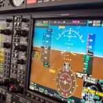
flight simAnother important challenge in neuroergonomics is determining which physiological measurements provide the most reliable indicators of mental workload. A collaborative team of researchers from France investigated how physiological measurements can be used to establish standardized levels of mental workload during aviation-inspired tasks. Using the Multi-Attribute Task Battery (MATB-II), participants performed simulated flight-related tasks under varying levels of demand while researchers measured EEG, eye-tracking, electrodermal activity, and cardiac activity. ECG and EDA signals were acquired using a BIOPAC data acquisition and analysis system running AcqKnowledge software. The study found that several cardiac, ocular, and electroencephalographic measures successfully distinguished high-workload conditions and correlated with subjective workload ratings. The researchers found that several physiological measures, particularly EEG, eye tracking, and cardiovascular metrics, reliably distinguished between different workload levels and closely tracked participants’ subjective assessments of task difficulty. Their findings suggest that multimodal physiological monitoring could support the development of adaptive systems that detect cognitive overload in real time, improving both performance and safety in demanding operational environments.

These studies highlight the increasingly multimodal nature of neuroergonomics research. Eye tracking provides insight into attention and visual processing, cardiovascular measures reveal how the autonomic nervous system responds to mental demands, and brain-imaging techniques such as EEG and fNIRS offer direct windows into neural workload. By combining these complementary perspectives, researchers are moving beyond simple measures of task performance toward a deeper understanding of how people think, adapt, and perform in complex environments. As workplaces continue to evolve alongside automation and intelligent technologies, neuroergonomics is helping researchers design systems that better support human capabilities, improve safety, and reduce the hidden costs of cognitive overload.


Are you planning a multimodal neuroergonomics study? Our team of experts can help you plan your research and identify the tools that will provide the best possible data.

Under the Skin: Digging Deeper into the Physiology of Fear

From the thrill of cinematic jump scares to debilitating phobias that can turn daily life for some into a waking nightmare, our sense of fear can feel like both a blessing and a curse. Fear is one of the most fundamental human emotions, serving as a necessary safeguard for recognizing and responding to danger. Despite its primal origins, the mechanisms of fear remain complex. For decades, psychophysiologists have studied fear using measures such as the startle reflex and skin conductance, building a deep understanding of how the body reacts to threats. Today, however, researchers are beginning to look beyond these traditional markers. Technologies such as virtual reality (VR) expand how researchers can stimulate the fear response. At the same time, a broad range of signals and techniques can measure not only the body’s autonomic responses, but also how people allocate attention to threats and how the brain processes fear-related information.

One reason fear remains such a compelling topic is that it sits at the intersection of cognition, emotion, memory, and physiology. Fear responses can be observed through changes in sweating, heart activity, breathing patterns, pupil size, neural activity, and even social behavior. As researchers seek to gain a better understanding of anxiety disorders, phobias, post-traumatic stress disorder (PTSD), and related conditions, they increasingly rely on multiple synchronized signals to capture a more complete picture of how fear is learned, remembered, and generalized.

Among the most widely used measures is electrodermal activity (EDA), often recorded as skin conductance response (SCR), which reflects changes in sympathetic nervous system arousal. More recently, eye tracking and pupillometry have emerged as valuable tools for measuring vigilance and attention, while functional near-infrared spectroscopy (fNIRS) has enabled monitoring of changes in cortical activity during fear-related tasks in more naturalistic settings. Together, these approaches help researchers move beyond viewing fear as a single physiological reaction and toward understanding it as a coordinated brain-body process.

A team of researchers at South China Normal University in China explored how the duration of memory retrieval influences the reconsolidation and extinction of fear memories. Using a four-day fear-conditioning paradigm, participants were conditioned to associate visual cues with mild electric shocks. Simultaneously, skin conductance responses were recorded using an MP36 running Biopac Student Lab System. The experiment was designed and implemented with E-Prime stimulus presentation software. The team found that briefly reactivating a fear memory before training participants to view the stimulus as safe was most effective at preventing the fear from returning. Longer periods of memory reactivation produced weaker effects. The study highlights how electrodermal activity can reveal subtle changes in fear-memory processing and may ultimately contribute to improved behavioral interventions for anxiety-related disorders.

fingers and faceA study conducted at the University of Würzburg in Germany examined how anxiety sensitivity influences attention and memory during threat and safety learning. Participants learned associations between neutral faces and aversive outcomes while researchers recorded both electroencephalography (EEG) and pupil diameter. Pupillometry data were acquired using a Tobii eye tracker, allowing researchers to measure moment-to-moment changes in attention. Individuals with higher anxiety sensitivity showed larger pupil responses and enhanced neural markers of attention when viewing both threatening and safe cues, reflecting a generalized pattern of hypervigilance. Interestingly, this heightened attentional engagement did not translate into better memory performance. The findings suggest that pupillometry provides a valuable window into how anxious individuals process potential threats and may help researchers better understand mechanisms underlying pathological anxiety.
Another perspective on fear comes from research at Sichuan Normal University in China, where investigators look at how trait anxiety influences observational fear learning. Unlike traditional conditioning studies, participants learned about danger by watching another person experience threat-related events. Researchers measured skin conductance using a BioNomadix wireless EDA plus data acquisition and analysis system and monitored prefrontal brain activity using fNIRS. The study found that individuals with high trait anxiety exhibited stronger physiological fear responses and increased medial prefrontal cortex activation, even when observing threats indirectly. The work demonstrates that fear can be acquired socially and that neural measures such as fNIRS can reveal aspects of fear processing that are not readily captured by autonomic measures alone. These findings may have important implications for understanding how fear and anxiety spread through social environments and how vulnerability to anxiety disorders develops.

These studies illustrate how fear research leverages multimodal approaches to incorporate multiple physiological perspectives. Skin conductance continues to provide valuable information about autonomic arousal, while pupillometry reveals how attention is divided between threat and safety cues, and fNIRS offers insight into the neural processes involved in fear learning and regulation. As multimodal research platforms become increasingly accessible, researchers are gaining unprecedented insight into how fear is acquired, maintained, and modified. By combining signals from the body, the eyes, and the brain, modern psychophysiology is shining a light on the shadowy labyrinth that is one of humanity’s most powerful and important emotions. To learn more about how to construct a fear stimulation lab for your next fear-conditioning research project, check out our webinar on the subject.


If you are planning a multimodal fear conditioning study, our staff can help identify the right equipment and software for stimulus presentation and data recording and analysis.


Muscles in Motion: Electromyography in Physiological Research

When we think about the human muscular system, movement often comes to mind. Activities such as running, lifting, and maintaining posture all rely on coordinated skeletal muscle activity, while cardiac muscle drives circulation and smooth muscle supports essential functions like digestion. Yet muscles do more than move us from point A to B; they also reflect what is happening inside the body. Patterns of muscle activation can reveal information about physical effort, fatigue, coordination, and recovery, as well as emotional expression, stress, and cognitive engagement. Electromyography (EMG) is a widely used technique for measuring the electrical signals generated by muscles during contraction, providing researchers with a direct and objective means to study both physiological function and the links among the nervous system, behavior, and mental state. By capturing muscle activity that may be too subtle to observe visually, EMG enables precise investigation of processes ranging from motor control and performance to emotional and stress-related responses.

There are several types of EMG, each suited to different research needs. Surface EMG (sEMG) uses small sensors placed on the skin to detect activity from nearby muscles and is the most common approach in behavioral and human performance studies. Intramuscular EMG involves inserting a fine-needle electrode directly into a muscle to record activity from specific muscle fibers, often used in clinical or specialized research settings. Facial EMG (fEMG), a form of surface EMG, focuses on facial muscles. By measuring muscle activity associated with smiling or frowning, fEMG allows researchers to study emotional responses, even when those responses are subtle or not consciously expressed.

One of the key strengths of EMG is its ease of integration with other physiological measurements. In many studies, EMG is recorded alongside electrocardiography (ECG), which measures heart activity; electroencephalography (EEG), which records electrical activity in the brain; functional near-infrared spectroscopy (fNIRS), which tracks changes in brain oxygenation; and eye tracking, which monitors visual attention and gaze patterns. By synchronizing these signals in time, researchers can better understand how different systems in the body work together. For example, combining EMG with EEG can reveal how brain activity leads to muscle movement, while pairing EMG with ECG can show how physical effort relates to heart responses during stress or exercise. This multimodal approach is central to advancing research in areas such as emotion, cognition, and human performance.

EMG also has important real-world applications. In rehabilitation, it helps clinicians monitor muscle recovery after injury. In ergonomics, it provides objective data about muscle strain in the workplace. In sports science, EMG helps optimize training and performance by identifying how and when muscles are activated. Increasingly, EMG is also used in human–machine interfaces, such as systems that allow people to control prosthetic limbs using their muscle signals. Its ability to detect fine-grained changes in muscle activity makes EMG a valuable tool across both research and applied settings.

Best practices for EMG data quality begin with careful electrode placement and attention to potential sources of noise. For sEMG and fEMG, electrodes should be placed over the “belly” of the target muscle, aligned with the muscle fibers, and spaced consistently to capture clean, interpretable signals. Gently cleaning the skin and, when appropriate, lightly abrading the surface helps reduce impedance and improve signal quality. In fEMG studies, precise and repeatable placement over well-defined facial muscles is critical, as these muscles are small and closely spaced. To minimize noise and artifacts, researchers should secure electrodes and cables to prevent movement, ask participants to limit unnecessary body motion, and ensure proper grounding of the recording system. Electrical interference from nearby equipment, power lines, or poor cable management can also contaminate EMG signals, so shielded cables and a controlled recording environment are recommended. Finally, synchronizing EMG with other physiological signals using a common acquisition system helps prevent timing errors and reduces the need for later corrections, resulting in cleaner data and more reliable conclusions.

hand mechanicsRecent studies demonstrate how EMG continues to expand into new and innovative areas. In a study published in Results in Engineering, a team of researchers from China developed a low-computational method for recognizing hand gestures from EMG signals to improve prosthetic device control. The researchers extracted key features from EMG recordings and used a lightweight machine-learning model, a multilayer perceptron (MLP), to classify gestures in real time. EMG data were collected using a four-channel BIOPAC data acquisition system running Biopac Student Lab (BSL) software. Surface Ag/AgCl electrodes were placed on four forearm muscles to capture relevant muscular activity. The study showed that accurate gesture recognition can be achieved without heavy computational requirements, making the system more practical for everyday use. This work highlights how EMG can support the development of more responsive, accessible assistive technologies.
Another study, conducted by a team of researchers from Mahidol University in Thailand and presented in the Institute of Electrical and Electronics Engineers (IEEE) proceedings, explored how combining EMG with motion sensors can improve ergonomic assessments. Focusing on occupational health, the study integrated surface EMG with data from an inertial measurement unit (IMU), a sensor that tracks movement and orientation. The researchers used the IMU to calculate Rapid Upper Limb Assessment (RULA) scores—a common method for evaluating postural risk—while EMG data collected via a BIOPAC four-channel research system provided insight into muscle activity. By combining these measures, the team found that posture alone did not always reflect actual muscle strain. The inclusion of EMG revealed additional muscle stress that might otherwise go unnoticed, suggesting that multimodal approaches can lead to more accurate workplace assessments and better injury-prevention strategies.

Researchers from Louisiana State University and the Pennington Biomedical Research Center in Baton Rouge, Louisiana, examined how training experience influences muscle activation and fatigue during resistance exercise performed with and without blood flow restriction (BFR) in young men. The study, published in Physiological Reports, compared trained and untrained participants while they completed knee extension exercises under low-, medium-, and high-load conditions. The team used sEMG to measure muscle excitation and total muscle activation in the vastus lateralis, with participants fitted with a portable BIOPAC BioNomadix Wireless EMG system. EMG signals were sampled at 2000 Hz and analyzed using a data acquisition system with AcqKnowledge software, allowing the researchers to quantify both peak muscle excitation and overall activation across repetitions. The results showed that low-load exercise with blood flow restriction produced lower peak muscle excitation but substantially greater total muscle activation compared to traditional high-load exercise, regardless of training status. These findings suggest that low-load BFR training may provide an effective alternative for stimulating muscle adaptations while reducing mechanical stress, with potential applications in rehabilitation and strength training programs.

Taken together, these examples illustrate the versatility and effectiveness of EMG in neuroscience, psychophysiological research, and beyond. Whether used to study emotion, improve physical training and workplace safety, or develop assistive technologies, EMG provides a direct link between muscle activity and human behavior. When combined with other physiological measures, it offers a more comprehensive understanding of how the brain and body interact. As research tools and techniques continue to advance, EMG will remain an essential method for exploring brain-body connections. For additional information on how to design studies around EMG, best practices for gathering EMG data, and how to integrate EMG with other signals, check out our webinar on EMG acquisition and analysis.


If EMG will be part of your next physiological or psychophysiological research project, reach out to our knowledgeable staff, who are standing by to help you with each stage of the planning and design process.

Hands-on Learning: How Real‑Time Physiological Data Deepens Clinical Education

Physicist Albert Einstein noted, “The only source of knowledge is experience.” This principle is particularly relevant in healthcare education, where hands-on learning is vital for cultivating the skills needed to care for patients effectively. The practice of recording and analyzing physiological signals in the learning lab serves as a powerful teaching tool, enabling students to immerse themselves in real-world scenarios and reveal autonomic responses, cardiovascular patterns, and emotional states that are often hidden from view.

For students in nursing, dentistry, and pharmacology, engaging with these signals transforms abstract concepts into concrete, measurable experiences. By analyzing heart activity, visual attention, stress markers, and pharmacodynamic effects, students not only enhance their critical thinking skills but also prepare themselves for clinical challenges with greater confidence.

Physiological signals across the health sciences teach learners to interpret subtle features embedded in physiological waveforms. Modern teaching laboratories increasingly incorporate multimodal systems to help students better understand the complexity of human physiology.

Nursing programs emphasize accurate patient monitoring and interpretation of vital signs, including electrocardiograms (ECG) and heartrate variability (HRV). A 2025 study published in Sensors evaluated the validity of Apple Watch Series 6 HRV data against laboratory‑grade ECG recordings using a BIOPAC 3‑lead ECG system to provide a comparative baseline for measurement. By synchronizing event markers between the Apple Watch and BIOPAC timeline, researchers ensured precise temporal alignment for comparing R‑R intervals and HRV indices. The results showed near-perfect agreement in R-R interval timing under resting conditions, indicating that such wearable devices can serve as a reasonable tool for certain wellness-oriented assessments. At the same time, the BIOPAC system remains the gold standard for precise physiological analysis.

For nursing students, this study demonstrated that consumer wearables must be validated against clinical-grade systems before being relied upon for assessment or decision-making. By examining the comparative data, students can explore how artifacts, sampling frequencies, and sensor placement influence the fidelity of HRV measurements. Moreover, because HRV relates directly to autonomic balance and stress physiology, the study provides a meaningful touchpoint for nursing courses in pathophysiology and patient assessment. Through hands‑on lab sessions using BIOPAC ECG amplifiers, students can visualize waveform morphology, measure beat‑to‑beat variability, and learn how cardiovascular signals reflect underlying homeostatic regulation.

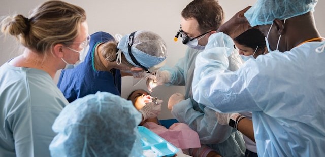
Dental students should understand not only the mechanics of clinical procedures but also the emotional and perceptual experiences of their patients, especially children. A 2025 cross-sectional pilot study in the European Archives of Paediatric Dentistry used an eye tracking and  data acquisition and analysis system from BIOPAC to measure children’s gaze behavior and physiological stress when viewing images of dentists, dental chairs, and operatory designs.

Using eye-tracking metrics such as mean dwell time, fixation count, time to first fixation, and total fixation time, researchers found that study participants stared longer at dentists wearing white coats, indicating a perception of compitance and safety, but also indimidation. In addition, gender matching mattered, as children showed longer fixation on dentists of the same gender. The study also found that warm‑colored dental chairs (especially yellow) attracted greater visual attention, particularly among younger children. Simultaneously, HRV and ECG signals recorded with the BIOPAC system revealed that younger children exhibited greater autonomic arousal than older peers when exposed to dental images.

These findings offer dental students actionable insights into clinic design, caregiver attire, and emotional perception, all of which influence pediatric patient comfort. The study demonstrates how combining eye tracking with physiological monitoring can reveal otherwise hidden psychological responses. For dental educators, integrating such physiological signal analysis into coursework helps students develop empathetic, patient‑centered care strategies.

colored pillsIn pharmacology courses, students learn how drugs influence physiological systems. A study published in Brain Sciences investigated the lingering cardiovascular effects of caffeine, challenging the assumption that its effects dissipate within six hours. Using BIOPAC respiratory transducers, noninvasive blood pressure modules, bioimpedance amplifiers, and ECG amplifiers connected to a data acquisition and analysis system researchers monitored participants before and after consumption of caffeinated and decaffeinated coffee. Results showed measurable changes in heart rate, respiratory patterns, and thoracic impedance well beyond the expected metabolic window, suggesting caffeine’s physiological effects outlast its pharmacokinetic half‑life.

For pharmacy students, these findings reinforce critical distinctions between pharmacokinetics (drug metabolism) and pharmacodynamics (physiological response). By analyzing real‑world signal data, learners gain a more nuanced understanding of how stimulants affect autonomic regulation. Such insights prepare future pharmacists to counsel patients on medication timing, drug interactions, and individual responses to common agents such as caffeine.

Across nursing, dentistry, and pharmacy, physiological signals provide potent educational value. They bridge the gap between theory and clinical application by giving students concrete, measurable access to physiological processes such as autonomic reactivity, cardiovascular regulation, and perceptual attention. Using both research and education-based lab systems, learners can visualize real-time physiological data, develop analytical skills, and cultivate a deeper understanding of the human body under stress, stimulation, and clinical observation.

As healthcare grows more data‑driven, integrating hands-on experience with signal acquisition into professional training helps future clinicians interpret complex physiological information with confidence. Whether validating wearable devices, improving pediatric patient experiences, or exploring drug‑induced autonomic effects, physiological signals empower students to connect scientific principles with evidence‑based, care. Check out our webinar, Advancing Practical Learning: Biosignal Essentials, to learn more.


If you’re exploring ways to incorporate physiological signals into your curriculum or simulation lab, our team is ready to help identify the best tools and workflows for your program.

Mind Over Matter: Cognitive Neuroscience and Aging

“Age is an issue of mind over matter. If you don’t mind, it doesn’t matter.”  —  Mark Twain.

While American author and humorist Mark Twain suggested that mental attitude could make aging less daunting, modern science shows that the changes our bodies and brains undergo over time are very real. Yet researchers are discovering that aging doesn’t have to mean inevitable cognitive decline. In fact, the growing field of cognitive neuroscience is revealing ways to track, understand, and even influence how our brains change with age, offering hope that we can preserve mental acuity, independence, and quality of life well into our later years.

When studying how aging affects the brain, cognitive neuroscience researchers use a variety of tools. In addition to standard behavioral tests like the Stroop test, n-back test, and verbal fluency exercises, which track reaction times, accuracy, and problem-solving strategies, they also rely on neuroimaging and physiological measurements to gather deeper insights. Neuroimaging techniques include electroencephalography (EEG), which monitors real-time electrical activity in the brain; functional near-infrared spectroscopy (fNIRS), which measures blood oxygen levels in specific cortical regions during cognitive tasks; and magnetic resonance imaging (MRI), which provides detailed structural and functional maps of the brain.

Other physiological signals play an essential role in this field, including heart rate variability (HRV), a key indicator of the autonomic nervous system’s ability to balance various physiological functions and handle stress. Electrodermal activity (EDA) allows researchers to track arousal among participants undergoing cognitive exercises. Electromyography (EMG) measures muscle activation during fine and gross motor tasks. Eye tracking and electrooculography (EOG) provide researchers with data on how aging impacts attention and the ability to process visual cues. Respiration and blood pressure provide vital information on vascular health, which is often linked to cognitive performance.

Taiwanese researchers from National Yang Ming Chiao Tung University in Taipei turned to HRV and fNIRS to uncover how aging and, more specifically, mild cognitive impairment (MCI), affect both brain and heart responses during cognitive tasks. Healthy young adults, cognitively healthy older adults, and older adults with MCI were asked to perform cognitive function and executive tests. Researchers recorded the participants’ prefrontal cortex (PFC) activation through changes in oxyhemoglobin using fNIRS and autonomic responses via HRV. The team gathered ECG data using a BIOPAC data acquisition module running AcqKnowledge data acquisition and analysis software, and equipped with an ECG amplifier. The study found that older adults who did not exhibit MCI showed lower global and executive cognitive performance, reduced HRV, but increased prefrontal activation. The research team interpreted this to be the result of compensatory neural effort. However, older adults with MCI showed even greater cognitive decline, but no further differences in HRV or prefrontal activation. The findings suggest that HRV and PFC activation patterns could serve as biomarkers for distinguishing healthy aging from early pathological decline.

To better understand how learning processes develop and change with age, researchers from Tilburg University in the Netherlands investigated how younger adults and older adults differ in their brain and body responses while learning a visuomotor sequence task. To capture these responses, the team measured EEG, skin conductance (EDA), heart rate, HRV, breathing rate, and eye movements during the task. Physiological data were collected using BIOPAC’s BioNomadix wireless dual ECG/respiration amplifier and PPG/EDA amplifier, which continuously recorded three-lead ECG, respiration, and EDA at 2,000 Hz, with signals processed and analyzed in AcqKnowledge software. This setup allowed the researchers to precisely synchronize physiological activity with learning performance. Both younger and older adults improved with practice, but older adults needed more trials to reach the same precision. Brainwave activity showed apparent age differences, while measures like HRV, respiration, and skin conductance did not. This suggests that EEG is a more sensitive marker of age-related learning changes and could help guide training programs for older adults.

readingA long-term study by researchers from Radboud University Medical Center in the Netherlands, together with UK collaborators, tracked a group of healthy older adults over nearly 11 years to see how their brain blood-flow regulation changed by their 80s. Using transcranial Doppler ultrasound to measure blood velocity in the middle cerebral arteries, the team also monitored control signals including heart rate, blood pressure, and breathing gases. Physiological data were recorded with a BIOPAC data acquisition and analysis system, with end-tidal CO₂ (EtCO₂) measured using capnography via a BIOPAC carbon dioxide measurement amplifier. The researchers found a slight decline in CO₂ reactivity but, surprisingly, an improvement in dynamic cerebral autoregulation (the brain’s ability to maintain steady blood flow despite blood pressure changes). These effects were most apparent in participants who later reported memory complaints, suggesting that while aging may reduce vascular responsiveness, the autoregulatory system may help compensate for early cognitive challenges. The findings show how vascular health in the brain relates to aging and memory, providing valuable insight for detecting early risks of dementia and shaping preventive strategies.

These studies highlight how combining brain imaging with physiological measurements is helping scientists reveal the complex relationship between aging, cognition, and overall health. Through cognitive neuroscience, researchers are identifying early signs of decline and gaining a better understanding of how the brain and body adapt over time. Research is paving the way to more effective screening, personalized interventions, and lifestyle strategies that may help people maintain mental sharpness and independence well into older age. For more information on the tools and techniques being applied to cognitive neuroscience research, see our Webinars and visit our page on Cognitive State Analysis.


Are you planning a study on how aging affects the brain or just looking to expand your cognitive neuroscience toolbox? Our customer service team is ready to help you find what you need to obtain the best possible data.

Pour One Out: The Physiology of Alcohol Consumption

“Dry January,” the cutting out of alcohol consumption during the first month of the calendar year, entered the public vernacular a little over a decade ago. The idea was first promoted in 2013 by a British charity to encourage alcohol abstinence, or at the very least, healthy moderation. The idea took off as a counterbalance to the excesses of indulgence that often characterize the end-of-year holiday season. As if on cue, the U.S. surgeon general released a report earlier this month linking increased cancer risk to even moderate alcohol use and called for alcohol products to carry a warning label like those found on tobacco products.

Since the early 1990s, the prevailing belief among some in the medical community held that moderate amounts of alcohol may not necessarily be detrimental to overall health and may even have beneficial effects. Despite the surgeon general’s report, the consensus in the benefits versus risks debate remains elusive, a fact summarized well in a recent article in The Atlantic and an accompanying podcast by journalist Derek Thompson. Without wading further into the controversy, it is worth pointing out that there is a rich and growing body of research into the effects of alcohol on human physiology, cognitive and psychological well-being, and longevity, all of which rely on some form of life science metrics to measure these effects.

Physiological signals offer a powerful window into the biological and neurological effects of alcohol consumption, providing objective, real-time insights into the body’s and brain’s response. By measuring electrical, chemical, and physical signals generated by the body, researchers can examine changes in heart rate, brain activity, muscle tone, and more. Commonly used signals in alcohol studies include electroencephalography (EEG) to monitor brain activity, electromyography (EMG) for muscle function, electrocardiography (ECG) for heart rhythms, and electrodermal activity (EDA) and galvanic skin response (GSR) for stress and arousal levels. Each tool captures aspects of how alcohol influences the human system, from cognitive processing impairments to autonomic regulation shifts. In research settings, these signals are applied to explore both short- and long-term effects of alcohol. For example, EEG studies can track alterations in brainwave patterns, shedding light on reduced attention, memory disruption, or impaired decision-making during intoxication. ECG and GSR data, meanwhile, reveal how alcohol modulates cardiovascular activity and stress responses, highlighting its impact on the autonomic nervous system (ANS).

A 2021 study by researchers at Wake Forest University and John Hopkins University School of Medicine used heart rate variability (HRV) to examine the relationship between alcohol abstinence, ANS function, and whole brain functional brain networks among everyday drinkers. Whole brain functional brain networks demonstrate how different parts of the brain work together as a connected system. This study combined functional brain wave mapping generated with resting-state MRI scans during three-day periods of alcohol consumption and abstinence. The effects of abstinence were measured using HRV. Alcohol craving in patients with an alcohol use disorder (AUD) has been associated with high-frequency heart rate variability (HF-HRV) with acute consumption being related to decreasing HF-HRV. During the study, ECG data was collected via a BIOPAC data collection and analysis system. The study concluded that “while brain networks do differ across drinking states in risky drinkers, the change is primarily driven by HRV.”woman drinking

A current study by a team of German researchers explores the effectiveness of Virtual Reality (VR) in Cue Exposure Therapy (CET), a strategy in Cognitive Behavioral Therapy (CBT) used for treating AUD. Research participants are exposed to VR scenarios with alcohol-related cues. VR with integrated eye tracking offers advantages over real-life scenarios when developing new treatment options, according to the study authors. Participants wore head-mounted displays (HMD) to immerse them in three different scenarios while heart rate, HRV, pupillometry, and EDA were measured. Respiration and ECG data are gathered via a wireless BioNomadix BN-RSPEC while PPG and EDA are gathered with a BN-PPGED. Physiological signal data is then fed to a BIOPAC data acquisition system running AcqKnowledge software for analysis. While the study is ongoing, researchers believe it will lead to a better understanding of the induction of craving in VR cue exposure…” and that it could offer “new diagnostic and therapeutic perspectives…providing a basis for future biofeedback training in VR exposure therapy.”

These are a few of the ways physiological signals are being applied in alcohol-related research. For more information on integrating HRV analysis into your next research study, see our webinar on the topic. Better yet, get hands-on training on a full range of research equipment and techniques at our biannual conference on Tools, Trends, Techniques, and Technologies in Human Physiology, 4T|Phys.


Are you planning an alcohol or addiction-related study? BIOPAC has the tools and our regional sales staff is ready to help you with everything you need.

Keep Your Eye on the Ball: Eye Tracking in Exercise Research

When a coach tells a player to keep their eye on the ball, they are addressing the importance of attention and focus to success, both figuratively and literally. Whether hitting a baseball, shooting a basket, or maintaining focus in an exercise routine, how we process visual information is linked to physical and mental performance, and eye tracking is the key to unlocking this relationship.

Modern eye tracking technology is revolutionizing how researchers understand human behavior, perception, and performance during physical activity. With the rise of wearable and mobile technology, eye tracking has been freed from the confines of the lab, offering real-world insights into our exercise habits and mindset. Thanks to innovators like BIOPAC partner Argus, researchers can now access eye tracking information recorded directly from the putting green, ski slope, and even the mountain biking trail.

One of the primary applications of eye tracking in exercise research lies in understanding visual attention and gaze behavior. By tracking where individuals look while engaging in physical activity, researchers can better understand how attentional focus influences performance. For instance, studies have revealed that elite athletes exhibit more focused and efficient gaze patterns, directing their attention to critical cues that enhance their performance. A 2023 study by researchers at North Carolina State University assessed the viability of using an array of mobile physiological measurement tools, including eye tracking, to measure performance optimization techniques for rock climbers. Tools included BIOPAC monitors and transmitters for wireless tracking of skeletal muscle electromyography (EMG) and eye tracking glasses to record gaze. The study affirmed that such technologies could accurately monitor multiple athletic performance metrics in even the most challenging environments. basketball

Beyond performance enhancement, eye tracking technology holds promise in understanding the psychological aspects of exercise behavior. By examining visual attention during exercise-related stimuli such as advertisements or motivational cues, researchers can unravel the underlying mechanisms driving exercise motivation and adherence.

The same principles of eye tracking research used in fitness and physical performance-related exercise studies can be applied in other fields, such as those that examine job performance in highly stressful and physically demanding occupations, like law enforcement. A study published in the 2023 edition of the Journal of Motor Behavior explored the relationship between gaze control and decision-making by police officers in highly stressful situations. The goal was to assess how a participant’s levels of experience and training affected tactical decisions. Participants wore eye tracking glasses during a simulated traffic incident. The participants’ respiration and electrocardiogram (ECG) signals were monitored via a BIOPAC BioNomadix dual-signal wireless transmitter.  Gaze-tracking data, ECG, and respiration data were fed into a BIOPAC data acquisition system for analysis. The study found that higher levels of training “led to increased processing efficiency and effective attentional control.”

The integration of eye tracking technology into exercise physiology and psychology research offers a nuanced understanding of human behavior and performance during physical activity. With wearable and mobile solutions, this technology not only expands the possibilities of research but also holds the potential for personalized programs and the enhancement of exercise experiences for individuals across a wide range of circumstances. For more information on integrating mobile eye tracking into your exercise study, see our webinars on Recording Great Data from Exercising Subjects and Eye Tracking Glasses and AcqKnowledge Integration.


Are you planning to employ eye tracking in your next exercise study? Contact our experts for assistance in selecting the best tools for gathering and analyzing exercise data with eye tracking.

VR Integration: Enhancing Research with Virtual Environments

Measuring the human body and brain’s response to real-world conditions and stimuli has long challenged researchers. This problem has usually been dealt with in one of two ways. The first approach is to take research participants into the field—not impossible with today’s mobile data recording technology but not always safe or convenient. The second option is to imitate real-world conditions in the lab using screens, audio equipment, and even scent delivery, but in the end, it is difficult to completely remove the lab setting from the participant’s experience—that is, until the introduction of Virtual Reality (VR) technology in research.

Just as it has revolutionized everything from entertainment to training, VR has provided researchers with the opportunity to create immersive experiences in the lab that mimic the real world. Advances in technology have not only made these simulated environments more realistic, but they have also made it easier to integrate a wide range of signals and stimulus delivery to enhance the recording of physiological and psychophysiological data.

BIOPAC, through cooperation with several partner companies, offers a range of data recording options that integrate seamlessly with third-party VR devices. fNIR Devices, for example, designs solutions for recording functional near-infrared spectroscopy (fNIRS) signals that can easily be incorporated with standard VR head-mounted displays (HMD) to measure neural activity and hemodynamic response in the prefrontal cortex while participants experience VR environments. BIOPAC partner Seenel has created MedelOpt, a line of whole-head wearable devices that combine electroencephalography (EEG) and fNIRS, including a VR option that integrates an HMD. Finally, partner company WorldViz offers a full spectrum of VR solutions from complete VR kits to full-room Projection VR technology to software that can be combined with both wired and wireless data recording technologies to provide a complete array of bio-signal data, from eye tracking to electrodermal activity.

A Canadian study published in 2023 used a combination of BIOPAC and WorldViz products to investigate how sensory feedback and arousal influence neuromusVR Shadowcular control and posture. Researchers measured arousal via electrodermal activity (EDA) and psychosocial questionnaires, rating perceived fear and anxiety in both real-world situations in VR environments that simulated standing both at ground level and on a raised platform. Participants wore a commercial HMD running WorldViz’s Vizard VR toolkit software. Electrical nerve stimulation was provided via a BIOPAC linear isolated stimulator. The study found the “physiological and perceptual indicators of fear and anxiety increased” in the VR platform simulation, providing “novel evidence that VR can be used to modulate upright postural control.”

A similar study published in the 2022 issue of the Journal of Sport Rehabilitation examined the effect non-physical mental training has on balance skills. The study used fNIRS technology to measure hemodynamic response in the prefrontal cortex of participants experiencing virtual environments through VR HMDs. Participants were instructed to imagine themselves imitating balance exercises viewed through an Oculus VR HMD while also fitted with an fNIR Devices Imager headband. Data was gathered using Cobi Studio software and analyzed via fNIRSoft software. The study identified several advantages of virtual reality mental training (VRMT) over conventional mental training (CMT).

These are just a few examples of how VR technology can be integrated with a variety of methodologies for gathering research data. For additional information on the subject, check out our Virtual Reality Resources, which include an array of online training on integrating VR into research.


Are you planning a research product and need help integrating VR with other data acquisition and analysis equipment? Contact your local BIOPAC sales rep to find the right solutions for you.

Eye-Catching: Mobile Tech Enhances Immersive Experiments

Freed from the limitations of stationary eye tracking technology, mobile eye tracking enables researchers to study their subjects while they perform a wide range of immersive tasks in natural settings.

Sixteenth-century pediatrician Thomas Phaer mused that “the eyes are the windows to the soul.” These sensory organs and their movements certainly provide a wealth of information to researchers studying the interaction of mind and body. Eye tracking has long been a key tool of physiological and psychophysiological research, and recent developments in mobile eye tracking take this valuable data resource out of the confines of the lab and into the real world.

Freed from the limitations of stationary eye tracking technology, mobile eye tracking enables researchers to study participants while they perform a wide range of immersive tasks in natural settings. These self-contained devices come in a variety of form factors including wearable glasses that provide untethered eye tracking data acquisition in real time that can be combined with other physiological data collected during live experiments.

Today’s lightweight mobile devices can record a broad spectrum of eye tracking data, including pupil size and position, gaze path, vergence angle, and areas of interest (AOI). The latter connects stimuli to eye-movement metrics, providing data on the participant’s psychophysiological response to stimuli within their field of vision. In previous blog posts and webinars, we have explored how mobile eye tracking technology gathers this data. Numerous studies have been published that demonstrate its potential in physiological and psychophysiological research. Settings and applications include user experience, sports and physical therapy, public safety and law enforcement, driving and equipment operation, and work environments from the office to the factory floor.

Mobile eye tracking technology allowed researchers at The University of Sheffield to take their experiments to the city streets for a 2020 study into how different lighting affects cyclists navigating urban environments at night. Mobile eye tracking provided data on the gaze behavior of nocturnal cyclists in parallel with skin conductance response (SCR) to better understand how obstacle detection is influenced by variations in type, location, and level of lighting. Bicycles and their riders were equipped with a BIOPAC data acquisition unit and audio stimulation units along with a BioNomadix EDA transmitter and electrodes to record the participants’ SCR. Eye tracking data, including AOI, were synchronized with SCR data and analyzed using AcqKnowledge software to determine how the riders reacted to a variety of lighting conditions. Researchers concluded that lighting plays a significant role in riders’ comfort levels at night and that improved lighting could increase urban bicycle use.

VGM playerLess intrusive by design, mobile eye tracking can provide indoor experiments with a heightened degree of immersion. A study at the University of British Columbia used mobile eye tracking glasses to create a more natural environment to investigate how the immersive effects of electronic gaming machines (EGM) impact gambling addiction. The study looked at how machine design triggered psychophysiological responses in participants by assigning various AOI to EGM screens. ECG data were recorded along with respiration effort via a BIOPAC respiration effort transducer and amplifier, which were fed to a data acquisition unit and synchronized with AOI data recorded via mobile eye tracking glasses. The study was able to successfully identify several key markers for game immersion using AOI that suggest behavioral links between immersion and gambling addiction.

Possible applications for mobile eye tracking in research continue to expand with the evolution of the technology. To learn more about recent developments in mobile eye tracking, check out our latest webinar on mobile eye tracking glasses with AcqKnowledge integration.


Learn more about how to integrate mobile eye tracking into your next research project by contacting your local BIOPAC sales representative.

error: Content is protected !!