Imaging is Everything: Data Acquisition in the MRI Environment

The marriage of physiological data recording and magnetic resonance imaging (MRI) has allowed scientists to unravel intricate connections between the mind and body. However, this synergy comes with its unique set of challenges that researchers must be aware of to ensure the integrity of both data and the well-being of study participants.

One of the foremost challenges in recording physiological data within the MRI lab is the strong magnetic field. Traditional monitoring equipment can be rendered useless or, worse, pose extreme safety risks due to their ferromagnetic components. Another critical consideration is the impact of radiofrequency (RF) pulses used in the imaging process. RF pulses can introduce artifacts in physiological recordings, affecting the quality of data collected. In previous posts we have discussed the basics of integrating data collection in the functional MRI (fMRI) environment as well as techniques for avoiding artifacts and signal noise while recording data in the MRI.

Researchers must also contend with challenges related to the confined space and noise levels within an MRI environment. A 2021 study conducted by the Lab for Autonomic Neuroscience, Imaging and Cognition (LANIC) at Jena University Hospital in Jena, Germany, focused on how the functional MRI (fMRI) environment itself affected outcomes in autonomic research. Specifically, the study investigated the influence of a loud and cramped environment during MRI on resting heart rate variability (HRV) measures and whether a familiarization session with MRI equipment might lower anxiety levels among participants. The research team recorded an electrocardiogram (ECG) and a photoplethysmogram (PPG) over 15 minutes from participants using a BIOPAC data acquisition and analysis system connected to finger-attached PPG sensors during non-MRI recording. For recording in the fMRI, researchers used a separate BIOPAC data acquisition system equipped with an MR-Conditional PPG module. While the study did establish a link between participant anxiety and HRV levels in the lab and fMRI environment, it did not find that the familiarization sessions had a significant impact on HRV outcomes.

gas maskDespite the challenges, the recording of physiological signals in the MRI environment provides unique opportunities for gathering critical data. A study published in the journal Military Medicine in 2020 tested the effectiveness of F MRI (not to be confused with fMRI) scans in identifying cases of War Lung Injury (WLI) in Iraq and Afghanistan war veterans. WRI refers to reductions in lung function resulting from exposure to environmental factors such as sandstorms and burn pits. F MRI employs magnetic resonance imaging with perfluorinated gases as inhaled contrast agents to evaluate various pulmonary diseases. If successful, the approach would provide an alternative to surgical biopsy for diagnosing suspected WLI. The researchers measured respiratory waveforms throughout the imaging sessions, during both normal room air breathing and PFP-oxygen gas mixture breathing using a BIOPAC MR-Conditional pneumotachometer. The study found that “F MRI provides better insight regarding lung function abnormalities” than other standard methods and that “F MRI could provide a functional tool to assist in identifying subjects with the disorder and potentially could aid in the evaluation of novel therapeutic approaches.”

BIOPAC provides a wealth of tools and information resources for gathering data in the MRI environment. This includes multiple webinars on recording data in both MRI and fMRI settings. As the field continues to evolve, BIOPAC’s innovative solutions pave the way for groundbreaking discoveries at the intersection of physiological signal recording and medical imaging.


If you are interested in recording and analyzing physiological signals in your next MRI-based research study, BIOPAC sales representatives are standing by to help you choose the right tools and resources for the job.
Practical Tips for Recording Physiological Data in MRI

The MRI system incorporates a VERY strong magnetic field and both emits and records radio frequency (RF) energy. This has impacts on subject and experimenter safety and on data accuracy.

By Alan Macy, BIOPAC Systems, Inc.

When recording physiological signals from subjects in MRI systems, two issues are very important:

  1. The MRI system incorporates a VERY strong magnetic field. It is extremely important that no significant magnetically susceptible objects are taken into the room that houses the MRI System.  All materials have some degree of magnetic susceptibility. However, materials with mass magnetic susceptibility values, similar to or have magnitude less than that of water (-0.91E-8 m^3/kg), are typically permitted to be used in the vicinity of MRI Systems. No electrodes, electrode leads, transducers or cabling, that contain significant magnetically susceptible components, should be used when recording data from a subject in the MRI.
  2. The MRI System both emits and records radio frequency (RF) energy. This energy is typically in the range of 5 to 500 MHz. For a 3T MRI, the characteristic proton precession frequency will be 3T * 42.7 MHz or 128.1 MHz.  MRI generated RF energy will, at least, partially reflect from encountered conductors and induce current flow in those conductors. Signal reflections and induced currents can disrupt sensitive imaging operations by creating signal artifacts. Also, RF induced currents, if not properly controlled, can cause local heating.

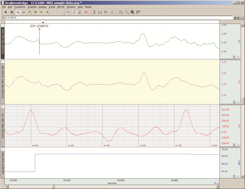
Care must be taken to limit the flow of RF energy both in and out of the room that houses the MRI. RF energy from the MRI superimposes with the very small biopotentials and transducer signals in the vicinity of the subject. Alternatively, RF energy from recording or other equipment in the MRI Control Room (e.g. digital clocks and power supply switching noise) can leak into the MRI chamber, via subject cabling, and cause degradation of the image created by the MRI. It can be complicated to record biopotentials, from a subject’s body, during an MRI scan. These signals are very tiny and the RF energy, and associated magnetic field shifting, generated by the MRI corrupts the recording. Accordingly, successful biopotential recording in the MRI involves the use of specialized amplifiers, optimized cabling, patch-panel filtering, advanced signal processing and/or synchronization with MRI scanning processes to perform clean measurements.

When RF energy is pulsed into the subject, this energy spreads throughout the subject because the subject is a volume conductor. As the MRI magnetic gradient shifts in synchrony with RF energy pulsing, the gradient shift induces a current flow in conductors that intersect with the field. From a practical standpoint, RF energy pulsing and gradient-induced current flows often manifest themselves as repetitive artifact signals directly in the band of the physiological generated biopotentials. This is because RF energy pulsing and magnetic field gradient switching are performed synchronously for imaging and the repetition rates for image slice generation usually fall within the range of 1-100 Hz. Depending upon the biopotential frequency range (from lowest to highest: EGG, EOG, ECG, EEG and EMG), different amplification and signal processing techniques can be used to isolate the signal of interest. Furthermore, it’s often helpful to control imaging processes so that the MRI artifact signal spectrum does not unduly impact the measured physiological signal spectrum.

For more information on BIOPAC’s MR Safe and MR Conditional solutions, visit BIOPAC’s MRI page or view BIOPAC’s full line of electrodes, amplifiers, and wearable, wireless transmitters and loggers.

Download a related and important application note that describes the relationship between MRI imaging sequences and associated physiological measurements, when subjects are monitored in the MRI. In particular, this note addresses newfound insights on the impact that imaging sequence types have upon simultaneously recorded electrocardiography (ECG) and impedance cardiography (ICG) subject measurements.

BIOPAC Systems, Inc. provides life science researchers and educators with data acquisition and analysis systems that inspire people and enable greater discovery about life.  Visit us at www.biopac.com.

error: Content is protected !!