2025: The Year in Review

As 2025 winds to a close, there is much to celebrate both here at BIOPAC and in the ever-expanding frontiers of research that our products engage with every day. Let’s take a quick look at where we have been over the past 12 months and what the future holds.

Popular Posts

Speaking of engagement, this is the time of year when we do a bit of self-assessment and revisit what topics our readers were most inspired by and most interested in. Apparently, many of our readers are closet gamers at heart, because our blog on research on game design and gaming impacts drove more eyes to our site than any other topic this year, followed by posts on cognitive neuroscience and aging and the human impact of isolation.

New Products and Updatesclimbing kid
On the heels of the 2024 release of our flagship MP200 data acquisition and analysis system and a significant upgrade to AcqKnowledge software, 2025 concludes with another milestone: the imminent release of AcqKnowledge 6.0.2. This latest version features a host of improvements and additions, including full integration of MedelOpt+, which blends whole-head functional near-infrared spectroscopy (fNIRS) with electroencephalography (EEG) in a single, highly flexible device.

BIOPAC in the Lab

Every year, BIOPAC products continue to drive the most exciting research in physiology, psychology, and neurology, and 2025 was no different. Each month, we highlight breakthrough studies in which BIOPAC played a role in gathering and analyzing key data. These are usually centered around a theme and feature research published in the last few years. To close out the year, we like to spotlight a couple of significant studies from 2025 that didn’t get mentioned.

A collaborative study by researchers from the University of Georgia and Beijing Normal University examined how mothers and children coordinate their physiological responses during stressful interactions. Using BIOPAC data acquisition systems coupled with wireless ECG transmitters, the team simultaneously recorded cardiac activity from each participant pair as they completed structured challenge tasks. This setup provided the temporal precision needed to track changes in heart rate and moment-to-moment autonomic synchrony, revealing how caregivers and children influence one another biologically during shared stress. The study found that greater synchrony was linked to both emotional dynamics and regulatory behaviors during the tasks, highlighting the deep coupling between social relationships and physiological responses. The wireless BIOPAC configuration enabled naturalistic interaction while capturing high-quality dyadic data, a persistent challenge in synchrony research.

In its first full year of availability, the MP200 is already making waves in the research community. At Guangdong University of Science and Technology in China, researchers examined how head-shoulder posture and cervical muscle activation differ between college students with chronic neck pain (CNP) and healthy individuals. The team employed surface electromyography (sEMG) recorded with a BIOPAC MP200 data acquisition system and AcqKnowledge software. Researchers placed surface electrodes over targeted neck muscles to record raw EMG signals during various strength tests and physical tasks. Students with CNP demonstrated significantly poorer head and shoulder alignment, reduced neck extensor strength, and altered EMG activity, indicating neuromuscular dysfunction associated with pain. Findings showed the connection between altered posture and impaired muscle function in chronic neck pain. The study demonstrates the effectiveness of surface EMG to support clinical diagnosis and rehabilitation.

For more inspiration as we head into 2026, explore our webinar library for tips, tricks, and methods directly from experts across a range of research fields.


If you are planning to launch a study for 2026, now is the time to reach out to your nearest BIOPAC customer service representative for help finding the right tools to get started.

Brain and Body: SPA-fNIRS Solves Physiological Signal Noise

Functional near-infrared spectroscopy (fNIRS) offers researchers a powerful noninvasive tool for studying brain activity by measuring changes in blood oxygen levels. These changes reflect how various brain regions respond to stimuli such as tasks that involve thinking, emotion, movement, and social interaction. These are often in natural, real-world settings that are not possible with other brain imaging methods. However, fNIRS is not without its own limitations. Systemic fluctuations in the body, such as breathing patterns, cardiovascular changes, and autonomic responses, can result in signal noise that is misinterpreted as brain activity by traditional fNIRS. The solution: a visit to the SPA.

Systemic Physiology Augmented fNIRS (SPA-fNIRS) is an advanced approach to fNIRS that goes beyond measuring local changes in cerebral oxygenation. While standard fNIRS measures cortical hemodynamics by tracking oxygenated (HbO) and deoxygenated (HbR) hemoglobin, noise originating from systemic physiological changes can corrupt those signals. Combining data from both neural and systemic physiological signals, SPA-fNIRS provides researchers with a more diverse and accurate view of how the brain and body interact in real time.

The method is being applied across various fields, including psychophysiology, cognitive neuroscience, exercise physiology, mental health, and human performance research. Studies have utilized SPA-fNIRS to investigate a range of topics, including meditation and emotional regulation, movement control, and social interaction. The broader goal is to move beyond studying the brain in isolation and to understand it as part of an integrated mind-body system. This holistic view is transforming how scientists interpret brain-function data, opening new paths for applied research in education, rehabilitation, and human-machine interface design.

soundResearchers in India investigated the impact of a traditional sound-based meditation technique, known as the Mind-Sound Resonance Technique (MSRT), on both brain and heart activity. Using fNIRS to track changes in oxygen levels in the prefrontal cortex, and a BIOPAC data acquisition and analysis system with AcqKnowledge software to record and analyze heart rate variability (HRV), the study employed a SPA-fNIRS design, integrating neural and physiological data in a single, synchronized dataset. HRV analysis allowed the researchers to quantify shifts in autonomic balance during and after meditation. The results showed increased oxygenation in the right prefrontal cortex and enhanced low-frequency HRV components, reflecting greater parasympathetic regulation. Together, these findings suggest that sound-based meditation not only improves brain oxygenation but also promotes calmer, more balanced heart-brain dynamics, illustrating how SPA-fNIRS methods can reveal the comprehensive physiological impact of mindfulness practices.

In her doctoral research at the Université de Lille in France, neuroscientist Aurore Guérin examined how the brain controls movement speed using a multimodal combination of fNIRS and physiological data recording systems. Participants performed repetitive arm movements at fast, natural, and slow tempos. At the same time, the study captured oxygenation in the prefrontal and motor cortices using fNIRS, alongside heart rate and respiration signals collected with a BIOPAC data acquisition system. A respiratory effort transducer measured breathing patterns, while a BioNomadix ECG module recorded cardiac activity for HRV analysis. All physiological signals were synchronized and analyzed in AcqKnowledge software, allowing the team to identify and remove systemic influences (e.g., heartbeat and breathing rhythms) from the fNIRS signals, following a SPA-fNIRS approach. The results revealed that faster movements primarily engaged motor regions, while slower, more deliberate pacing resulted in higher prefrontal activation, reflecting greater cognitive control. By integrating physiological monitoring with fNIRS, the study provided a clearer picture of how the brain and body coordinate timing and control during movement.

These studies illustrate how SPA-fNIRS is reshaping the way scientists investigate the interaction between brain and body. By combining neural and physiological data in a single synchronized system, researchers can separate brain activity from systemic noise and uncover the interplay between cognition, emotion, and autonomic regulation. For more information on this topic, watch our webinar on Multimodal SPA-fNIRS and check our our latest multimoldal fNIRS research products.


Are you planning a study that blends fNIRS with other physiological signals? Contact your local BIOPAC customer service representative to learn more about how SPA-fNIRS can benefit your research.

Mind Over Matter: Cognitive Neuroscience and Aging

“Age is an issue of mind over matter. If you don’t mind, it doesn’t matter.”  —  Mark Twain.

While American author and humorist Mark Twain suggested that mental attitude could make aging less daunting, modern science shows that the changes our bodies and brains undergo over time are very real. Yet researchers are discovering that aging doesn’t have to mean inevitable cognitive decline. In fact, the growing field of cognitive neuroscience is revealing ways to track, understand, and even influence how our brains change with age, offering hope that we can preserve mental acuity, independence, and quality of life well into our later years.

When studying how aging affects the brain, cognitive neuroscience researchers use a variety of tools. In addition to standard behavioral tests like the Stroop test, n-back test, and verbal fluency exercises, which track reaction times, accuracy, and problem-solving strategies, they also rely on neuroimaging and physiological measurements to gather deeper insights. Neuroimaging techniques include electroencephalography (EEG), which monitors real-time electrical activity in the brain; functional near-infrared spectroscopy (fNIRS), which measures blood oxygen levels in specific cortical regions during cognitive tasks; and magnetic resonance imaging (MRI), which provides detailed structural and functional maps of the brain.

Other physiological signals play an essential role in this field, including heart rate variability (HRV), a key indicator of the autonomic nervous system’s ability to balance various physiological functions and handle stress. Electrodermal activity (EDA) allows researchers to track arousal among participants undergoing cognitive exercises. Electromyography (EMG) measures muscle activation during fine and gross motor tasks. Eye tracking and electrooculography (EOG) provide researchers with data on how aging impacts attention and the ability to process visual cues. Respiration and blood pressure provide vital information on vascular health, which is often linked to cognitive performance.

Taiwanese researchers from National Yang Ming Chiao Tung University in Taipei turned to HRV and fNIRS to uncover how aging and, more specifically, mild cognitive impairment (MCI), affect both brain and heart responses during cognitive tasks. Healthy young adults, cognitively healthy older adults, and older adults with MCI were asked to perform cognitive function and executive tests. Researchers recorded the participants’ prefrontal cortex (PFC) activation through changes in oxyhemoglobin using fNIRS and autonomic responses via HRV. The team gathered ECG data using a BIOPAC data acquisition module running AcqKnowledge data acquisition and analysis software, and equipped with an ECG amplifier. The study found that older adults who did not exhibit MCI showed lower global and executive cognitive performance, reduced HRV, but increased prefrontal activation. The research team interpreted this to be the result of compensatory neural effort. However, older adults with MCI showed even greater cognitive decline, but no further differences in HRV or prefrontal activation. The findings suggest that HRV and PFC activation patterns could serve as biomarkers for distinguishing healthy aging from early pathological decline.

To better understand how learning processes develop and change with age, researchers from Tilburg University in the Netherlands investigated how younger adults and older adults differ in their brain and body responses while learning a visuomotor sequence task. To capture these responses, the team measured EEG, skin conductance (EDA), heart rate, HRV, breathing rate, and eye movements during the task. Physiological data were collected using BIOPAC’s BioNomadix wireless dual ECG/respiration amplifier and PPG/EDA amplifier, which continuously recorded three-lead ECG, respiration, and EDA at 2,000 Hz, with signals processed and analyzed in AcqKnowledge software. This setup allowed the researchers to precisely synchronize physiological activity with learning performance. Both younger and older adults improved with practice, but older adults needed more trials to reach the same precision. Brainwave activity showed apparent age differences, while measures like HRV, respiration, and skin conductance did not. This suggests that EEG is a more sensitive marker of age-related learning changes and could help guide training programs for older adults.

readingA long-term study by researchers from Radboud University Medical Center in the Netherlands, together with UK collaborators, tracked a group of healthy older adults over nearly 11 years to see how their brain blood-flow regulation changed by their 80s. Using transcranial Doppler ultrasound to measure blood velocity in the middle cerebral arteries, the team also monitored control signals including heart rate, blood pressure, and breathing gases. Physiological data were recorded with a BIOPAC data acquisition and analysis system, with end-tidal CO₂ (EtCO₂) measured using capnography via a BIOPAC carbon dioxide measurement amplifier. The researchers found a slight decline in CO₂ reactivity but, surprisingly, an improvement in dynamic cerebral autoregulation (the brain’s ability to maintain steady blood flow despite blood pressure changes). These effects were most apparent in participants who later reported memory complaints, suggesting that while aging may reduce vascular responsiveness, the autoregulatory system may help compensate for early cognitive challenges. The findings show how vascular health in the brain relates to aging and memory, providing valuable insight for detecting early risks of dementia and shaping preventive strategies.
These studies highlight how combining brain imaging with physiological measurements is helping scientists reveal the complex relationship between aging, cognition, and overall health. Through cognitive neuroscience, researchers are identifying early signs of decline and gaining a better understanding of how the brain and body adapt over time. Research is paving the way to more effective screening, personalized interventions, and lifestyle strategies that may help people maintain mental sharpness and independence well into older age. For more information on the tools and techniques being applied to cognitive neuroscience research, see our Webinars and visit our page on Cognitive State Analysis.


Are you planning a study on how aging affects the brain or just looking to expand your cognitive neuroscience toolbox? Our customer service team is ready to help you find what you need to obtain the best possible data.

A System of Systems: Network Physiology Connects the Dots

In the field of physiology, it is tempting to see the human body as a collection of independent systems performing a range of functions—converting food to energy, protecting us from harm and disease, and helping us react to and interact with our environments, to name a few. It is easy to overlook that these systems often work together and are interconnected. The field of network physiology does just that—look at how these systems (nervous, cardiovascular, muscular, etc.) work together, seeing them as part of a dynamic network rather than a collection of independent parts.

The author of the article linked above, Boston University research professor of physics Plamen Ivanov, first coined the term “network physiology” in 2006. Through his work as director of the Keck Laboratory of Network Physiology, Ivanov and his team pioneered research in this new and rapidly growing field. The latest research methods, breakthroughs, and ideas in network physiology will take center stage this summer at the Fourth International Summer Institute on Network Physiology (ININP) hosted at the Lake Como School of Advanced Studies in Italy.

Researchers are exploring various ways to apply the concepts of network physiology, including how stress affects both the brain and the body. Medical researchers are looking into how to identify early warning signs of future health problems in how various physiological systems interact when developing treatments that address multiple bodily systems. While this may sound like the multimodal approach to physiology—collecting data simultaneously and synchronously from multiple systems—the two terms are not synonymous. Network physiology uses multimodal methods to collect data, but goes further by connecting the dots between the various systems.

A study by a team of researchers from the University of New York and the University of Houston demonstrated how combining data from different body systems can give a more complete picture of how the brain and body work together, specifically in response to music and mental effort. The study explored how different types of music—either calming or exciting—affect a person’s working memory (the ability to retain and use information for short periods) and overall brain and body arousal. During the experiment, participants completed memory tasks while listening to either calming or exciting music. Researchers collected detailed data from the body using a set of BIOPAC sensors and electrodes, including general-purpose electrodes for gathering electrocardiogram (ECG) signals, a respiratory effort transducer, a wireless skin temperature sensor, a wireless pulse transducer for photoplethysmogram (PPG) data, and electrodes to measure electrodermal activity (EDA), blood flow in the finger, and muscle activity in the shoulders. These multimodal signals were gathered using a BIOPAC data acquisition and analysis system. The data was collected at 2 kHz to capture even small changes in the body’s responses. The research team found that while the difficulty of the memory tasks didn’t significantly impact arousal levels, the type of music did. Exciting music helped participants feel more alert and even improved their performance on some tasks.

connected manA 2024 South Korean study investigated how the autonomic nervous system (ANS) responds differently to social pain (such as emotional distress from losing a loved one) versus physical pain (like discomfort from a pressure cuff). Researchers used a BIOPAC 16-channel data acquisition and analysis system running AcqKnowledge software to collect a range of signals from 73 healthy young adult participants. The following signals were gathered synchronously using a variety of sensors and transducers: ECG electrodes placed on both wrists and the left ankle to monitor heart activity, EDA sensors attached to the index and middle fingers to assess skin conductance (indicating stress levels), PPG sensor on the thumb to measure blood volume changes, chest-wrapped sensor to track breathing patterns, and a temperature sensor on the ring finger to monitor peripheral temperature changes. Participants experienced social pain via a 60-second grief-themed film clip and physical pain through a blood pressure cuff inflated to 250 mmHg. Social pain increased heart rate and skin conductance, while lowering blood volume pulse, pulse transit time, breathing rate, and finger temperature, reflecting a mix of sympathetic and parasympathetic nervous system activity. Physical pain, on the other hand, raised heart rate variability and skin conductance, reduced blood volume pulse and pulse transit time, but left finger temperature unchanged, suggesting activation of the body’s stress response system. These distinct patterns show how different types of pain trigger unique responses across interconnected body systems.
A multimodal approach was employed in a study by researchers from several institutions including the University of Southern California, University of California, Irvine, Rutgers University, Vanderbilt University, and Wayne State University School of Medicine to examine how daily heart rate variability (HRV) biofeedback training influences brain function, emotional regulation, and physiological responses in both younger and older adults. In the study, 193 participants were randomly assigned to either increase or decrease their heart rate oscillations through daily biofeedback sessions over five weeks. Before and after the intervention, participants underwent various assessments, including MRI scans, psychological evaluations, and sleep monitoring using wristband sensors. During the MRI sessions, physiological data such as blood pressure, pulse, respiration (using a BIOPAC MRI-safe respiration transducer and module), and end-tidal CO₂ were continuously recorded. During stress tasks, blood pressure and hemodynamic measurements were taken using a noninvasive blood pressure monitor and transferred to a BIOPAC data acquisition and analysis system. The study’s findings suggest that HRV biofeedback can positively impact both brain function and physiological health, highlighting the interconnected nature of bodily systems, a central concept in network physiology.

These are just a few examples of how network physiology is being applied in research today. For more information on the topic, including links to related webinars, citations, and events, see our Network Physiology applications page.


Are you planning to integrate a range of signals for your next network physiology study? Our customer service specialists can help identify the best equipment and strategies for the synchronous collection of high-quality multimodal data.

Free Your EEG: Wireless and Wearable Systems for the Lab and Beyond

The evolution of wearable technology for recording physiological signals represents a transformative leap for research, particularly when it comes to electroencephalography (EEG). These advancements allow researchers to capture brain activity data in real-world settings, moving beyond the confines of laboratory-based experiments. Mobile and wearable EEG devices utilize wireless technology to transmit data, offering significant advantages over traditional methods.

Everyone aspires to more freedom—to speak, think, or move about as one pleases. Should researchers be any different? Why be shackled to the static wires and cables of the lab, when one can release study participants with the help of mobile and wearable technology?

The evolution of wearable technology for recording physiological signals represents a transformative leap for research, particularly when it comes to electroencephalography (EEG). These advancements allow researchers to capture brain activity data in real-world settings, moving beyond the confines of laboratory-based experiments. Mobile and wearable EEG devices utilize wireless technology to transmit data, offering significant advantages over traditional methods.

EEG is a cornerstone in the study of brain activity, providing insights into the electrical patterns generated by neuronal firing. EEG has traditionally been used in physiology and psychophysiology research to understand brain function, monitor cognitive states, and investigate neural responses to various stimuli. The conventional EEG setup typically involves a stationary system with numerous electrodes affixed to the scalp using a conductive gel, which can limit the subject’s movement and impact the naturalistic settings of studies.

One of the primary benefits of mobile and wearable EEG systems is their ability to record brain activity during dynamic, everyday activities. This capability provides a more comprehensive understanding of how the brain responds to real-world stimuli and stressors, as opposed to controlled, static environments. For instance, researchers can now study cognitive processes in natural settings like workplaces or homes, enhancing the ecological validity of their findings.

A study by the School of Architecture at the University of Liverpool provides an example in which researchers leveraged wearable EEG technology when comparing subjective and physiological responses to noise within wooden buildings. The research team conducted listening tests using EEG data recorded via the B-Alert X-24 wireless headset system, which allowed participants to “sit comfortably in a simulated living room” while listening to a variety of sounds that were both airborne and resonating through the floors. Participants rated their level of noise annoyance on an 11-point scale and results were compared to EEG data. In discussing the design of their study, the authors highlighted the importance of incorporating noninvasive, wearable technology, stating, “Among the several options available for measuring physiological reactions to auditory stimuli, contemporary technologies made possible the recording of EEG in a relatively easy way by means of portable devices. These offer the precious opportunity of acquiring data directly from the scalp, where brain activity is detectable almost instantaneously in synchrony with the stimulus presentation.”

Similarly, when studying occurrences of hyperventilation among a participant group with a range of anxiety-related disorders, researchers from Yale University turned to a BIOPAC BioNomadix wireless receiver to gather EEG signal data. By attaching individual electrodes to a third-party cap that connected wirelessly to a BIOPAC data acquisition and analysis system, they minimized the discomfort and restriction of movement that may have resulted from a wired solution, thereby influencing participants’ response to stimuli.

Researchers at the Transylvania University of Brașov in Romania faced similar demands that required a flexible noninvasive means to record surface EEG signals from participants as they responded to a variety of colors and patterns with the goal of better understanding the complexity of human color and texture perception. The research team compared participants’ subjective responses to EEG responses via Event-Related Potentials and computational measurements of color and texture.  EEG data was gathered with a cap-based BioNomadix wireless system and analyzed using AcqKnowledge software. The study’s authors explained their choice of wireless EEG recording devices as follows: “Since human users are the target, it is important to focus the technology development towards small-scale, wearable, wireless systems that provide flexibility, easy use and comfort to the user side, while at the same time maintaining high signal quality.”

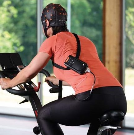
APEX cycleAdditional wearable EEG options available through BIOPAC include the MedelOpt Mobility EEG system, combining wearable EEG and functional near-infrared spectroscopy (fNIRS) technology with advanced signal processing and robust data transmission capabilities. APEX wireless EEG systems further enhance the landscape with their ability to record high-quality EEG data with flexibility for a range of applications from cognitive neuroscience to mental workload studies. Learn more about recording EEG data by watching our webinar: A Beginner’s Guide to EEG.


Are you interested in unlocking the benefits of mobile and wireless EEG for your next research project? Let our staff of experts find the right tools for you.

Mind and Machine: Exploring Brain-Computer Interfaces

In neuroscience, the frontier of brain-computer interfaces (BCI) holds the promise of bridging the gap between the human brain and external devices, offering revolutionary applications in fields ranging from robotics and prosthetics to communication and beyond. In the lab, scientists studying and developing new BCI technology leverage the human brain’s electrical and physiological signals.

Electroencephalography (EEG), functional near-infrared spectroscopy (fNIRS), and steady-state visually evoked potentials (SSVEP) are among the primary physiological and psychophysiological signals utilized in these investigations. Multimodal integration of two or more signal types allows researchers and engineers to take advantage of multiple data streams in their applications and studies.

EEG, which can measure electrical activity in the brain, is a cornerstone of BCI research. Researchers can decode various mental states and intentions by analyzing EEG patterns, enabling users to control external devices through mere thought. fNIRS complements EEG by measuring changes in blood oxygenation levels, offering insights into brain function with superior spatial resolution. SSVEP, on the other hand, capitalizes on the brain’s response to visual stimuli, providing a means for precise control in BCI applications.

The practical implications of BCI research extend far beyond the laboratory setting. In robotics, BCIs facilitate direct brain-to-machine communication, empowering users to manipulate robotic arms or navigate autonomous vehicles with their thoughts alone. A study published in the Journal of Information Science Engineering explored ways that an SSVEP-based BCI could be used to control assembly line robots via a headset measuring the operator’s EEG signals. The signals would communicate instructions to robots while operators identified defective parts, which the robots would pick out. During the experiment, EEG signals were obtained using a B-Alert X24 headset. The EEG-based BCI produced a 93.33% success rate in correctly identifying and removing defective parts from the assembly line process, allowing the operator to effectively “collaborate with the robot and communicate defective parts without manually operating the robot.”

Prosthetics benefit immensely from BCI advancements, enabling seamless integration with the user’s neural signals for intuitive control and enhanced functionality. A 2023 study by a team of researchers from the University of California Irvine explored the viability of creating low-cost cortical stimulators for bi-directional BCI (BD-BCI) research. BD-BCI takes brain-computer-interfaces to the next level with the brain not only providing motor commands to an artificial limb but also allowing it to receive sensory input from such a device. A BIOPAC data acquisition and analysis system was used to validate the charge balancing of subcortical electrocorticographic (ECoG) implants used in a stimulator. The charge balancing test was performed on phantom tissue designed to simulate real brain tissue. The resulting study successfully demonstrated “clinical validation of a fully-programmable electrical stimulator, integrated into an embedded BCI system.”

As researchers continue to refine BCI technologies, the possibilities for real-world applications are boundless. BIOPAC is among the companies on the cutting edge of BCI research, partnering with other innovators to provide the latest in multimodal BCI technologies such as Axon-R. For additional information on Axon-R, check out our recent webinar on the device.


Are you planning to explore the frontiers of BCI in the lab? Our regional customer reps have all the knowledge you need to integrate BIOPAC products into your BCI study.

VR Integration: Enhancing Research with Virtual Environments

Measuring the human body and brain’s response to real-world conditions and stimuli has long challenged researchers. This problem has usually been dealt with in one of two ways. The first approach is to take research participants into the field—not impossible with today’s mobile data recording technology but not always safe or convenient. The second option is to imitate real-world conditions in the lab using screens, audio equipment, and even scent delivery, but in the end, it is difficult to completely remove the lab setting from the participant’s experience—that is, until the introduction of Virtual Reality (VR) technology in research.

Just as it has revolutionized everything from entertainment to training, VR has provided researchers with the opportunity to create immersive experiences in the lab that mimic the real world. Advances in technology have not only made these simulated environments more realistic, but they have also made it easier to integrate a wide range of signals and stimulus delivery to enhance the recording of physiological and psychophysiological data.

BIOPAC, through cooperation with several partner companies, offers a range of data recording options that integrate seamlessly with third-party VR devices. fNIR Devices, for example, designs solutions for recording functional near-infrared spectroscopy (fNIRS) signals that can easily be incorporated with standard VR head-mounted displays (HMD) to measure neural activity and hemodynamic response in the prefrontal cortex while participants experience VR environments. BIOPAC partner Seenel has created MedelOpt, a line of whole-head wearable devices that combine electroencephalography (EEG) and fNIRS, including a VR option that integrates an HMD. Finally, partner company WorldViz offers a full spectrum of VR solutions from complete VR kits to full-room Projection VR technology to software that can be combined with both wired and wireless data recording technologies to provide a complete array of bio-signal data, from eye tracking to electrodermal activity.

A Canadian study published in 2023 used a combination of BIOPAC and WorldViz products to investigate how sensory feedback and arousal influence neuromusVR Shadowcular control and posture. Researchers measured arousal via electrodermal activity (EDA) and psychosocial questionnaires, rating perceived fear and anxiety in both real-world situations in VR environments that simulated standing both at ground level and on a raised platform. Participants wore a commercial HMD running WorldViz’s Vizard VR toolkit software. Electrical nerve stimulation was provided via a BIOPAC linear isolated stimulator. The study found the “physiological and perceptual indicators of fear and anxiety increased” in the VR platform simulation, providing “novel evidence that VR can be used to modulate upright postural control.”

A similar study published in the 2022 issue of the Journal of Sport Rehabilitation examined the effect non-physical mental training has on balance skills. The study used fNIRS technology to measure hemodynamic response in the prefrontal cortex of participants experiencing virtual environments through VR HMDs. Participants were instructed to imagine themselves imitating balance exercises viewed through an Oculus VR HMD while also fitted with an fNIR Devices Imager headband. Data was gathered using Cobi Studio software and analyzed via fNIRSoft software. The study identified several advantages of virtual reality mental training (VRMT) over conventional mental training (CMT).

These are just a few examples of how VR technology can be integrated with a variety of methodologies for gathering research data. For additional information on the subject, check out our Virtual Reality Resources, which include an array of online training on integrating VR into research.


Are you planning a research product and need help integrating VR with other data acquisition and analysis equipment? Contact your local BIOPAC sales rep to find the right solutions for you.

‘Tis the Season for Stress: Research Tools for Anxious Times

Holiday-related stress is as much of a seasonal tradition as family gatherings, frantic shopping forays, and over-indulgence in food and drink. Add to this potent mixture nearly two years of pandemic-induced tension and we have a volatile cocktail of emotions and anxiety awaiting us in November and December. What better time to study the causes and effects of stress?

Research has established strong links between stress and our moods and sense of well-being, not to mention overall mental and physical health. While certain types of stress during development can teach individuals to adapt to life’s many challenges, excessive and prolonged stress is shown to contribute to chronic disease and a range of psychological disorders. According to the American Psychological Association, which conducts an annual survey monitoring stress, the COVID pandemic has had an unprecedented impact on stress levels across all demographics, particularly among young people.

The pandemic is just one of many factors driving demand for new approaches to stress research. Thankfully, researchers now have access to a myriad of technologies to support any experiment design. In a recent webinar on techniques for measuring physiological changes related to stress, BIOPAC head of European Sales, Alex Dimov, outlined a range of stress-connected physiological responses that can be measured in the lab with available technologies. These include heart rate, heart rate variability (HRV), trans-radial-ulnar electrical bioimpedance velocimetry (TREV), cardiac output, blood pressure, EDA, pulse, respiration, EMG, EEG, and functional near infrared spectroscopy (fNIRS).

One of the challenges of measuring stress in a natural environment is finding the right data gathering tools to support the mobility of test participants. A 2019 study tackled this by examining a variety of techniques for measuring stress in day-to-day life using mobile phones and wearable sensors. Settings included the lab, automobile, office, school, and other daily life locations. Along with consumer products like smartphones, smartwatches, and fitness bands, researchers also employed wearable sensors, including the BioNomadix wireless PPG transmitter to measure blood volume pulse (BVP) using a photo plethysmogram (PPG) signal. Researchers found that the BIOPAC device provided a high level of accuracy (80%) when employed outside of the traditional lab environment.

Smelling RosesStress research technologies are not limited to data gathering. Stimulus presentation tools offer researchers options for both inducing and alleviating stress in the lab. A 2019 University of Wisconsin study investigating the role scents can play in reducing stress utilized EDA, heart rate, and respiratory rate to measure how rose scent reduced anxiety among students taking a difficult test. Respiratory rate was measured using a BIOPAC respiratory transducer while EDA data was collected with an EDA finger transducer. Data was collected and analyzed via the Biopac Student Lab System. While rose scent was delivered using scented lotion, it should be noted that BIOPAC offers a new modular scent delivery system that is capable of delivering up to eight scents per experiment, with more than 120 scent options.
With so many data gathering tools available, planning your next experiment should not leave you feeling on edge. Let your BIOPAC sales rep help you take the stress out of stress research.


BIOPAC offers a wide array of wired and wireless equipment that can be used in your research. To find more information on solutions for recording and analyzing signals such as ECG, heart rate, respiration, and more using any platforms mentioned in this blog post, you can visit the individual application pages on the BIOPAC website.

fNIRS and the Brain: How cognition is defined and recorded

As the most crucial organ of the human body that quintessentially defines humanity, the brain is still our greatest puzzle to solve. Medical doctors, neuroscientists, physiological scientists, and others have been trying to understand how its functions affect our actions, dating all the way back to the dark days of phrenology. In our study of the brain, the popular research is directed towards conscious thought, thinking, or the scientific term, cognition. In physical terms, the brain is separated into different areas of grey matter for different tasks, and like an electricity operator supplies a power grid with different levels of electricity on demand, regions of the brain gearing up for critical thought are saturated with fresh, oxygenated blood to boost performance: again a sign of an active cognitive state.

In modern scientific research, one of the popular means to measure this oxygenation is through Spectroscopy—more specifically, a unique field called functional near infrared spectroscopy. How does fNIRS work in measuring human cognition? These measurements are performed with optical diodes and sensors measuring a near infrared wave pattern.

Dedicated fNIRS software displaying tissue oxygenation
A graphical display of oxygenation in the prefrontal cortex

Near infrared light is sent through the skull in a spectrum that can pass through bone and tissue, and the sensors record the plots of photons absorbed by tissue or reflected back. Naturally with such a delicate subject such as the human brain, this process is both safe for the participant and painless.

The practical benefits of this spectroscopic measurement are numerous—continuous and real time values on changes in hemoglobin levels in the prefrontal cortex (measured as oxyhemoglobin [Hb] and deoxygenated hemoglobin [HbO2]) as subjects perform different tasks, either in a stationary recording position or in a mobile experiment environment. fNIRS technology offers standalone and field-deployable technology able to determine localized brain activity. Modern recording tools such as the fNIR103M offer a wireless recording platform, so research subjects can react naturally in real-world situations outside of the lab environment while continuously recording spectroscopic data.

fNIRS is a popular signal to record simultaneously with other physiological signals such as heart rate, ECG, HRV, EMG, respiration, and more to get a clear picture of a subject’s physiological reactions. Application areas for this signal typically include pain assessment, geriatrics, attention, sports science, Human-Computer Interface, virtual reality, stress, workload assessment, and learning. Thousands of scholarly citations have been written featuring functional near-infrared spectroscopy, as studies visualize how the brain works through cognitive problems and processes. To further your physiological research, BIOPAC’s AcqKnowledge software can record multiple physiological signals at once and provide automated analysis tools for event related potentials and ensemble averaging. fNIRS easily integrates with other physiological and neurobehavioral methods of assessing brain activity, including eye tracking, pupil reflex, respiration, and electrodermal activity.

BIOPAC Systems, Inc. provides life science researchers and educators with data acquisition and analysis systems that inspire people and enable greater discovery about life. Visit us at www.biopac.com. We invite you to look at our fNIRS webinars from BIOPAC. BIOPAC also offers a line of wired and wireless fNIRS imagers for research and education.

Eye Tracking Insights

Try Not To Blink—This Won’t Hurt

What exactly, is eye tracking? Do I have to put on a helmet or stick something unpleasant into my eye? No, nothing so invasive, and not the least bit uncomfortable. Eye tracking is the process by which eye movements are recorded for the purpose of tracking where a subject is looking, what he/she is looking at, and the duration of the subject’s gaze on a given area. Eye tracking differs from electrooculogram (EOG) in that it records where and at what the subject is actually looking, whereas EOG primarily records the movements of the eye muscles. Rather than merely recording eye movement and position, eye tracking forges an invisible yet accurate connection between the subject and the objects within the subject’s field of vision. These “visual connections” of where the subject was looking during the study are clearly visible in the resulting data, as dramatic as tire tracks on an impressionable surface.

So how does eye tracking work? Using invisible near-infrared light combined with high-definition cameras, the eye tracking system projects invisible light onto the eye and records the direction from which it is reflected off the cornea. Eye tracking software is used to calculate the exact position and focal point of the eye. The sophisticated combination of eye tracking hardware and software allows for extremely accurate measuring and analyzing of visual behavior and fine eye movements. This makes it possible to create a detailed map of a subject’s field of vision and the specific visual elements honed in on by the subject. Eye tracking data is commonly acquired in tandem with other body signals for the purpose of determining the effects of what we are looking at has on our physiology. Common signals acquired during eye tracking studies are:

  • EEG (cognitive processes)
  • EDA (alertness)
  • ECG (anxiety)
  • EMG (muscle activity, conditioning)
  • EGG (pulse, respiration, temperature)

What are researchers looking for when conducting eye tracking studies? Eye tracking data is useful in a variety of different research fields including interface usability, reading analysis, psychological and emotional response, marketing research, training, clinical vision diagnosis, and more.

Eye tracking hardware mainly comes in two flavors. Screen based eye tracking involves having a subject sit in front of a computer monitor while being presented with various types of visual stimuli. A “bar type” eye tracker records the subject’s responses and interfaces with eye tracking software to present the stimulus and store the resulting data for analysis. BIOPAC’s AcqKnowledge software version 5.0.6 and higher, along with the BIOPAC MP160, MP36R, or Wireless Smart Center system, supports fully-integrated eye tracking functionality*. Rather than having to learn a second software program, the familiar AcqKnowledge interface has been extended to incorporate the EYE-TRACK-BAR and EYE-TRACK-BAR-XL remote screen eye tracking systems.

Mobile or “wearable” eye tracking systems utilize a pair of glasses with two types of built-in cameras. One camera faces the subject and tracks eye movements, gaze path, and other parameters, and a second camera (called a scene camera) records what the subject is looking at during the study. Mobile eye tracking allows the subject to engage in independent activities outside the lab, such as driving an automobile. Additional sensors record and log data from other body signals in real time, which can be loaded into the software for subsequent analysis.

For more information about eye tracking, we invite you to sign up for our free “Eye Tracking Fundamentals” webinar to be broadcast live on November 21, 2019 at 8:00 AM PST. Did you miss it or can’t attend? No problem. Follow the same link to watch it on demand.

*Eye Tracking Integration License for AcqKnowledge required.

Please also visit the following links for information about BIOPAC Eye Tracking products.

Eye Tracking Systems

Eye Tracking License


BIOPAC offers a wide array of wired and wireless equipment that can be used in your research. To find more information on solutions for recording and analyzing signals such as ECG, heart rate, respiration and more using any platforms mentioned in this blog post, you can visit the individual application pages on the BIOPAC website.

Photo by Marina Vitale on Unsplash

error: Content is protected !!