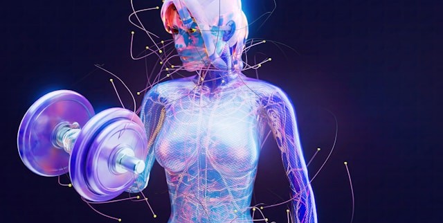
Muscles in Motion: Electromyography in Physiological Research

When we think about the human muscular system, movement often comes to mind. Activities such as running, lifting, and maintaining posture all rely on coordinated skeletal muscle activity, while cardiac muscle drives circulation and smooth muscle supports essential functions like digestion. Yet muscles do more than move us from point A to B; they also reflect what is happening inside the body. Patterns of muscle activation can reveal information about physical effort, fatigue, coordination, and recovery, as well as emotional expression, stress, and cognitive engagement. Electromyography (EMG) is a widely used technique for measuring the electrical signals generated by muscles during contraction, providing researchers with a direct and objective means to study both physiological function and the links among the nervous system, behavior, and mental state. By capturing muscle activity that may be too subtle to observe visually, EMG enables precise investigation of processes ranging from motor control and performance to emotional and stress-related responses.

There are several types of EMG, each suited to different research needs. Surface EMG (sEMG) uses small sensors placed on the skin to detect activity from nearby muscles and is the most common approach in behavioral and human performance studies. Intramuscular EMG involves inserting a fine-needle electrode directly into a muscle to record activity from specific muscle fibers, often used in clinical or specialized research settings. Facial EMG (fEMG), a form of surface EMG, focuses on facial muscles. By measuring muscle activity associated with smiling or frowning, fEMG allows researchers to study emotional responses, even when those responses are subtle or not consciously expressed.

One of the key strengths of EMG is its ease of integration with other physiological measurements. In many studies, EMG is recorded alongside electrocardiography (ECG), which measures heart activity; electroencephalography (EEG), which records electrical activity in the brain; functional near-infrared spectroscopy (fNIRS), which tracks changes in brain oxygenation; and eye tracking, which monitors visual attention and gaze patterns. By synchronizing these signals in time, researchers can better understand how different systems in the body work together. For example, combining EMG with EEG can reveal how brain activity leads to muscle movement, while pairing EMG with ECG can show how physical effort relates to heart responses during stress or exercise. This multimodal approach is central to advancing research in areas such as emotion, cognition, and human performance.

EMG also has important real-world applications. In rehabilitation, it helps clinicians monitor muscle recovery after injury. In ergonomics, it provides objective data about muscle strain in the workplace. In sports science, EMG helps optimize training and performance by identifying how and when muscles are activated. Increasingly, EMG is also used in human–machine interfaces, such as systems that allow people to control prosthetic limbs using their muscle signals. Its ability to detect fine-grained changes in muscle activity makes EMG a valuable tool across both research and applied settings.

Best practices for EMG data quality begin with careful electrode placement and attention to potential sources of noise. For sEMG and fEMG, electrodes should be placed over the “belly” of the target muscle, aligned with the muscle fibers, and spaced consistently to capture clean, interpretable signals. Gently cleaning the skin and, when appropriate, lightly abrading the surface helps reduce impedance and improve signal quality. In fEMG studies, precise and repeatable placement over well-defined facial muscles is critical, as these muscles are small and closely spaced. To minimize noise and artifacts, researchers should secure electrodes and cables to prevent movement, ask participants to limit unnecessary body motion, and ensure proper grounding of the recording system. Electrical interference from nearby equipment, power lines, or poor cable management can also contaminate EMG signals, so shielded cables and a controlled recording environment are recommended. Finally, synchronizing EMG with other physiological signals using a common acquisition system helps prevent timing errors and reduces the need for later corrections, resulting in cleaner data and more reliable conclusions.

hand mechanicsRecent studies demonstrate how EMG continues to expand into new and innovative areas. In a study published in Results in Engineering, a team of researchers from China developed a low-computational method for recognizing hand gestures from EMG signals to improve prosthetic device control. The researchers extracted key features from EMG recordings and used a lightweight machine-learning model, a multilayer perceptron (MLP), to classify gestures in real time. EMG data were collected using a four-channel BIOPAC data acquisition system running Biopac Student Lab (BSL) software. Surface Ag/AgCl electrodes were placed on four forearm muscles to capture relevant muscular activity. The study showed that accurate gesture recognition can be achieved without heavy computational requirements, making the system more practical for everyday use. This work highlights how EMG can support the development of more responsive, accessible assistive technologies.

Another study, conducted by a team of researchers from Mahidol University in Thailand and presented in the Institute of Electrical and Electronics Engineers (IEEE) proceedings, explored how combining EMG with motion sensors can improve ergonomic assessments. Focusing on occupational health, the study integrated surface EMG with data from an inertial measurement unit (IMU), a sensor that tracks movement and orientation. The researchers used the IMU to calculate Rapid Upper Limb Assessment (RULA) scores—a common method for evaluating postural risk—while EMG data collected via a BIOPAC four-channel research system provided insight into muscle activity. By combining these measures, the team found that posture alone did not always reflect actual muscle strain. The inclusion of EMG revealed additional muscle stress that might otherwise go unnoticed, suggesting that multimodal approaches can lead to more accurate workplace assessments and better injury-prevention strategies.

Researchers from Louisiana State University and the Pennington Biomedical Research Center in Baton Rouge, Louisiana, examined how training experience influences muscle activation and fatigue during resistance exercise performed with and without blood flow restriction (BFR) in young men. The study, published in Physiological Reports, compared trained and untrained participants while they completed knee extension exercises under low-, medium-, and high-load conditions. The team used sEMG to measure muscle excitation and total muscle activation in the vastus lateralis, with participants fitted with a portable BIOPAC BioNomadix Wireless EMG system. EMG signals were sampled at 2000 Hz and analyzed using a data acquisition system with AcqKnowledge software, allowing the researchers to quantify both peak muscle excitation and overall activation across repetitions. The results showed that low-load exercise with blood flow restriction produced lower peak muscle excitation but substantially greater total muscle activation compared to traditional high-load exercise, regardless of training status. These findings suggest that low-load BFR training may provide an effective alternative for stimulating muscle adaptations while reducing mechanical stress, with potential applications in rehabilitation and strength training programs.

Taken together, these examples illustrate the versatility and effectiveness of EMG in neuroscience, psychophysiological research, and beyond. Whether used to study emotion, improve physical training and workplace safety, or develop assistive technologies, EMG provides a direct link between muscle activity and human behavior. When combined with other physiological measures, it offers a more comprehensive understanding of how the brain and body interact. As research tools and techniques continue to advance, EMG will remain an essential method for exploring brain-body connections. For additional information on how to design studies around EMG, best practices for gathering EMG data, and how to integrate EMG with other signals, check out our webinar on EMG acquisition and analysis.


If EMG will be part of your next physiological or psychophysiological research project, reach out to our knowledgeable staff, who are standing by to help you with each stage of the planning and design process.

Healing Hands: EDA and Affective Touch

“Nothing eases suffering like human touch.” — Bobby Fischer

The gentle caress of a mother to her child. A reassuring pat on the shoulder from a friend. The touch of another human is one of the first forms of communication we experience in life, preceding the development of spoken language. Touch can have a remarkable effect on our emotional, psychological, and physiological states. It shapes our relationships with others and helps us build social connections.

The positive effects of touch are primarily associated with affective touch, characterized by comforting, caress-like stroking that activates C-tactile afferent (CT-afferent) neurons in the skin of mammals (including humans). Affective touch has attracted the interest of researchers seeking to decode how the sympathetic nervous system confronts and manages a variety of challenges, including pain regulation, stress management, and social interactions. The subject has drawn attention in the study of neurodevelopmental disorders, such as Autism Spectrum Disorders (ASD), where it is playing an essential role in understanding the sensory and social challenges facing individuals with atypical neurocognitive function.

While studies on touch, and affective touch specifically, utilize a range of physiological signals to gather data, electrodermal activity (EDA) has proven particularly useful in this area. Also referred to as Galvanic Skin Response (GSR), EDA provides a direct window into the sympathetic nervous system, which is responsible for emotional or affect-related arousal. When someone experiences pleasant or meaningful touch (e.g., a gentle stroke or supportive hand), the brain triggers subtle changes in sweat gland activity manifested as shifts in skin conductance. These changes occur when people aren’t fully aware of their emotional response. This makes EDA a sensitive, objective marker of how the body reacts to social and tactile experiences. In studies of affective touch, researchers track EDA to see whether a touch feels calming, engaging, or stressful, and how these responses differ across individuals or clinical groups such as autistic youth. Because EDA reflects genuine physiological arousal tied to emotional processing, it helps scientists understand the underlying autonomic mechanisms that shape how we experience social touch.

Researchers at the University of Tours in France examined how children with ASD respond to gentle, affective touch to determine whether they exhibit physiological patterns that differ from those of their neurotypical peers under the same conditions. The team measured multiple aspects of the autonomic nervous system, including EDA and electrocardiogram (ECG). These signals were collected using a BIOPAC MP36 data acquisition and analysis system to capture subtle changes in sympathetic arousal. Researchers collected EDA data using two 8 mm Ag/AgCl cup electrodes placed on the second phalanges of the index and middle fingers of the right hand. ECG was collected via two disposable vinyl electrodes placed on the sternum and on the right shoulder.  The study found that autistic children exhibited atypical EDA responses, showing either blunted or inconsistent autonomic reactions relative to controls, even when their outward behavior appeared typical. These results suggest that affective touch may not regulate internal arousal in autistic children the same way it does in others, providing an essential insight for therapies and sensory-based interventions that rely on touch.

In a similar study, a team from the University of Turin in Italy explored how autistic adults process affective touch by comparing their subjective ratings of pleasantness with their physiological responses. The researchers used a BIOPAC data acquisition research system with an EDA amplifier module to record EDA. At the same time, participants received gentle, slow stroking intended to activate the brain’s affective touch pathways. The goal was to determine whether autistic adults show a mismatch between what they say they feel and how their bodies respond. The findings revealed an apparent dissociation. While autistic participants often rated the touch similarly to neurotypical participants, their EDA responses differed significantly, indicating altered sympathetic arousal. This physiological-subjective mismatch provides compelling evidence that affective touch is processed differently in ASD, which may help explain challenges with social touch and sensory integration in everyday interactions.hands

Researchers from the University of Liverpool and Manchester Metropolitan University examined how brief, naturalistic social touch alters brain activity, autonomic physiology, and emotion recognition performance. Although this multimodal study did not focus on an autism-only sample, it is relevant to social-touch research more broadly and provides methodological examples that apply to ASD studies. To capture changes in prefrontal cortex function, the team employed a BIOPAC fNIRS imager system, with signals sent to a data acquisition unit. Further analysis of fNIRS signals was handled via fNIRS visualization software. Both EDA and photoplethysmography (PPG) were recorded at 2 kHz, using shielded Ag/AgCl electrodes on the index and fourth fingers and a PPG sensor on the middle finger, and were acquired and analyzed in AcqKnowledge. The results showed that social touch increased prefrontal activation, modulated cardiac and vascular dynamics captured via PPG, and produced measurable changes in sympathetic arousal, as indexed by EDA. Participants also demonstrated improved emotion-recognition accuracy following touch. Together, these findings suggest that social touch engages coordinated central and peripheral systems in ways that may support social cognition and emotional understanding.

These are just a few examples of how EDA is being used in research into affective touch. For additional information on integrating EDA in your study, see our multiple webinars on the subject.


Are you designing a study into affective touch or just planning a research project that will focus on EDA? Our customer service staff can help you streamline the process to produce best possible data.

Facing Fear: Measuring What Scares Us

In many countries, October ushers in the season of ghosts, goblins, and all manner of frightening things that inhabit our deepest fears. While the popularity of Halloween reflects the thrills many derive from a good scare, facing one’s fears is not always fun and games. Our fears can protect us from danger, but they can also hamper individuals from performing the simplest tasks and negatively impact one’s quality of life and mental health.

The study of fear conditioning and fear learning sheds light on how humans and animals develop responses to threatening stimuli. Research often relies on psychophysiological measurements—techniques that capture the body’s physical responses to fear. Key among these measurements are skin conductance response (SCR), electrodermal activity (EDA), heart rate variability (HRV), respiration, and electromyography (EMG).

SCR and EDA track changes in sweat gland activity, providing a direct measure of emotional arousal when a person experiences fear. HRV and respiration rate reveal the autonomic nervous system’s reaction to stress, indicating shifts between sympathetic (fight-or-flight) and parasympathetic (rest-and-digest) states. EMG measures muscle activity, especially facial muscles, to detect subtle expressions or physical tension associated with fear. Understanding these mechanisms has practical applications in understanding and treating anxiety disorders, phobias, and post-traumatic stress disorder (PTSD). By analyzing the body’s automatic fear responses, scientists can develop targeted therapies to help individuals recondition or dampen these reactions.

A study published in the Journal of Behavior Therapy and Experimental Psychiatry used a range of indices to assess learned fear (unconditioned stimulus expectancy or US-expectancy) in individuals. These included both self-reporting and measurements of autonomic response such as skin conductance. Researchers using E-Prime Stimulus Presentation Systems, paired various neutral visual stimuli with unconditioned stimuli in the form of electrical shocks delivered via a constant voltage stimulator coupled with a BIOPAC STM100C amplifier. After a brief break, both neutral stimuli were administered without shock. Skin conductance response data was gathered and analyzed during each phase with a BIOPAC data acquisition and analysis system and software. Results showed a positive correlation between US-expectancy ratings and skin conductance response.

facing fearResearchers at the University of Bologna in Italy took a different approach to measuring the psychophysiological mechanisms of fear learning by focusing on HRV. In their study, published in the journal Psychophysiology, they explained that “understanding transient dynamics of the autonomic nervous system during fear learning remains a critical step to translate basic research into treatment of fear-related disorders.” Researchers gathered SCR and electrocardiogram (ECG) data. As with the previous study, mild electrical shocks were applied as unconditioned stimuli and paired with neutral or conditioned stimuli (CS). SCR signals were acquired using a skin conductance transducer attached to the participants’ fingertips connected to a BIOPAC EDA amplifier. ECG data was collected using disposable surface electrodes connected to an ECG100C amplifier. EDA and ECG signals were processed with a BIOPAC data acquisition and analysis system running AcqKnowledge software, which calculated HRV from the ECG data. Researchers concluded that “learned fear elicited a transient heart rate deceleration in anticipation of noxious stimuli.” They added that the study’s findings “provide a proximal measure of the involvement of cardiac vagal dynamics into the psychophysiology of fear learning and extinction, thus offering new insights for the characterization of fear in mental health and illness.”

Another study published in the journal Psychophysiology examines fear learning by applying a broad range of physiological signals and responses to determine which provides the “optimal measurement of human trace fear conditioning.” For this study, researchers compared SCR, pupil size response, heart period response (via ECG), respiration amplitude response, and electromyography (EMG) to measure participants’ responses to electrical current, audio, and visual stimulation. EDA data was gathered via a BIOPAC EDA amplifier, while ECG, EMG, and respiration data were fed to BIOPAC ECG100C, EMG100C, and RSP100C amplifiers, respectively. All data was processed using BIOPAC data acquisition and analysis systems and software. Results of the experiment suggested that startle eye-blink response through EMG provided the most sensitive metric for measuring fear conditioning.

Fear is just one of the emotions that can be studied through the body’s affective response to stimuli. Read more about the relationship between affect and emotion or watch our webinar on Emotion and Objective Physiological Measures.


Are you facing the reality of putting together your own fear-based study? Don’t be afraid to contact your local BIOPAC customer service representative for help finding the right tools for the job.

Mind and Body: Signals to Enhance Your Psychophysiology Research

The human body and mind share an intrinsic link, both influencing and reflecting the health and general well-being of the other. Within the field of psychophysiology, researchers gain a wealth of insight into mental processes and disorders by studying physiological signals recorded from both the brain and body. Signals such as electrodermal activity (EDA), skin temperature (SKT), electroencephalogram (EEG), heart rate variability (HRV), and functional near-infrared spectroscopy (fNIRS) provide just a few of the tools helping us to better understand these mind-body interactions.

A wide range of research technologies has made these signals increasingly accessible in the lab as well as in everyday environments, allowing studies to place participants in increasingly naturalistic settings while gathering critical physiological data. In this way, researchers can provide a more accurate picture of how study participants react to stress and arousal, experience emotion, or interact within group environments.

One recent study used EDA and heart rate (HR) to examine the role of emotion in student success within computer-based learning environments (CBLE). As discussed in previous posts, a range of signals can be used to measure the body’s physiological response to emotion, otherwise known as “affect.” By gaining a better understanding of learners’ emotional responses to CBLE stimuli, researchers in this study hope to apply their data to improvements in CBLE design. EDA data was gathered from participants via SS57LA EDA leads while HR signals were collected with SS2LB shielded lead sets, both of which were connected to a BSL MP36 data acquisition and analysis system. Participants were shown imagery designed to evoke a negative emotional reaction before being asked to read a complex academic paper. The study was able to establish a link between the participants’ emotions and their comprehension of the reading material.

head with brain exposedIn another study, researchers used EDA and skin conductance response (SCR) to measure participants’ reactions to conditioned stimuli (CS) and unconditioned stimuli (US). The goal was to determine if fear-inducing memories could be modified either pharmacologically or through behavior modification to overcome fear. An MP36R System with AcqKnowledge software allowed analysis of SCR signals gathered via SS3LA finger transducers across a range of experiments designed to trigger fear-inducing memories. While the overall results were inconclusive, the SCR signals provided valuable feedback regarding the participants’ response to stimuli.

While EDA is a common thread through many of these psychophysiology studies, other signals provide researchers with a more complete and nuanced data set from which to draw their conclusions. Researchers exploring the factors influencing musical preferences combined EDA data with ECG and facial electromyography (fEMG) signals to assess how listeners reacted to familiar and unfamiliar musical stimuli. Data from all three signals were fed to an MP36 data acquisition and analysis system. The study found a strong correlation between musical preferences and familiarity with material as reflected by the subjects’ emotional responses.

These examples only scratch the surface of how psychophysiological research is being aided by an array of signals, all made accessible with available technology. To learn more about how to implement these signals in your next study, see BIOPAC’s Psychophysiology Applications webpage.


Contact your BIOPAC sales representative to learn more about the wide range of data-gathering tools available to help you construct your next psychophysiology study.

Guilt Trips: Taking Measure of Our Moral Compass

“So full of artless jealousy is guilt, It spills itself in fearing to be spilt”
—Gertrude, William Shakespeare’s “Hamlet”

In Act 4 of Shakespeare’s famous tragedy, Hamlet’s mother Gertrude points out that the harder one tries to conceal guilt, the easier it is to expose. Little does she realize that she may have hit upon how future researchers approach the study of guilt in the lab. For practical purposes, it is a primary motivator in moral action and impacts how an individual responds to certain stimuli.

Psychologists have developed a myriad of tests to gauge guilt in a clinical setting such as the Test of Self-Conscious Affect (TOSCA), a multiple-choice test that tracks responses to various moral scenarios. While such techniques have been useful in linking guilt with conditions such as depression and anxiety, they have also shown their limitations in providing a deeper understanding of guilt’s influence on behavior, development, and psychological health.

In a recent BIOPAC webinar, UCSB professor Dr. Hongbo Yu addressed these limitations. Scenario-based tasks, he points out, are non-committal in examining the causes of guilt and do a poor job of measuring behavioral tendencies linked to guilt. Interaction-based research methods, however, allow participants to experience emotions in real-time, allow direct control of the causes of guilt in the lab, and allow for direct measurement of responses to stimuli.

As discussed in prior posts on affect, the body’s physiological manifestation of emotion, there is a range of methods and physiological signals available to researchers for measuring affect and gaining a better understanding of emotional states like guilt. Dr. Yu utilized eye-tracking, electrodermal activity (EDA), and functional magnetic resonance imaging (fMRI) in his examples. These are just a few of the signals currently being used in the lab to measure guilt.

One of the practical applications of guilt research is detecting when a subject is concealing the truth, as one would with a polygraph test. A recent study conducted by researchers at the University of Wisconsin-Madison’s Department of Psychology sought to gauge deception among research participants taking the Concealed Information Test (CIT). The goal was to improve upon traditional polygraph test signals such as heart rate and respiratory rate. Respiration data was gathered using a respiration transducer belt while ECG signals were collected with BIOPAC electrodes. In addition, researchers gathered EDA data using a transducer and finger electrodes. The study found that thGuilty Girle EDA signal was significantly more effective in accurately measuring physiological responses during the test procedure.

A 2018 study by researchers at Liverpool Hope University looked at the link between guilt and aggression in children by examining activity in the autonomic nervous system during so-called “bad behavior.” The study measured respiratory sinus arrhythmia (RSA) and skin conductance in children 4–8 years old while they imagined engaging in antisocial activities. EDA and ECG signals were gathered via wireless amplifiers and fed to an MP150 data acquisition unit (superseded by the MP160) running AcqKnowledge software. The study found a linkage between physiological responses to aggression and guilt that may provide a better understanding of the mechanisms behind guilt and related behavior in early and middle childhood.

These are just a few examples of how physiological signals can be applied to measuring guilt in research. BIOPAC offers a wide range of data acquisition, stimulus presentation, and VR options suitable for creating custom guilt research environments in the lab.


For additional information, contact your BIOPAC representative to learn more about the range of signals that can be utilized to gather data on guilt and other emotional states in the lab.

Measuring Moods: Signals for Insight into Emotion

“Do you ever look at someone and wonder what is going on inside their head?”

Joy, an anthropomorphized manifestation of happiness, delivers this line at the opening of Pixar’s 2015 animated hit “Inside Out,” a film that explores the emotional struggles of its young protagonist. She could have been speaking for generations of researchers who have plumbed the depths of the human psyche for a better understanding of the mechanisms behind our feelings, moods, and emotions.

When today’s researchers talk about emotion, they typically do so in terms of affect, the underlying physiological and psychological experience of feelings and emotions as a reaction to stimuli. Affect and emotion are not the same thing, per se, but are fundamentally connected. While the feelings we describe as fear, joy, love, hate, etc. remain abstract, affect is more concrete. BIOPAC co-founder and head of R&D describes it as “the measurable quality or manifestation of emotion in the human body.”

This manifestation can be measured in the human nervous system’s response to stimuli. Depending on the nature of the stimuli and how this sensory data is interpreted by the central nervous system, an array of physiological responses can take place: heart rate changes, the stomach and muscle groups tense, skin temperature responds, triggering sweat glands, among others.

The signals corresponding to these reactions provide researchers with valuable data to help them better understand affect and emotion. These signals—electroencephalogram (EEG), electrodermal activity (EDA), electromyogram (EMG), heart rate, and respiratory rate, to name a few—can be recorded and measured using a range of currently available sensing technologies.

As an example, a 2019 study published through the Queensland University of Technology used respiration signals as a means of classification for human emotions such as happiness, sadness, surprise, disgust, fear, and anger. In the study, breathing signals, including airflow rate and volume, were acquired and recorded using a BIOPAC MP36 data acquisition unit while participants were shown seven film clips, each associated with a different emotion. The study concluded that the respiratory data was 80% effective in accurately classifying affective responses to the film stimuli.

A team of researchers at the Khwaja Fareed University of Engineering and Information Technology in Pakistan took their investigation into the connection between physiological signals and emotion a step further. They used a similar approach by measuring participant response to movie clips, however this time, four types of physiological signals were acquired, including ECG, EMG, respiration, and skin conductivity (SC). The data was acquired with a wide range of signal sensors and analyzed via a BIOPAC MP160 DAQ System.Signals for Emotion

Signal sensing technology also allows researchers to study emotion in multiple participants simultaneously, as was the case in a 2020 dyadic study of how a parent’s stress and emotional suppression affects their children. The study measured physiological responses of both parents and children using ECG and impedance cardiography (ICG) via a BIOPAC MP150 data acquisition unit.

The mysteries of human emotion are being unlocked through a growing understanding of the affective response to stimuli. For more information on how physiological signals can be gathered and analyzed for this purpose, see our app note on emotional state physiology as well as our webinar Emotion and Objective Physiological Measures.


Contact BIOPAC for the latest information on how we can help you measure affect in the lab.

error: Content is protected !!