Only the Lonely: The Human Impact of Social Isolation

Humans by their nature are social creatures. A sense of community, daily interactions, and shared experiences are tied to our overall mental and physical well-being. Few events in modern history have shut down such interactions more thoroughly than the outbreak of COVID-19, which forced millions into prolonged isolation. As lockdowns and social distancing became the norm, an increasing number of people reported heightened feelings of loneliness, anxiety, and depression. Beyond the pandemic, other modern social factors contribute to isolation, including the growing reliance on social media and virtual interactions in place of face-to-face connections. While technology offers new ways to stay in touch, excessive screen time and digital communication can sometimes deepen feelings of loneliness by reducing meaningful, in-person social engagement. In the wake of these societal shifts, researchers are working to understand the long-term consequences of social isolation on both mental and physical health.

Studies suggest that loneliness isn’t just an emotional experience—it has measurable physiological effects on the body. Using advanced biometric signals, researchers are trying to uncover how isolation influences our nervous system, stress responses, and overall wellness. Heart rate variability (HRV) provides insights into autonomic nervous system balance, while electrodermal activity (EDA) reflects changes in stress and emotional arousal. Respiration patterns, electromyography (EMG) readings, and other physiological markers help paint a complete picture of how isolation affects both mind and body. By combining psychological assessments with physiological data, scientists aim to better understand how chronic loneliness can contribute to conditions such as cardiovascular disease, weakened immune function, and mental health disorders.

In one such study published in Britain’s Royal Society Open Science journal in 2024, researchers looked at the effect of social isolation on human adolescents, specifically their ability to learn to perceive threats. Study participants between the ages of 16 and 19 underwent sessions of complete isolation and isolation combined with virtual social interaction, as well as baseline sessions. Following each session, study participants reported their psychological state and completed a threat learning task (participants learned to associate neutral shapes with an aversive sound) to measure physiological response to learned threats and safety cues. During these sessions, researchers recorded participants’ EDA signals using BIOPAC EDA finger transducers connected to an MP36R data acquisition and analysis system running AcqKnowledge software. Results showed heightened EDA levels during threat extinction following isolation compared to the baseline. Researchers reported that their findings suggest that isolation and loneliness among adolescents might increase vulnerability and increase the instances of emotional disorders such as phobias, anxiety, and obsessive-compulsive disorder (OCD).

A team of researchers in Brazil compared how giving and receiving of social touch as well as heart rate variability serve as accurate predictors of loneliness in college-age adults. Participant loneliness levels were measured using the UCLA Loneliness Scale. Participants completed a psychiatric screening questionnaire that reported loneliness, social support, and social touch levels. Participant HRV measurements were taken via electrocardiographic (ECG) signal processing using a BIOPAC ECG amplifier connected to a 16-channel MP data acquisition and analysis system running AcqKnowledge. According to the researchers’ published findings, “social support and social touch, specifically receiving touch, accounted for a significant proportion of the variance in loneliness. However, neither giving touch nor resting HRV was a significant predictor of loneliness.”alone

A study by researchers from the University of Bonn examined the link between loneliness and how males and females process trauma and react to fear-inducing stimuli. Over three days, participants were exposed to experimental trauma and evoked intrusive thoughts. Researchers employed functional magnetic resonance imaging (fMRI) scans to assess neural habituation to fearful faces and fear learning (conditioning and extinction) before trauma exposure. Fear conditioning and extinction tasks used a BIOPAC MP system to deliver stimulus in the form of electric impulses. EDA and respiration data were recorded using the BIOPAC data acquisition and analysis system. Study results revealed “a significant interaction between loneliness and sex such that loneliness is associated with more intrusions in men, but not in women.” The implication is, according to the study’s authors, “that loneliness may confer vulnerability to intrusive memories after trauma exposure in healthy men and that this phenotype relates to altered limbic processing of fear signals.”

As the physiological and psychological effects of the COVID-19 pandemic continue to resonate and we grapple with the isolating influence of technology, increasing numbers of research studies are being launched to measure these impacts. Studies into human isolation and loneliness represent just one small corner of a broad spectrum of research on the topic. For information on how BIOPAC equipment is being used in such research areas, check out our on-demand webinars for additional ideas on how to deploy BIOPAC resources in your next study.


Are you planning a research project investigating the effects of isolation, loneliness, or other related subjects? Reach out to our team of experts to help you get started with the right resources and equipment.


A Remarkable 2024: Introducing Tools for a Better and Brighter Research Future

As 2024 winds down and we ready the confetti to ring in 2025, we at BIOPAC Systems Inc. want to toot our horn after wrapping up an exciting year of growth and innovation. This year brought some big wins for the research community, thanks to BIOPAC’s efforts to make physiological data acquisition and analysis more accessible and efficient than ever. Two major milestones were the launch of the MP200 system, the latest evolutionary step for our flagship data acquisition platform, and two feature-rich release updates to our AcqKnowledge data acquisition and analysis software.

Introducing the MP200: A New Achievement for Data Acquisition

MP200The MP system series has been the backbone of BIOPAC’s efforts to provide powerful and yet accessible data acquisition tools to researchers. Released in November, the newest incarnation of MP device, the MP200, ushers in a new era of precision, reliability, and flexibility for data acquisition. Designed to meet the evolving needs of researchers, the MP200 offers unparalleled scalability and compatibility with a wide range of physiological signals and measurements. Key enhancements include the following:

  • Enhanced Signal Quality: The MP200 uses advanced signal processing algorithms to ensure superior data integrity across diverse research applications, from neuroscience to exercise physiology.
  • Modular Design: The MP200’s modular architecture allows seamless integration with BIOPAC’s extensive sensors and amplifiers, creating near-limitless customization possibilities for complex experimental setups.
  • Future-Ready Connectivity: The MP200 facilitates real-time collaboration and analysis with high-speed data transfer protocols and cloud integration support.

Researchers will benefit from the MP200’s improved ability to streamline workflows and deliver insights with unmatched precision. This latest system exemplifies BIOPAC’s dedication to fostering groundbreaking discoveries in physiology and related fields.

AcqKnowledge Updates: Pioneering the Future of Data Analysis

AcqKnowledge data acquisition and analysis software has been a go-to tool for researchers for decades, and 2024 saw two major updates that brought exponential improvements: versions 5.0.10 and 6.0.0. These updates are packed with features that make data collection and analysis easier and more comprehensive.

One of the most impactful new licensed features is the integration of Lab Streaming Layer (LSL). This feature lets researchers sync and stream data from multiple sources in real time. Whether one is working on interdisciplinary projects or using several devices at once, LSL makes it simple to bring everything together for analysis. LSL also opens the door to a host of new products from our partners for which BIOPAC has added integration in AcqKnowledge. These include the Axon-R brain-computer interface (BCI) hub with augmented reality (AR) and artificial intelligence (AI) and the APEX wearable, wireless EEG system. Other new products have also been integrated into AcqKnowledge and are now available from BIOPAC, including new eye tracking products from Tobii and EyeLogic.

Another game-changing licensed addition, AcqKnowledge Cloud, allows users to securely store their data online, streamlining processes to access, share, and work on data from any location. Collaboration between teams is also easier, and users no longer need to worry about managing large files on their local system.

These updates are all about making work less complicated and more effective for researchers. With tools like these, diving into complex data, finding patterns, and pushing one’s research further has never been more straightforward.

Looking Ahead to 2025

More excitement is on the horizon for BIOPAC. Not only are several new product updates and releases in the works, but we will also be hosting our bi-annual 4T Human Physiology Conference on Tools, Trends, Techniques, and Technologies. Customers, educators, researchers, and other experts in Physiology and related fields will gather in Santa Barbara, California July 14-16. Participants will share knowledge, take part in hands-on workshops, and see what is shaping the future of research. Learn more and sign up today. Hopefully, we will be seeing you in 2025.

Facing Fear: Measuring What Scares Us

In many countries, October ushers in the season of ghosts, goblins, and all manner of frightening things that inhabit our deepest fears. While the popularity of Halloween reflects the thrills many derive from a good scare, facing one’s fears is not always fun and games. Our fears can protect us from danger, but they can also hamper individuals from performing the simplest tasks and negatively impact one’s quality of life and mental health.

The study of fear conditioning and fear learning sheds light on how humans and animals develop responses to threatening stimuli. Research often relies on psychophysiological measurements—techniques that capture the body’s physical responses to fear. Key among these measurements are skin conductance response (SCR), electrodermal activity (EDA), heart rate variability (HRV), respiration, and electromyography (EMG).

SCR and EDA track changes in sweat gland activity, providing a direct measure of emotional arousal when a person experiences fear. HRV and respiration rate reveal the autonomic nervous system’s reaction to stress, indicating shifts between sympathetic (fight-or-flight) and parasympathetic (rest-and-digest) states. EMG measures muscle activity, especially facial muscles, to detect subtle expressions or physical tension associated with fear. Understanding these mechanisms has practical applications in understanding and treating anxiety disorders, phobias, and post-traumatic stress disorder (PTSD). By analyzing the body’s automatic fear responses, scientists can develop targeted therapies to help individuals recondition or dampen these reactions.

A study published in the Journal of Behavior Therapy and Experimental Psychiatry used a range of indices to assess learned fear (unconditioned stimulus expectancy or US-expectancy) in individuals. These included both self-reporting and measurements of autonomic response such as skin conductance. Researchers using E-Prime Stimulus Presentation Systems, paired various neutral visual stimuli with unconditioned stimuli in the form of electrical shocks delivered via a constant voltage stimulator coupled with a BIOPAC STM100C amplifier. After a brief break, both neutral stimuli were administered without shock. Skin conductance response data was gathered and analyzed during each phase with a BIOPAC data acquisition and analysis system and software. Results showed a positive correlation between US-expectancy ratings and skin conductance response.

facing fearResearchers at the University of Bologna in Italy took a different approach to measuring the psychophysiological mechanisms of fear learning by focusing on HRV. In their study, published in the journal Psychophysiology, they explained that “understanding transient dynamics of the autonomic nervous system during fear learning remains a critical step to translate basic research into treatment of fear-related disorders.” Researchers gathered SCR and electrocardiogram (ECG) data. As with the previous study, mild electrical shocks were applied as unconditioned stimuli and paired with neutral or conditioned stimuli (CS). SCR signals were acquired using a skin conductance transducer attached to the participants’ fingertips connected to a BIOPAC EDA amplifier. ECG data was collected using disposable surface electrodes connected to an ECG100C amplifier. EDA and ECG signals were processed with a BIOPAC data acquisition and analysis system running AcqKnowledge software, which calculated HRV from the ECG data. Researchers concluded that “learned fear elicited a transient heart rate deceleration in anticipation of noxious stimuli.” They added that the study’s findings “provide a proximal measure of the involvement of cardiac vagal dynamics into the psychophysiology of fear learning and extinction, thus offering new insights for the characterization of fear in mental health and illness.”

Another study published in the journal Psychophysiology examines fear learning by applying a broad range of physiological signals and responses to determine which provides the “optimal measurement of human trace fear conditioning.” For this study, researchers compared SCR, pupil size response, heart period response (via ECG), respiration amplitude response, and electromyography (EMG) to measure participants’ responses to electrical current, audio, and visual stimulation. EDA data was gathered via a BIOPAC EDA amplifier, while ECG, EMG, and respiration data were fed to BIOPAC ECG100C, EMG100C, and RSP100C amplifiers, respectively. All data was processed using BIOPAC data acquisition and analysis systems and software. Results of the experiment suggested that startle eye-blink response through EMG provided the most sensitive metric for measuring fear conditioning.

Fear is just one of the emotions that can be studied through the body’s affective response to stimuli. Read more about the relationship between affect and emotion or watch our webinar on Emotion and Objective Physiological Measures.


Are you facing the reality of putting together your own fear-based study? Don’t be afraid to contact your local BIOPAC customer service representative for help finding the right tools for the job.

Free Your EEG: Wireless and Wearable Systems for the Lab and Beyond

The evolution of wearable technology for recording physiological signals represents a transformative leap for research, particularly when it comes to electroencephalography (EEG). These advancements allow researchers to capture brain activity data in real-world settings, moving beyond the confines of laboratory-based experiments. Mobile and wearable EEG devices utilize wireless technology to transmit data, offering significant advantages over traditional methods.

Everyone aspires to more freedom—to speak, think, or move about as one pleases. Should researchers be any different? Why be shackled to the static wires and cables of the lab, when one can release study participants with the help of mobile and wearable technology?

The evolution of wearable technology for recording physiological signals represents a transformative leap for research, particularly when it comes to electroencephalography (EEG). These advancements allow researchers to capture brain activity data in real-world settings, moving beyond the confines of laboratory-based experiments. Mobile and wearable EEG devices utilize wireless technology to transmit data, offering significant advantages over traditional methods.

EEG is a cornerstone in the study of brain activity, providing insights into the electrical patterns generated by neuronal firing. EEG has traditionally been used in physiology and psychophysiology research to understand brain function, monitor cognitive states, and investigate neural responses to various stimuli. The conventional EEG setup typically involves a stationary system with numerous electrodes affixed to the scalp using a conductive gel, which can limit the subject’s movement and impact the naturalistic settings of studies.

One of the primary benefits of mobile and wearable EEG systems is their ability to record brain activity during dynamic, everyday activities. This capability provides a more comprehensive understanding of how the brain responds to real-world stimuli and stressors, as opposed to controlled, static environments. For instance, researchers can now study cognitive processes in natural settings like workplaces or homes, enhancing the ecological validity of their findings.

A study by the School of Architecture at the University of Liverpool provides an example in which researchers leveraged wearable EEG technology when comparing subjective and physiological responses to noise within wooden buildings. The research team conducted listening tests using EEG data recorded via the B-Alert X-24 wireless headset system, which allowed participants to “sit comfortably in a simulated living room” while listening to a variety of sounds that were both airborne and resonating through the floors. Participants rated their level of noise annoyance on an 11-point scale and results were compared to EEG data. In discussing the design of their study, the authors highlighted the importance of incorporating noninvasive, wearable technology, stating, “Among the several options available for measuring physiological reactions to auditory stimuli, contemporary technologies made possible the recording of EEG in a relatively easy way by means of portable devices. These offer the precious opportunity of acquiring data directly from the scalp, where brain activity is detectable almost instantaneously in synchrony with the stimulus presentation.”

Similarly, when studying occurrences of hyperventilation among a participant group with a range of anxiety-related disorders, researchers from Yale University turned to a BIOPAC BioNomadix wireless receiver to gather EEG signal data. By attaching individual electrodes to a third-party cap that connected wirelessly to a BIOPAC data acquisition and analysis system, they minimized the discomfort and restriction of movement that may have resulted from a wired solution, thereby influencing participants’ response to stimuli.

Researchers at the Transylvania University of Brașov in Romania faced similar demands that required a flexible noninvasive means to record surface EEG signals from participants as they responded to a variety of colors and patterns with the goal of better understanding the complexity of human color and texture perception. The research team compared participants’ subjective responses to EEG responses via Event-Related Potentials and computational measurements of color and texture.  EEG data was gathered with a cap-based BioNomadix wireless system and analyzed using AcqKnowledge software. The study’s authors explained their choice of wireless EEG recording devices as follows: “Since human users are the target, it is important to focus the technology development towards small-scale, wearable, wireless systems that provide flexibility, easy use and comfort to the user side, while at the same time maintaining high signal quality.”

APEX cycleAdditional wearable EEG options available through BIOPAC include the MedelOpt Mobility EEG system, combining wearable EEG and functional near-infrared spectroscopy (fNIRS) technology with advanced signal processing and robust data transmission capabilities. APEX wireless EEG systems further enhance the landscape with their ability to record high-quality EEG data with flexibility for a range of applications from cognitive neuroscience to mental workload studies. Learn more about recording EEG data by watching our webinar: A Beginner’s Guide to EEG.


Are you interested in unlocking the benefits of mobile and wireless EEG for your next research project? Let our staff of experts find the right tools for you.

Speak Up: Physiological Signals for Speech Research

In the symphony of human communication, speech is the principal instrument. Behind every uttered word lies a complex interplay of physiological and psychological signals, each telling a unique story about the speaker’s state of mind and physical well-being. The critical data provided by these signals has become a pivotal focus in both speech research and therapy, offering useful insights into the mechanisms of verbal communication and its disorders while enhancing speech ability and overall health.

Electromyography (EMG), a technique used to measure muscle activity, plays an invaluable role in speech research by revealing the interaction of facial muscles involved in articulation. By monitoring EMG signals from muscles such as the lips, tongue, and jaw, researchers can elucidate the precise movements and coordination required for speech production, shedding light on the underlying mechanisms of speech disorders like dysarthria or apraxia.

A French study published in the 2024 edition of Science Reports used EMG to establish the role tongue reflex plays in speech posture control. The presence of a tongue reflex in maintaining tongue posture, the proper positioning of the tongue in the mouth, has been a matter of debate among speech researchers. The study team employed an unorthodox recording technique that involved attaching EMG electrodes to the base of the tongue “to record activation of the anterior genioglossus, which is responsible for the control of the anterior part of the tongue.” A robotic arm was used to rapidly stretch the tongue while researchers measured muscular reflex EMG data during the formation of various vowel sounds, which were fed to a BIOPAC EMG amplifier for signal amplification and filtering. According to the research team, the study results “represent the first physiological demonstration in humans that the tongue is driven by reflex in response to a sudden stretch.”

Electrocardiography (ECG), which measures the electrical activity of the heart, offers insights into the intricate relationship between emotion and speech. Research has shown that changes in heart rate variability (HRV) correlate with emotional arousal and cognitive load during speech tasks. By integrating ECG data with speech analysis, researchers can explore the complex connection between emotional states and linguistic expression, paving the way for emotion-aware speech synthesis systems or interventions for social communication disorders.microphone

A 2023 study by researchers at Vanderbilt University used ECG to examine the relationship between respiratory sinus arrhythmia (RSA) on the persistence of stuttering and, specifically, the role emotional regulation plays in stuttering. Researchers measured potential differences in RSA in preschool-age children with persistent stuttering, children who recovered from stuttering, and children who do not stutter. RSA, an index of parasympathetic nervous system activity, was measured during the viewing of both neutral (baseline) and emotionally stimulating video clips and during subsequent speaking tasks. To obtain RSA for each study participant, ECG data was gathered via a BIOPAC data collection and analysis system running AcqKnowledge. A low RSA baseline is often associated with difficulty regulating emotion and, according to the study’s main hypothesis, plays a role in speech disorders like stuttering. The study’s findings supported this hypothesis, with some caveats, but the overall result demonstrated that emotional regulation could provide a promising avenue for speech therapy.

Electroencephalography (EEG), a noninvasive method for recording brain activity, offers insight into neural underpinnings of speech processing and comprehension. By analyzing EEG signals, researchers can map the spatiotemporal dynamics of neural networks involved in language processing, from phonological encoding to semantic integration. EEG-based studies offer insights into the neural signatures of speech disorders like aphasia or developmental language disorders, informing targeted therapeutic interventions and neurorehabilitation strategies. EEG has presented challenges with regard to artifacts created by participant movement during speech, but recent studies have shown promise in EEG as a speech research tool.

This is just an introduction to the signals that are being used to provide researchers with a better understanding of speech and speech pathology. For additional information on how BIOPAC products are being integrated into the study of speech, see our application note on Speech Motor Control.


Are you planning a research project focusing on speech? Our regional sales representatives are ready to help you find the right tools for your study.

Measuring Mindfulness: Ancient Awareness for Modern Research

The concept of mindfulness has garnered significant attention in recent years, transcending its roots in ancient meditation practices to become a focal point in modern scientific research, particularly in the fields of physiology and psychophysiology. Understanding mindfulness involves delving into its essence: a state of heightened awareness and acceptance of the present moment, free from judgment or attachment to external stimuli. But how exactly does this concept intersect with the realms of physiology and psychophysiology, and how do researchers measure its impact on the human body and mind?

At its core, mindfulness entails being fully present and engaged in the current experience, whether it be a sensation, thought, or emotion. This mental state has been associated with numerous health benefits, including stress reduction, improved cognitive function, and enhanced emotional well-being. In the realm of physiology and psychophysiology, researchers are keen to explore how mindfulness practices influence the body’s physiological processes and how these changes can be quantified and understood.

One of the primary methods used to measure mindfulness in laboratory settings is through the analysis of physiological signals, such as electrodermal activity (EDA) and heart rate variability (HRV). During mindfulness practices, individuals often experience a shift in these physiological parameters. For example, studies have shown that mindfulness meditation can lead to decreased EDA, indicating a reduction in sympathetic nervous system activity and arousal levels. Similarly, practitioners may exhibit increased HRV, reflecting greater parasympathetic nervous system dominance and improved adaptability to stressors.

A collaborative study by a team of researchers from Wake Forest University, the University of Utah, and San Diego State University examined the role of HRV in mindfulness-based pain relief practices. Specifically, the study looked at the role of high-frequency heart rate variability (HF HRV), a marker of parasympathetic nervous system (PNS) activity, during mindfulness-based pain relief versus a sham-breathing exercise that served as a control. Respiration and electrocardiogram (ECG) data was fed into a BIOPAC data acquisition and analysis system. While the study results were inconclusive, researchers determined that further study was needed to explore “parasympathetic activation in mindfulness-based pain relief as compared to other meditative techniques.”

lotusA study published in the 2023 edition of the Journal of Psychiatric Research compared how rest and breath-focused mindfulness could be used to regulate and control various symptoms of post-traumatic stress disorder (PTSD) and dissociative symptoms related to past trauma in female study participants. Researchers used skin conductance response (SCR), one of the components of EDA, to measure autonomic function as it relates to PTSD symptoms. SCR data were collected during alternating resting control and breath-focused mindfulness conditions via BIOPAC EDA electrodes connected to a BIOPAC data acquisition and analysis system. The study found that rest and breathing exercises had produced positive results for individuals with low to moderate levels of trauma but were less effective with higher levels.

One notable feature of BIOPAC’s technology is its ability to integrate multiple physiological signals simultaneously, allowing researchers to better understand how mindfulness practices impact various bodily systems. An example is a study by a group of researchers in Israel that examined the effectiveness of mindfulness-based interventions (MBI) in improving mental well-being and resilience. MBIs are non-religious adaptations of contemplative Buddhist practices that are used to reduce suffering and increase well-being. Researchers used a BIOPAC BioNomadix system to wirelessly record a range of physiological signals, including ECG, electromyography (EMG), and EDA. Their goal was to determine whether mindfulness could positively influence how interoceptive signals from the autonomic nervous system regulate one’s sense of well-being. The study concluded that “mindfulness seems to increase the sensitivity of regulatory selections to interoceptive signals, and this is associated with subjective well-being.”

Mindfulness represents a fascinating intersection between ancient contemplative traditions and modern scientific inquiry. To learn more about how BIOPAC is helping researchers uncover new insights into the physiological aspects of mindfulness and its potential implications for health and well-being, check out our webinar on The Science of Mindfulness.


Are you looking for techniques and tools to measure mindfulness in your study? Contact your regional BIOPAC sales representative to integrate everything you will need to gather and analyze data.

VR Integration: Enhancing Research with Virtual Environments

Measuring the human body and brain’s response to real-world conditions and stimuli has long challenged researchers. This problem has usually been dealt with in one of two ways. The first approach is to take research participants into the field—not impossible with today’s mobile data recording technology but not always safe or convenient. The second option is to imitate real-world conditions in the lab using screens, audio equipment, and even scent delivery, but in the end, it is difficult to completely remove the lab setting from the participant’s experience—that is, until the introduction of Virtual Reality (VR) technology in research.

Just as it has revolutionized everything from entertainment to training, VR has provided researchers with the opportunity to create immersive experiences in the lab that mimic the real world. Advances in technology have not only made these simulated environments more realistic, but they have also made it easier to integrate a wide range of signals and stimulus delivery to enhance the recording of physiological and psychophysiological data.

BIOPAC, through cooperation with several partner companies, offers a range of data recording options that integrate seamlessly with third-party VR devices. fNIR Devices, for example, designs solutions for recording functional near-infrared spectroscopy (fNIRS) signals that can easily be incorporated with standard VR head-mounted displays (HMD) to measure neural activity and hemodynamic response in the prefrontal cortex while participants experience VR environments. BIOPAC partner Seenel has created MedelOpt, a line of whole-head wearable devices that combine electroencephalography (EEG) and fNIRS, including a VR option that integrates an HMD. Finally, partner company WorldViz offers a full spectrum of VR solutions from complete VR kits to full-room Projection VR technology to software that can be combined with both wired and wireless data recording technologies to provide a complete array of bio-signal data, from eye tracking to electrodermal activity.

A Canadian study published in 2023 used a combination of BIOPAC and WorldViz products to investigate how sensory feedback and arousal influence neuromusVR Shadowcular control and posture. Researchers measured arousal via electrodermal activity (EDA) and psychosocial questionnaires, rating perceived fear and anxiety in both real-world situations in VR environments that simulated standing both at ground level and on a raised platform. Participants wore a commercial HMD running WorldViz’s Vizard VR toolkit software. Electrical nerve stimulation was provided via a BIOPAC linear isolated stimulator. The study found the “physiological and perceptual indicators of fear and anxiety increased” in the VR platform simulation, providing “novel evidence that VR can be used to modulate upright postural control.”

A similar study published in the 2022 issue of the Journal of Sport Rehabilitation examined the effect non-physical mental training has on balance skills. The study used fNIRS technology to measure hemodynamic response in the prefrontal cortex of participants experiencing virtual environments through VR HMDs. Participants were instructed to imagine themselves imitating balance exercises viewed through an Oculus VR HMD while also fitted with an fNIR Devices Imager headband. Data was gathered using Cobi Studio software and analyzed via fNIRSoft software. The study identified several advantages of virtual reality mental training (VRMT) over conventional mental training (CMT).

These are just a few examples of how VR technology can be integrated with a variety of methodologies for gathering research data. For additional information on the subject, check out our Virtual Reality Resources, which include an array of online training on integrating VR into research.


Are you planning a research product and need help integrating VR with other data acquisition and analysis equipment? Contact your local BIOPAC sales rep to find the right solutions for you.

ENS: Signals for Studying the Body’s “Second Brain”

We have all heard the saying “thinking with one’s stomach,” a cliché that suggests basing one’s critical thought processes on the whims of their digestive system. However, there may be more than a little truth to this saying. Often referred to as the body’s “second brain,” the enteric nervous system (ENS) comprises a complex network of neurons and ganglia embedded within the walls of the gastrointestinal tract. In recent years, researchers have increasingly recognized the significance of the ENS and its application in physiological research.

The ENS is a division of the autonomic nervous system (ANS) responsible for regulating the digestive process. It consists of millions of neurons spread throughout the esophagus, stomach, small intestine, and large intestine. These neurons form an intricate network that can function independently of the central nervous system (CNS) but also communicates with it. The ENS controls essential gastrointestinal functions, such as peristalsis, secretion of digestive enzymes, and blood flow regulation within the gut.

Several effective techniques are available to researchers to evaluate and measure ENS activity for their studies. Electrogastrography (EGG) provides a noninvasive approach to measuring the electrical activity of the stomach muscles. EEG can help researchers understand the rhythm and coordination of stomach contractions, aiding in the diagnosis of motility disorders.

In addition to EGG, a range of other physiological signals may be applied to ENS research. A recent study published in the June 2023 edition of Nature Communication investigated the gut-brain relationship by measuring physiological response to stimulation of the digestive system via different noninvasive techniques: EGG, electroencephalogram (EEG), skin conductance response (SCR), heart rate (HR), and heart rate variability (HRV). Research participants ingested a small vibrating probe that provided varying levels of stimulation as it passed through the lower digestive tract. EGG, EEG, and SCR signals were recorded along with hemodynamic signals via a BIOPAC data acquisition and analysis system. Researchers concluded that these techniques provided a viable alternative to more invasive approaches to measuring the body’s response to sensory stimulation of the digestive tract with, according to the published report, “implications for understanding gut feelings and gut-brain interactions in healthy and clinical populations.”

ENS ResearchMore invasive techniques for studying the ENS can still provide researchers with valuable data. A 2023 study by a team of South Korean researchers investigated how radial stretching affects intestinal contractions. The study also examined how drugs targeting the ENS affect such contractions to develop treatments for gastrointestinal disorders. Samples of intestinal tissue were taken from participants undergoing aggressive treatment for colorectal cancer. Samples were preserved in a tissue chamber and connected to a BIOPAC isometric force transducer to measure contractions, with data recorded and analyzed with a BIOPAC MP160 system running AcqKnowledge software. The study was able to detect regional differences in the stretch reflex of colon tissue via electrophysiological recordings of intestinal smooth muscles.

These are some of the ways in which researchers are currently using BIOPAC technologies to better understand the relationship between the ENS and other neurophysiological processes. For additional information on this topic, see our page on AcqKnowledge’s Gastric Myoelectric Activity feature and our EGG citations.


If you would like to know more about how BIOPAC can help you plan and equip your ENS study, reach out to your regional sales representative today.

4T|Phys 2023: BIOPAC Shares Tools, Trends, Techniques & Technology at UCSB

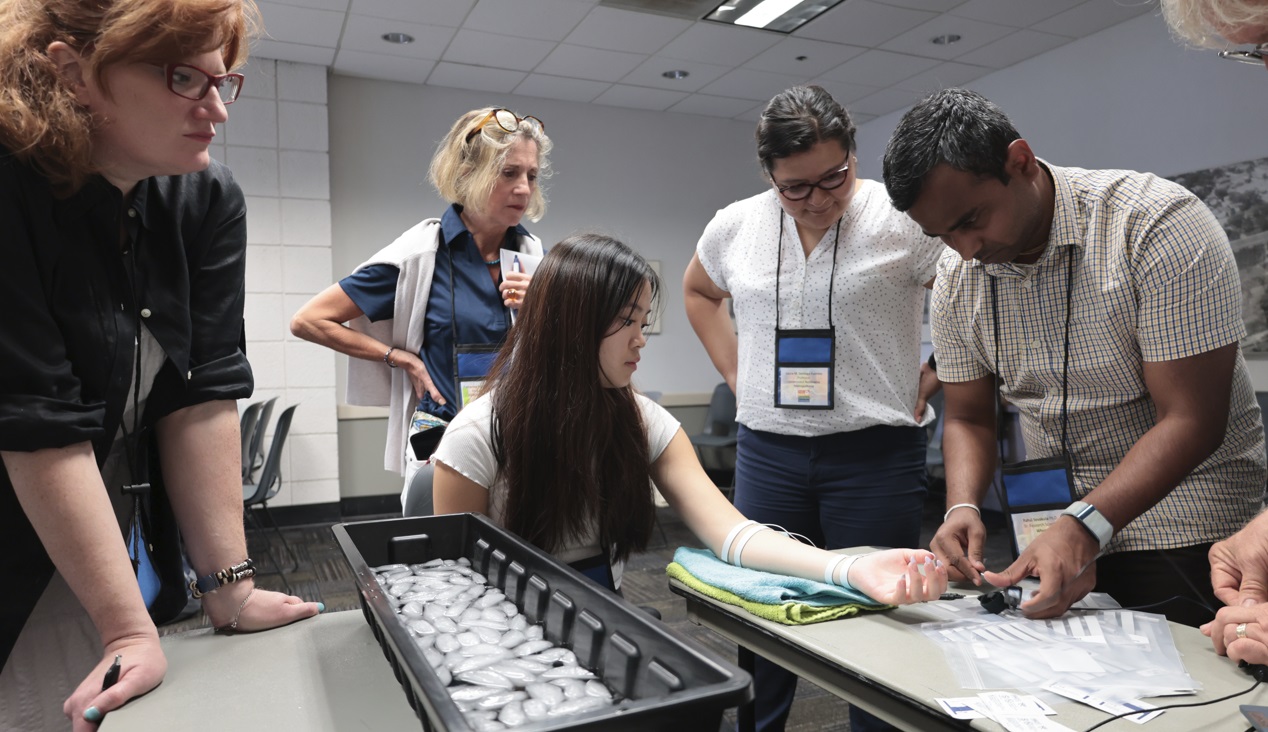
They hailed from across the globe, from Arkansas to Zanzibar and all points between. Students, teachers, researchers, and innovators gathered on the campus of UC Santa Barbara, overlooking the Pacific Ocean. They came to learn, share, and explore breakthroughs and best practices in physiology and related fields. 4T|Phys kicked off July 17 under clear California summer skies, an idyllic setting for discovering the Tools, Trends, Techniques, and Technology driving physiology education and exploration.

4T|Phys 2023 was the third installment in BIOPAC’s highly successful series of biennial industry conferences that began in 2017. After a two-year hiatus during the global Covid-19 pandemic, 4T came roaring back this summer with more workshops, more tracks, more sponsors, more presenters, and more countries represented than ever before. Over the course of three days, nearly 100 attendees participated in hands-on sessions with some of the top minds in physiology research, experiencing the latest tools for gathering and analyzing data.

BIOPAC’s in-house experts were joined by an array of authorities from the fields of physiology, psychology, imaging (fNIRS and fMRI), neuroscience, cardiovascular research, eye tracking, virtual reality (VR), biology, and engineering. Participants took part in workshop schedules drawn from five separate tracks that included fundamentals, which focused on the basics of data acquisition and analysis. The research track took attendees on a deeper dive into techniques and technologies, including recording evoked potentials, screen-based eye tracking, impedance cardiography using trans-radial bioimpedance velocimetry (TREV), stimulus presentation, exercise physiology, and many others. Participants were also able to tour UC Santa Barbara’s state-of-the-art Brain Imaging Lab to see up close how BIOPAC products are being used in both functional MRI (fMRI) and TREV studies.

“It was an incredible experience that left a lasting impact on me by helping me to understand more about physiological data collection and analysis,” said Dr. Parth Trivedi, a 4T|Phys attendee. “The choice of presenters and the topics covered were truly inspiring, offering valuable knowledge and fresh perspectives that enriched my understanding.”

4T|Phys 2023 Attendee MapThis year’s conference featured several new tracks such as a dedicated VR track led by partner company WorldViz. The WorldViz team showcased virtual reality solutions in a series of workshops that introduced how VR is being integrated in physiology and psychology research, explained how to create effective and immersive VR experiments, and demonstrated their latest PRISM technology for creating fully immersive projected 3D virtual environments.

Several other partner companies introduced their technologies and products both in the 4T exhibition hall and in workshop sessions. Argus Science presented their ETVision eye-tracking glasses that synch seamlessly with AcqKnowledge. Cognixion demonstrated the Cognixion One Axon-R—Research Edition wearable BCI-AR computing platform, which is being used to help individuals with neurodegenerative conditions and severe motor impairments communicate and interact with their environments assisted by augmented reality. Seenel Imaging showed off their MedelOpt Systems for whole-head EEG with fNIRS, including new VR integration. Participants also got a chance to try on the Research Ring by Senstream, a new user-friendly wearable research ring, which allows easy recording of EDA, PPG, Temperature, and ECG signals. CNSystems showed off the company’s NIBP continuous noninvasive blood pressure (NIBP) and hemodynamics devices.

If you missed the educational and networking opportunities at 4T|Phys 2023, don’t despair. We will be back again in a few years, bigger and better than ever. Watch this space for updates on our next installment. In the meantime, see our Video Tutorials and Webinars On Demand for trainings and seminars on our products and partners.


Make sure you never miss a BIOPAC event and keep up with the latest Tools, Trends, Techniques, and Technologies by contacting your local BIOPAC sales representative.

Effort Rewarded: Tools for Measuring Motivation

A job well done may be its own reward, but the mechanisms that drive humans to produce better results are far more complex. Much research has been devoted to the study of motivation and the psychophysiological processes that drive our behavior in pursuit of a goal, whether in relationships, on the job, or in education.

The nature of motivation is controversial, and a variety of competing theories have been put forward regarding what a motivational state comprises. Some researchers have argued that motivation is driven by specific human needs, like food, safety, or respect from one’s peers. Motivation may be categorized as intrinsic, meaning an individual pursues an activity because the activity itself is enjoyable or interesting, or extrinsic, which is when the pursuit offers some external reward.

Research into motivation has drawn interest across a wide range of fields, particularly in the areas of business and education. In the realm of business, corporations are especially focused on the motivation to work, which impacts productivity. In an era where buzzwords such as “quiet quitting” and “bare minimum Mondays” have entered the lexicon, companies are keen to know what motivates workers. Similarly, educators are looking to what types of intrinsic and extrinsic motivational factors are most effective in helping students achieve their learning goals. A key metric for measuring motivation is effort, which can be measured with a range of tools readily available to researchers.

work harderA 2020 Harvard University study published by the American Psychological Association investigated the differences in how adolescents and adults translated motivation into action based on the physical effort exerted for financial reward. Participants age 12 to 23 exerted force by squeezing a BIOPAC hand dynamometer in response to visual cues indicating monetary rewards from $.05 to $.75. The dynamometer, connected to a DA100C amplifier, measured grip force, which the participant saw displayed on a screen as a vertical bar. These tools provided the research team with a means of accurately demonstrating quantitative variations in motivational behavior across various age groups in pursuit of effort-based rewards.

A similar approach was used in a 2019 study from the Netherlands that examined eating behavior by “quantifying the dynamic aspects of appetitive motivation.” As with the above example, researchers relied on a BIOPAC device to measure grip effort, in this case, a bulb transducer connected to an MP36 data acquisition system running Biopac Student Lab software. By squeezing the bulb, participants were able to self-administer sips of chocolate milk via a tube in response to stimuli while experiencing various states of hunger and satiation. The process developed for this study, termed the “Grab-to-Eat Task”, or GET, successfully showed that handgrip effort provided an accurate metric for “evaluating motivational aspects of food consumption” with implications for further research into eating behavior and weight control.

Techniques for measuring motivation are not limited to grip effort. A 2019 study published by the National Institute of Health relied on cardiovascular signals to measure the willingness of older adults to engage in cognitively demanding tasks. Blood pressure and heart rate data were collected using a CNAP monitor connected to a BIOPAC MP150 system, which provided a detailed measure of effort exerted through the participants’ physiological response to each task.

These examples just begin to touch on the ways various psychophysiological measures are being applied to the study of motivation and effort. For additional insight, view the BIOPAC webinar on tools for measuring effort and motivation in the lab.


For more information on how to gather and integrate effort-related data into your motivation study, contact your regional BIOPAC sales representative.

error: Content is protected !!