Capturing More Than Physiology: Multi-Camera and Multimodal Research

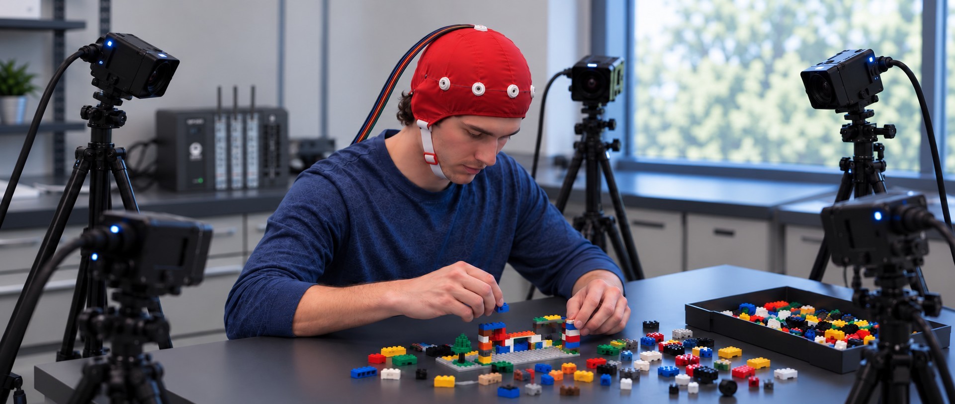
In the early 20th century, Cubist painters like Pablo Picasso and Georges Braque argued that traditional linear perspective on a subject was an illusion. Something was always hidden from the viewer, distorted, or lost altogether. Just as a Cubist painter might fracture their subject to reveal its true shape, a physiology lab uses multiple synchronized camera angles to bypass a single lens’ limitations.

The same principle applies to modern physiological and psychophysiological research. Physiological signals such as electrocardiogram (ECG), electroencephalogram (EEG), electrodermal activity (EDA), respiration, and blood flow provide valuable insight into how the body responds to stimuli, environments, and social interactions. Yet physiological data alone often leaves important questions unanswered. What was the participant doing? What was happening in the environment? Did social interaction, movement, or behavioral events coincide with a change in the recorded signal?

To answer these questions, many studies now combine physiological recordings with multiple video streams, eye tracking, audio, environmental monitoring, and other forms of media capture. This multimodal approach lets investigators examine physiological measurements alongside the events occurring around them, revealing a richer, more complete picture of human and animal behavior. As these data streams multiply, so does the need to capture, synchronize, and review them together.

Researchers at Macquarie University in Australia and collaborating institutions recently described a multimodal laboratory designed to study face-to-face communication and social interaction. The platform centered on a BIOPAC data acquisition system, amplifiers, and amplifier input module that acquired ECG, EDA, photoplethysmography (PPG), respiration, and skin temperature. The researchers combined physiological measurements with a Tobii eye tracker, wearable sensors, audio recording, motion analysis systems, and six video cameras positioned throughout the laboratory. The project showed that adding more recording sources often complicates data collection coordination, and that integrating multiple acquisition systems requires careful synchronization and workflow design. The camera system played a particularly important role. They used multiple viewing angles for behavioral coding, gesture analysis, participant observation, and synchronization verification. Rather than serving only as a record of the experiment, the video data became an important part of the analysis itself. For researchers studying communication, teamwork, education, or human-computer interaction, the work illustrates how behavioral observations can enhance interpretation of physiological measurements.

A team from the Swedish University of Agricultural Sciences (SLU) explored similar ideas outside the traditional laboratory by developing the SLU Multisensory Outdoor Laboratory (SENSOLA). The researchers used BIOPAC data acquisition and analysis systems with AcqKnowledge software and BioNomadix wireless devices to collect respiration and PPG measurements as participants moved through natural and built environments. Their platform combined those recordings with eye tracking, stimulation, video, audio, GPS data, participant feedback, and environmental measurements. One key observation was that physiological measurements become much more insightful when paired with information about a participant’s surroundings and experiences. Video proved especially valuable because it documented environmental conditions, participant behavior, and unexpected events that might otherwise be difficult to identify during later analysis. As physiological research moves beyond controlled laboratory settings, projects such as SENSOLA demonstrate the value of capturing physiological and observational data together.
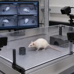
ratPreclinical research is increasingly applying the same principles. In a recent toxicology study from Japan, investigators developed the ToxQb platform around a BIOPAC data acquisition system and AcqKnowledge software with NDT to collect EEG and ECG data from laboratory rats. The physiological measurements were integrated with overhead, front, and side-view cameras, along with thermal imaging systems. The goal was to monitor behavioral and physiological changes simultaneously over extended periods. By reviewing camera footage alongside EEG and ECG recordings, researchers could relate changes in movement, posture, and activity to physiological events. The authors concluded that combining these diverse data streams provided deeper insight than any single measurement could. The study demonstrates that multimodal acquisition extends beyond human research, across a wide range of life-science applications.

Although these studies span communication research, environmental psychology, and toxicology, they point to a common trend: researchers are capturing much more than physiology. Like the artists who challenged the limitations of a single viewpoint, today’s researchers increasingly rely on multiple synchronized perspectives to reveal a more complete picture of physiology, behavior, and environment. Multiple cameras, video recordings, eye tracking, environmental sensors, and behavioral observations can provide valuable context for interpreting physiological measurements. Learn how using BIOPAC’s MultiCapture feature in AcqKnowledge 6 to integrates multi-camera video and other media into research via our webinar on the topic.


Are you planning to integrate multiple camera perspectives in your next study? Our sales team can help find the right equipment and show you how to leverage AcqKnowledge‘s MultiCapture feature to achieve the best possible results.

Under the Skin: Digging Deeper into the Physiology of Fear

From the thrill of cinematic jump scares to debilitating phobias that can turn daily life for some into a waking nightmare, our sense of fear can feel like both a blessing and a curse. Fear is one of the most fundamental human emotions, serving as a necessary safeguard for recognizing and responding to danger. Despite its primal origins, the mechanisms of fear remain complex. For decades, psychophysiologists have studied fear using measures such as the startle reflex and skin conductance, building a deep understanding of how the body reacts to threats. Today, however, researchers are beginning to look beyond these traditional markers. Technologies such as virtual reality (VR) expand how researchers can stimulate the fear response. At the same time, a broad range of signals and techniques can measure not only the body’s autonomic responses, but also how people allocate attention to threats and how the brain processes fear-related information.

One reason fear remains such a compelling topic is that it sits at the intersection of cognition, emotion, memory, and physiology. Fear responses can be observed through changes in sweating, heart activity, breathing patterns, pupil size, neural activity, and even social behavior. As researchers seek to gain a better understanding of anxiety disorders, phobias, post-traumatic stress disorder (PTSD), and related conditions, they increasingly rely on multiple synchronized signals to capture a more complete picture of how fear is learned, remembered, and generalized.

Among the most widely used measures is electrodermal activity (EDA), often recorded as skin conductance response (SCR), which reflects changes in sympathetic nervous system arousal. More recently, eye tracking and pupillometry have emerged as valuable tools for measuring vigilance and attention, while functional near-infrared spectroscopy (fNIRS) has enabled monitoring of changes in cortical activity during fear-related tasks in more naturalistic settings. Together, these approaches help researchers move beyond viewing fear as a single physiological reaction and toward understanding it as a coordinated brain-body process.

A team of researchers at South China Normal University in China explored how the duration of memory retrieval influences the reconsolidation and extinction of fear memories. Using a four-day fear-conditioning paradigm, participants were conditioned to associate visual cues with mild electric shocks. Simultaneously, skin conductance responses were recorded using an MP36 running Biopac Student Lab System. The experiment was designed and implemented with E-Prime stimulus presentation software. The team found that briefly reactivating a fear memory before training participants to view the stimulus as safe was most effective at preventing the fear from returning. Longer periods of memory reactivation produced weaker effects. The study highlights how electrodermal activity can reveal subtle changes in fear-memory processing and may ultimately contribute to improved behavioral interventions for anxiety-related disorders.

fingers and faceA study conducted at the University of Würzburg in Germany examined how anxiety sensitivity influences attention and memory during threat and safety learning. Participants learned associations between neutral faces and aversive outcomes while researchers recorded both electroencephalography (EEG) and pupil diameter. Pupillometry data were acquired using a Tobii eye tracker, allowing researchers to measure moment-to-moment changes in attention. Individuals with higher anxiety sensitivity showed larger pupil responses and enhanced neural markers of attention when viewing both threatening and safe cues, reflecting a generalized pattern of hypervigilance. Interestingly, this heightened attentional engagement did not translate into better memory performance. The findings suggest that pupillometry provides a valuable window into how anxious individuals process potential threats and may help researchers better understand mechanisms underlying pathological anxiety.

Another perspective on fear comes from research at Sichuan Normal University in China, where investigators look at how trait anxiety influences observational fear learning. Unlike traditional conditioning studies, participants learned about danger by watching another person experience threat-related events. Researchers measured skin conductance using a BioNomadix wireless EDA plus data acquisition and analysis system and monitored prefrontal brain activity using fNIRS. The study found that individuals with high trait anxiety exhibited stronger physiological fear responses and increased medial prefrontal cortex activation, even when observing threats indirectly. The work demonstrates that fear can be acquired socially and that neural measures such as fNIRS can reveal aspects of fear processing that are not readily captured by autonomic measures alone. These findings may have important implications for understanding how fear and anxiety spread through social environments and how vulnerability to anxiety disorders develops.

These studies illustrate how fear research leverages multimodal approaches to incorporate multiple physiological perspectives. Skin conductance continues to provide valuable information about autonomic arousal, while pupillometry reveals how attention is divided between threat and safety cues, and fNIRS offers insight into the neural processes involved in fear learning and regulation. As multimodal research platforms become increasingly accessible, researchers are gaining unprecedented insight into how fear is acquired, maintained, and modified. By combining signals from the body, the eyes, and the brain, modern psychophysiology is shining a light on the shadowy labyrinth that is one of humanity’s most powerful and important emotions. To learn more about how to construct a fear stimulation lab for your next fear-conditioning research project, check out our webinar on the subject.


If you are planning a multimodal fear conditioning study, our staff can help identify the right equipment and software for stimulus presentation and data recording and analysis.


Projection VR: Virtual Rooms Turn Science Fiction into Reality

Fans of Star Trek have long marveled at the technological innovations spawned from the imaginations of its creators—from warp drives that bend space to transporters beaming individuals from point A to B. Few of these speculative innovations inspired more wonder than the “holodeck,” a room that creates a simulated sensory experience nearly indistinguishable from reality, where crew members can train, study, or engage in recreation within the space’s artificial environments.

The border between science fiction and fact can be fluid, as proven by the proliferation of virtual reality (VR) technology in recent years, providing an immersive experience like that promised by the fictional holodeck. When thinking about VR in the modern sense, one usually envisions a person fitted with a head-mounted display (HMD) that delivers visual stimuli directly to the eyes, with movement and views controlled by directional sensors in the HMD and hand controllers.

While this can certainly deliver an immersive experience, HMD systems have their limitations. HMDs and controllers are often tethered via cables to the high-end computers needed to generate realistic imagery. This can limit the user’s ability to move and interact naturally in a virtual environment. A variation is something much closer to the holodeck of science fiction—a room in which a virtual environment is projected onto walls rather than displayed on a headset screen. This is the world of projection VR.

Virtual reality innovators like WorldViz are taking projection VR to the next level by creating highly-customizable virtual environments that can be scaled and adapted to a variety of tasks. One application in which VR projection rooms are being used is to train emergency medical technicians (EMT) and other first responders in realistic environments, such as on a virtual roadside. Trainees can move about performing first aid as cars seem to rush by, safe from real traffic hazards.

Outside of training and educational applications, projection VR and VR rooms have proven to be affective in a variety of research studies. These technologies have been used in machine learning and behavioral studies focusing on children with autism spectrum disorders (ASD). In such applications, HMD systems may cause motion sickness or discomfort for the participants with ASD. Projection rooms, however result in no significant negative impacts on the participants’ comfort while still providing an immersive experience.

VR Driver StudyAs mentioned, projection VR experiences allow for additional technologies to be introduced seamlessly into the virtual experience. A 2020 study of driver gaze habits integrated driving environments created in Vizard 5.0 and projected on a screen with eye tracking to determine how drivers process visual information behind the wheel. Participants were able to interact with real driving controls while their “vehicle” moved through the virtual environment as they focused on visual cues.

VR projection rooms are no longer restricted to fictional flights of fancy. Their real world use is limited only by the imaginations of those applying these potent tools in the classroom and research lab.


For more information on how projection VR can benefit research, education, and training, join a webinar hosted by BIOPAC as we explore this virtual frontier where science fiction becomes science reality.

T4 Conference Wrap-up

August is a month of barbecues, baseball, and last-minute vacations against the backdrop of a fading summer, and the eighth month of 2019 proved to be an equally exciting time at BIOPAC, with something of a pleasant vacation thrown in. On August 12-14, BIOPAC hosted its 2nd Biennial “Tools, Trends, Techniques, and Technology” Human Physiology Conference, known as “T4” to our friends and partners around the world. This three-day event, first held in 2017, featured lively, informative sessions hosted by BIOPAC staff, partners, noted academics, and industry experts, and covered a wide range of physiological techniques and disciplines. Over 150 attendees joined BIOPAC at the 2019 T4 Conference staged once again at the beautiful University of California at Santa Barbara campus. Participants and instructors winged in from China, Australia, Germany, Sweden, Italy, Spain, U.K., Ireland, and Egypt, as well as from wide regions of the U.S and Canada to soak up the idyllic surroundings of this ocean-side campus.

The focus of BIOPAC’s T4 Conference was to provide a fun, relaxed environment while hosting nearly 40 hands-on, human physiology training workshops chaired by over 20 distinguished instructors from around the globe. One of the many conference highlights was the “Mindfulness, Meditation, and Measurement: Mindfulness Exercises in Physiological Monitoring” session, hosted by Dr. Allan Weiss of the University of Southern California and  BIOPAC Co-founder and Director of R & D Alan Macy. These sessions, conducted in two parts over a two day period were booked beyond capacity, even though the early morning fog hadn’t yet lifted from the campus skyline.

The varied additional sessions covered a range from the basics, “Setting Up Your Lab, Getting Great Data and Troubleshooting,” (hosted by Dr. Terry Blumenthal of Wake Forest University, and BIOPAC’s Alan Macy and Tim Cook), and “Creating Your Own Lessons Using BSL PRO,” (hosted by Dr. Taffeta Elliot of the New Mexico Institute of Mining and Technology), to exciting new technologies such as “Integrating Screen-based Eye Trackers with AcqKnowledge,” with Keith Jackson of BIOPAC partner company EyeTech, BIOPAC CEO Frazer Findlay, and BIOPAC Engineering Manager Ed Peterlin. Additional wide-ranging physiological disciplines were also covered, including MRI techniques, stimulation, and exercise physiology. All sessions were conducted using BIOPAC hardware and software systems, as well as platforms provided by BIOPAC partners such as Cedrus, fNIR, WorldViz, Psychology Software Tools, and CNSystems. At the end of each day, it was time to relax and unwind with a beverage of choice, a a walk along the beach, and some good conversation. Each evening featured excellent meals and camaraderie in the relaxed yet elegant environment of some of Santa Barbara and Goleta’s most stunning surroundings. After three great days of immersive sessions, it came time for everyone to part ways, having forged new collaborative partnerships and an enhanced knowledge about the full potential of BIOPAC and BIOPAC partner products.

You don’t have to wait for T4 2021 to learn more about life science data—watch Fundamentals of Physiological Data Recording now!

For a closer look at T4, and a preview of what is sure to be an exciting 2021 event, we invite you to visit our T4 Human Physiology Conference page.

Looking for additional resources, tips, tricks, and expert advice on BIOPAC and BIOPAC Partner products? Please also visit our Webinars and Video Tutorial pages.


BIOPAC offers a wide array of wired and wireless equipment that can be used in your research. To find more information on solutions for recording and analyzing signals such as ECG, heart rate, respiration and more using any platforms mentioned in this blog post, you can visit the individual application pages on the BIOPAC website.

Understanding Stimulus Presentation Studies

Stimulus presentation studies are an important aspect of modern research that record and organize a participant’s reactions and physiological data to various inputs. These studies are commonly used in neuromarketing or psychophysiological experiments. First off, what is stimulus exactly? Put simply, a stimulus is a sensory input delivered to a participant with the purpose of eliciting a particular response. These stimuli can consist of images, words, lights, sounds, virtual reality, scent, or even mild electrical impulses.

The idea is to present varying types of stimulus to a participant at expected or unexpected intervals and record his/her response. For example, a collection of images might be presented (one at a time) ranging from pleasant (say an impressive vista) to neutral (say a street scene) to unpleasant (possibly a striking snake or other “fight or flight” image). Programmable stimulus systems (such as E-Prime or SuperLab) to present the stimuli, and can be used with a Chronos or M-Pod response keypad that the participant presses, each key representing a different response (e.g., pleasant image = press Key 1, unpleasant = press Key 2, etc.). A physiological signal commonly gathered from the participant is electrodermal activity (EDA), which measures the perspiration generated in a participant’s palms or fingertips. This “eccrine activity” has a strong correlation with emotional responses. For studies involving visual stimuli, a camera may be used to record the participant’s facial expressions. The expressions are simultaneously recorded and categorized using software such as FaceReader.

Stimulus experiments are generally set up using the hardware and software of the stimulus system and the resulting physiological data and stimulus markers recorded using a data acquisition system, such as BIOPAC’s MP160 with STP100C, MP36, or Smart Center Systems. These systems include AcqKnowledge or Biopac Student Lab life science software and the MP data acquisition units that interface with the stimulus hardware.

In practice, setting up stimulus studies is easier than it sounds on paper. The parameters and stimulus types are set up via drag-and-drop-interfaces in the E-Prime or SuperLab software, and the hardware interface set up using an I/O or USB connection between the stim hardware and the computer. The stimulus and BIOPAC data acquisition software can easily reside on the same computer, as can all stimulus and BIOPAC data acquisition hardware.

Stimulus studies can range from a simple setup, such as a participant responding to a sound for reaction time studies, to more randomized studies utilizing different types of stimulus delivered at varying pre-programmed intervals.

When the study is completed, data can be analyzed in AcqKnowledge software, and the various stimulus/responses organized automatically using AcqKnowledge’s automated Stim-Response Analysis routine.

For more information about conducting Stimulus studies using E-Prime, SuperLab, and AcqKnowledge software, we invite you to view the following free webinars and video tutorials:

Stimulus Presentation and Event Mark Up with E-Prime

Stimulus Presentation and Event Mark Up with SuperLab

AcqKnowledge Stim Synchronization and Analysis

Focus Areas – Automatically Create Using Define Between Events

BIOPAC offers a wide array of wired and wireless equipment that can be used in your research. To find more information on solutions for recording and analyzing signals such as ECG, heart rate, respiration and more using any platforms mentioned in this blog post, you can visit the individual application pages on the BIOPAC website.

Electrical Stimulators for Physiological Research: Compliance

All stimulators incorporate a fundamental characteristic known as “compliance.” Compliance comes in two flavors, namely voltage compliance and current compliance. Voltage stimulators emulate a voltage source, up to a certain current compliance. Current stimulators emulate a current source,

By Alan Macy, BIOPAC Systems, Inc. 

All stimulators incorporate a fundamental characteristic known as “compliance.” Compliance comes in two flavors, namely voltage compliance and current compliance. Voltage stimulators emulate a voltage source, up to a certain current compliance. Current stimulators emulate a current source, up to a certain voltage compliance. The stimulator compliance limit is important information. Here are examples:

Current Compliance

A voltage stimulator is being used to establish a field stimulation in a tissue bath. The voltage pulse is 100 volts, with a width of 1 ms. The bath establishes a 1000 ohm impedance between stimulation electrodes. Therefore, 100 volts/1000 ohms is 100 ma. In this situation, the voltage stimulator must be able to provide a current compliance up to 100 ma for at least 1 ms. If the stimulator’s current compliance is limited to 50 ma, then the output voltage will be limited to 50 ma * 1000 ohms or 50 volts, even if the stimulator is being instructed to output 100 volts.

Voltage stimulators commonly operate with the help of high voltage DC-DC converters and storage capacitors to provide the needed compliance current. If the storage capacitors can’t store enough charge or are not sufficiently supplied, then voltage stimulators may exhibit a quality known as voltage droop. Voltage droop is the quality when the stimulator output voltage can’t be maintained over a specific time interval, due to lack of sufficient current compliance. If the above stimulator can only be current compliant to 100 ma for 0.5 ms, then the output voltage pulse will be 100 volts for 0.5 ms and then will decay in direct proportion to the current compliance limit.

Voltage Compliance 

A current stimulator is being used for neuromuscular stimulation between two small electrodes. The current pulse is 10 ma, with a width of 1 ms and there is a 10,000 ohm impedance between stimulation electrodes. Therefore, 10 ma * 10,000 ohms is 100 volts. In this situation, the current stimulator must be able to provide a voltage compliance up to 100 volts for at least 1 ms. If the stimulator’s voltage compliance is limited to 50 volts, then the output voltage will be limited to 50 volts / 10,000 ohms or 5 ma, even if the stimulator is being instructed to output 10 ma.

Current stimulators commonly operate with the help of high voltage DC-DC converters and storage capacitors to provide the needed compliance voltage. If the storage capacitors can’t store enough charge or are not sufficiently supplied, then current stimulators may exhibit a quality known as current droop. Current droop is the quality when the stimulator output current can’t be maintained over a specific time interval, due to lack of sufficient voltage compliance. If the above stimulator can only be voltage compliant to 100 volts for 0.5 ms, then the current pulse will be 10 ma for 0.5 ms and then will decay in direct proportion to the voltage compliance limit.

Example

In the case of transcranial Direct Current Stimulation (tDCS), a constant current of up to 2 ma (Is) may be applied through the cranium. Assuming a combined series electrode / skin junction resistance of 20 Kohms, a cranium resistance of 100 ohms and other loop series resistances considered negligible, then the total stimulation loop impedance (Zs) is 20.1 Kohms. Accordingly, in this situation, the voltage compliance (Vs) of the tDCS current stimulator must be at least:

Is * Zs = Vs
0.002 amp * 20,100 ohms = 40.2 volts

Given the variable nature of the combined series resistances in the stimulation loop, it’s generally helpful to double the expected compliance voltage requirement of the stimulator. Accordingly, in this case, a stimulator compliance voltage of 80 volts or higher is recommended. Because the series resistance in the loop is largely a function of the electrode / skin interface junctions, the choice of electrode type and contact area has a great effect on the voltage compliance requirements of the current stimulator. Other factors contributing to the voltage compliance requirement are the equilibrium potentials and over-potentials associated with the specified electrode / electrolyte / skin junctions. Furthermore, if high resistance electrode leads are used, the potential across the leads may become material.


BIOPAC Systems, Inc. provides life science researchers and educators with data acquisition and analysis systems that inspire people and enable greater discovery about life. BIOPAC lets you measure physiology anywhere, with innovative, compatible wireless, wearable solutions that can be used by anyone for meaningful discovery. We make high-quality scientific tools for physiology measurement and interpretation with superior compatibility and world-class customer service and support. Visit us at www.biopac.com.

Electrical Stimulators for Physiological Research

Highly-controlled electrical stimulation capability can provide a unique tool for the life science researcher to discern sensory function and associated physiological responses in living subjects. There are two types of electrical stimulators, voltage and current.

By Alan Macy, BIOPAC Systems, Inc.

Any physiological measurement is concerned with the subject of stimulus and response, because all physiological processes are subject to change with applied stimuli. The processes of life encapsulate the idea that environmental change precipitates sensory activity that results in stimulation. Because sensory activity is mediated by the flow of ions in the nervous system, highly controlled electrical stimulation capability can provide a unique tool for the life science researcher to discern sensory function and associated physiological responses in living subjects.

Computer-based systems can be used to define very exacting electrical stimuli and those signals can be directed to a subject via electrodes. The level of stimuli can be extremely wide and the shape of the stimuli can be finely controlled. Signals can be introduced directly into nerves or muscle fibers to evoke a specific response.

There are two types of electrical stimulators, voltage and current. A voltage stimulator behaves like a voltage source, in that it outputs a variable voltage with low output impedance. A current stimulator behaves like a current source, in that it outputs a variable current with high output impedance.

The primary physical expression that characterizes the behavior of electrical stimulators is:

Vs = Is * Zs

where:

Vs – stimulation voltage from stimulator
Is – stimulation current from stimulator
Zs – impedance of stimulation loop

Zs is the collective impedance of the complete stimulation loop. From a simplified view, Zs consists of the series combination of two electrode / skin junction impedances and the stimulated tissue volume. Zs has resistive and capacitive components, so the capacitive components charge in accordance to the expression:

Ics = Cs * (dVcs/dt)

where:

Ics – stimulation current through Cs
Cs – capacitive component associated with Zs
dVcs/dT – change in stimulus voltage, across Cs, as a function of time

Accordingly, electrical stimulation is associated with the charging of the stimulation loop capacitance. If fast electrical stimulation is required, then the stimulator should have high current level drive capability. For a given Cs, the speed of charging (dVcs/dt) will be higher (faster) as Ics is increased.

There are two major sub-impedances in the stimulation loop that define the portion of the stimulator output voltage (Vs) that is directly across the tissue volume (Vt):

Ze – electrode to site impedance (total of both sites)

Zt – stimulated tissue volume impedance

Vs = Is * Zs

Vs = Is * (Ze + Zt)

Vt = Vs * (Zt / (Ze + Zt))

Because of this relationship, the value of Vt might not be reliably specified, given a certain Vs.For any given tissue volume, electrical stimulation can be specified in terms of voltage across the tissue volume (Vt) or current through the tissue volume (It). In practice, because two terminal stimulation is simple to apply, current stimulation is often used because the current through the tissue volume (It) is the same as the sourced stimulation current (Is). However, if the stimulation current (It) is specified then Vt may be indeterminate. Sometimes, both (It) and (Vt) are required to be known, such as with bioimpedance measurements. In this case, voltage monitoring electrodes can be placed on the tissue volume so both (It) and (Vt) can be identified.

Compliance, an essential characteristic of electrical stimulators, is addressed in Electrical Stimulators for Physiological Research: Compliance.


BIOPAC Systems, Inc. provides life science researchers and educators with data acquisition and analysis systems that inspire people and enable greater discovery about life. BIOPAC lets you measure physiology anywhere, with innovative, compatible wireless, wearable solutions that can be used by anyone for meaningful discovery. We make high-quality scientific tools for physiology measurement and interpretation with superior compatibility and world-class customer service and support. Visit us at www.biopac.com.

error: Content is protected !!