Skin Deep: Using EDA to Measure Affective Response

We say something “gets under our skin” when it elicits an unpleasant or irritating emotional response. This connection between our skin and emotion runs deeper than a simple idiomatic expression, however. Along with performing several critical functions—protective, regulatory, and sensory—our skin provides a window into our physiological and emotional states as we respond to a range of stimuli. Electrodermal Activity (EDA) is the mechanism providing these insights.

EDA, also known as galvanic skin response (GSR), measures eccrine activity resulting from the sympathetic nervous system’s response to environmental stimulation. As a function of the autonomic nervous system, it is an involuntary response to a range of sensory inputs, including touch, sight, sound, odor, and taste. EDA regulates the rate of sweat produced by sweat glands in the extremities, particularly in the fingertips, palms, and soles of the feet.

EDA increases in conjunction with the level of stress or nervousness experienced by an individual. For this reason, it provides researchers with a valuable means of measuring affective response to external stimuli. As we explored in a previous post, affect is the measurable physiological expression of emotion. While there are numerous physiological signals that researchers can employ to measure affect, EDA in particular functions as an indicator of arousal. For this reason, EDA has become one of the most relied upon signals employed by researchers exploring psychophysiological phenomena.

Violence StudyA recent Slovakian study published in Adaptive Human Behavior and Physiology illustrates how EDA is commonly employed in such research. Researchers studying the influence of gender differences on reaction to violent imagery used skin conductivity response (SCR), the mechanism behind EDA, to measure levels of arousal when participants were shown images of war and sports. The goal of the study was to test the “male warrior hypothesis” (MWH). The theory suggests that men have evolved psychological mechanisms to form aggressive alliances with the goal of acquiring or protecting reproductive resources in war and that team sports stimulate that same impulse. EDA was measured using BIOPAC electrodes and leads feeding data to an MP36 data acquisition unit. While the study found that visual perception of team sports and war imagery stimulated different levels of excitement as measured by EDA in men and women, it was inconclusive in showing that sports provide a substitute for war in the sample of participants.

A study conducted at the University of Ottawa and published in the Journal of Behavior Therapy and Experimental Psychiatry used EDA to examine whether response-focused emotion regulation (RF-ER) strategies provided an effective way to control evoked emotions—specifically, does smiling make one feel better in times of heightened stress? EDA data was gathered from participants using a BIOPAC skin conductance transducer and amplifier connected to a BIOPAC data acquisition unit while participants were shown images designed to evoke both arousal and negative responses. Participants were asked to either suppress their emotional response to images or employ expressive dissonance (e.g., smile when disturbed). The results of the study shed doubt on previously held assumptions that RF-ER strategies were effective in controlling emotion.

These are just a sampling of the ways in which EDA is being used in psychophysiological research. For additional information on the background, tools, and techniques for using EDA in your studies, visit our EDA Resource Directory, read our EDA Introductory Guide. and watch Electrodermal Activity: Intro to Recording and Analysis or any of our EDA webinars.


For additional information on how BIOPAC can assist you with your research using EDA and a range of other physiological signals, contact your local sales representative.

Balancing Act: HRV Provides Critical Marker for Psychophysiological Health

Two college wrestlers square off on the mat awaiting the signal that will begin their match. To the casual observer, the outcome seems impossible to predict. Their coach, however, knows one competitor has a clear advantage—a heart rate variability level (HRV) that is higher than his opponent’s. Examining HRV data provides more than insight into athletic performance, it offers a window into a broad spectrum of functions that influence psychophysiological health.

HRV balanceIn simple terms, HRV is the natural variation in the intervals between each heartbeat over time. Unlike a ticking clock, our hearts do not beat at a constant rate. As such, variations in heart rate can provide a wide range of valuable information on our bodily systems. This is because HRV represents a balancing act between our sympathetic and parasympathetic nervous systems, which together make up the autonomic nervous system. As we have discussed in previous posts, the autonomic nervous system controls nearly all the body’s involuntary functions.

The sympathetic nervous system is activated in times of stress or danger (i.e., the fight-or-flight response), during which our bodies sweat, our heart rates increase, and the interval between beats decreases. Conversely, the parasympathetic nervous system acts as a sort of switch, shutting down the sympathetic nervous system and activating a relaxed state (i.e., rest and digest). To do this, it releases acetylcholine, a neurotransmitter that makes the heart beat slower and at greater intervals.

When these two systems are in balance, they produce higher HRV through a more variable heart rate. Greater variability has been associated with the body’s ability to switch between these systems. Thus, HRV is used as a health marker, such that high HRV is associated with better cardiovascular health, mental focus, emotional health, stress management, and ability to recover from exercise, while low HRV is associated with inflammation, chronic fatigue, increased stress, depression, and chronic pain.

HRV has also been connected to willpower and is a predictor of persistence and self-control. HRV increases during periods of self-regulation sometimes called the “pause and plan response.” As such, HRV has been particularly useful in research into the mechanism associated with addiction. A 2019 study used HRV to explore how chronic pain patients treated with opioids could regulate drug cravings and depression through dispositional mindfulness. Electrocardiogram (ECG) data were sampled with a BIOPAC data acquisition unit while HRV was calculated with AcqKnowledge software.HRV chart

Because HRV plays such a critical role in physical and psychological health, several studies have investigated how to self-regulate HRV, including examining how it is affected by posture. A 2020 study used a  2-channel BSL data acquisition unit to gather ECG data to calculate HRV for a variety of bodily positions to determine their impact.

A similar study examined the link between HRV and resilience, i.e., the individual’s ability to return to stable physical and psychological equilibrium after experiencing stress. HRV calculations were made from data gathered via a BIOPAC ECG100C ECG amplifier, RSP100C respiratory amplifier, and EDA/GSR amplifier. The study identified possible trends connecting resilience factors and HRV levels during specific stress-inducing simulations.

Calculating HRV with BIOPAC hardware and software solutions provides researchers with a myriad of psychophysiological insights for their lab work. Learn more about HRV from our Knowledge Base entry, as well as application notes on data preparation for analysis and statistical and geometric measures.


For additional information on how BIOPAC can assist you with a wide range of research and educational applications, contact your local sales representative.


Silent Partner: Autonomic Function Testing in Research

Like the inner workings of an intricate machine, most of our bodily functions operate with little or no conscious effort. That task falls to the autonomic nervous system, a sort of control system that regulates a wide range of processes, including digestion, heart rate, respiration, urination, and sexual arousal, to name a few. The autonomic nervous system also controls physiological responses to stimuli—the fight-or-flight response, and critical reflex actions like vomiting, swallowing, coughing, and sneezing.

The ability to effectively and accurately test autonomic function allows clinicians to diagnose and treat a range of ailments. Such tests can non-invasively evaluate the severity and distribution of autonomic failure, for example. Beyond these clinical applications, autonomic function testing provides valuable insight into how the body regulates and manages various critical functions and how the autonomic nervous system affects our physical and psychological health.

adolescent depressionAutonomic function testing has been employed to examine the impact childhood trauma has on cognitive function as it relates to adolescent depression. A 2020 study published in Development and Psychopathology looked at whether economic deprivation and threat influenced cognitive processes and autonomic nervous system function connected to the severity of depression. Researchers measured study participants’ electrodermal activity (EDA), respiratory sinus arrhythmia (RSA), and electrocardiogram (ECG) data via an MP150 system. EDA was gathered using a BioNomadix wireless BN-PPGED amplifier set, while ECG and respiration (RSP) recordings were obtained with a BioNomadix wireless BN-RSPEC amplifier set. The study’s findings supported the assumption that a history of trauma and economic deprivation can lead to risk factors for youth depression.

Continuing with the theme of applying autonomic function testing to measure the effects of trauma, a 2021 study publish in Neurobiology of Stress examines autonomic function related to sex differences among individuals with posttraumatic stress disorder (PTSD). Skin conductance, blood pressure (BP), heart rate (HR), and high-frequency heart rate variability (HF-HRV) were recorded with an MP150 data acquisition system among male and female emergency personnel following trauma exposure. The results, which showed notable sex differences in autonomic functioning among trauma-exposed individuals, have implications for predicting cardiovascular risk in those with PTSD.

These are just a few examples of how autonomic function testing is being used outside of the clinical setting to shed new light on several research areas. Check out Part 1 and Part 2 of our webinars demonstrating how to use BIOPAC equipment and software in autonomic function research.


For additional information on how BIOPAC can assist you with a wide range of research and educational applications, contact your local sales representative.

Guilt Trips: Taking Measure of Our Moral Compass

“So full of artless jealousy is guilt, It spills itself in fearing to be spilt”
—Gertrude, William Shakespeare’s “Hamlet”

In Act 4 of Shakespeare’s famous tragedy, Hamlet’s mother Gertrude points out that the harder one tries to conceal guilt, the easier it is to expose. Little does she realize that she may have hit upon how future researchers approach the study of guilt in the lab. For practical purposes, it is a primary motivator in moral action and impacts how an individual responds to certain stimuli.

Psychologists have developed a myriad of tests to gauge guilt in a clinical setting such as the Test of Self-Conscious Affect (TOSCA), a multiple-choice test that tracks responses to various moral scenarios. While such techniques have been useful in linking guilt with conditions such as depression and anxiety, they have also shown their limitations in providing a deeper understanding of guilt’s influence on behavior, development, and psychological health.

In a recent BIOPAC webinar, UCSB professor Dr. Hongbo Yu addressed these limitations. Scenario-based tasks, he points out, are non-committal in examining the causes of guilt and do a poor job of measuring behavioral tendencies linked to guilt. Interaction-based research methods, however, allow participants to experience emotions in real-time, allow direct control of the causes of guilt in the lab, and allow for direct measurement of responses to stimuli.

As discussed in prior posts on affect, the body’s physiological manifestation of emotion, there is a range of methods and physiological signals available to researchers for measuring affect and gaining a better understanding of emotional states like guilt. Dr. Yu utilized eye-tracking, electrodermal activity (EDA), and functional magnetic resonance imaging (fMRI) in his examples. These are just a few of the signals currently being used in the lab to measure guilt.

One of the practical applications of guilt research is detecting when a subject is concealing the truth, as one would with a polygraph test. A recent study conducted by researchers at the University of Wisconsin-Madison’s Department of Psychology sought to gauge deception among research participants taking the Concealed Information Test (CIT). The goal was to improve upon traditional polygraph test signals such as heart rate and respiratory rate. Respiration data was gathered using a respiration transducer belt while ECG signals were collected with BIOPAC electrodes. In addition, researchers gathered EDA data using a transducer and finger electrodes. The study found that thGuilty Girle EDA signal was significantly more effective in accurately measuring physiological responses during the test procedure.

A 2018 study by researchers at Liverpool Hope University looked at the link between guilt and aggression in children by examining activity in the autonomic nervous system during so-called “bad behavior.” The study measured respiratory sinus arrhythmia (RSA) and skin conductance in children 4–8 years old while they imagined engaging in antisocial activities. EDA and ECG signals were gathered via wireless amplifiers and fed to an MP150 data acquisition unit (superseded by the MP160) running AcqKnowledge software. The study found a linkage between physiological responses to aggression and guilt that may provide a better understanding of the mechanisms behind guilt and related behavior in early and middle childhood.

These are just a few examples of how physiological signals can be applied to measuring guilt in research. BIOPAC offers a wide range of data acquisition, stimulus presentation, and VR options suitable for creating custom guilt research environments in the lab.


For additional information, contact your BIOPAC representative to learn more about the range of signals that can be utilized to gather data on guilt and other emotional states in the lab.

Measuring Moods: Signals for Insight into Emotion

“Do you ever look at someone and wonder what is going on inside their head?”

Joy, an anthropomorphized manifestation of happiness, delivers this line at the opening of Pixar’s 2015 animated hit “Inside Out,” a film that explores the emotional struggles of its young protagonist. She could have been speaking for generations of researchers who have plumbed the depths of the human psyche for a better understanding of the mechanisms behind our feelings, moods, and emotions.

When today’s researchers talk about emotion, they typically do so in terms of affect, the underlying physiological and psychological experience of feelings and emotions as a reaction to stimuli. Affect and emotion are not the same thing, per se, but are fundamentally connected. While the feelings we describe as fear, joy, love, hate, etc. remain abstract, affect is more concrete. BIOPAC co-founder and head of R&D describes it as “the measurable quality or manifestation of emotion in the human body.”

This manifestation can be measured in the human nervous system’s response to stimuli. Depending on the nature of the stimuli and how this sensory data is interpreted by the central nervous system, an array of physiological responses can take place: heart rate changes, the stomach and muscle groups tense, skin temperature responds, triggering sweat glands, among others.

The signals corresponding to these reactions provide researchers with valuable data to help them better understand affect and emotion. These signals—electroencephalogram (EEG), electrodermal activity (EDA), electromyogram (EMG), heart rate, and respiratory rate, to name a few—can be recorded and measured using a range of currently available sensing technologies.

As an example, a 2019 study published through the Queensland University of Technology used respiration signals as a means of classification for human emotions such as happiness, sadness, surprise, disgust, fear, and anger. In the study, breathing signals, including airflow rate and volume, were acquired and recorded using a BIOPAC MP36 data acquisition unit while participants were shown seven film clips, each associated with a different emotion. The study concluded that the respiratory data was 80% effective in accurately classifying affective responses to the film stimuli.

A team of researchers at the Khwaja Fareed University of Engineering and Information Technology in Pakistan took their investigation into the connection between physiological signals and emotion a step further. They used a similar approach by measuring participant response to movie clips, however this time, four types of physiological signals were acquired, including ECG, EMG, respiration, and skin conductivity (SC). The data was acquired with a wide range of signal sensors and analyzed via a BIOPAC MP160 DAQ System.Signals for Emotion

Signal sensing technology also allows researchers to study emotion in multiple participants simultaneously, as was the case in a 2020 dyadic study of how a parent’s stress and emotional suppression affects their children. The study measured physiological responses of both parents and children using ECG and impedance cardiography (ICG) via a BIOPAC MP150 data acquisition unit.

The mysteries of human emotion are being unlocked through a growing understanding of the affective response to stimuli. For more information on how physiological signals can be gathered and analyzed for this purpose, see our app note on emotional state physiology as well as our webinar Emotion and Objective Physiological Measures.


Contact BIOPAC for the latest information on how we can help you measure affect in the lab.

Baby Blues: Neonatal, Infant, and Toddler Research Challenges

The first five years of a child’s life are marked by considerable change—both physiological and psychological. Young children undergo incremental development in their ability to think and solve problems, their emotions, and their linguistic abilities, to name just a few developmental changes. Over time, the rapidity and extent of this development vary by individual, even among children of the same age. For these reasons, research techniques and tools must be adopted that are suitable for the participant’s age group. In this constantly evolving segment of the physiological and psychophysiological field, new approaches and technologies are being implemented with the needs of our youngest participants in mind.

Different Needs, New Approaches

A recent article published by researchers at the University of California Irvine outlined some of the challenges in conducting research within the neonatal to toddler age groups. Chief among these was the higher levels of attrition among these groups compared to older test subjects. Much of this can be linked to the general characteristics of the test group—reduced ability to sit still for long periods of time, shorter attention span, and lack of motivation to participate. While these factors may not surprise an experienced parent or childcare provider, they can throw a wrench into any study that has not taken such challenges into account.

Tools for Toddlers

Choosing the right tools and applying them appropriately in the lab setting can not only significantly improve attrition rates, but also the quality of data. Receiving a clear, uninterrupted signal when gathering data from a research participant is challenging under the best of conditions and can seem insurmountable when it involves a potentially fussy, fidgety young child or infant. Thankfully, research tools and technologies have been developed with our youngest participants in mind. These include electrodes for infants that are not only sized appropriately but also designed not to irritate sensitive skin, as well as infant-sized EEG caps that are comfortable enough to be worn for extended periods and secure to prevent excess movement and signal loss.

Combining Technique with Technology

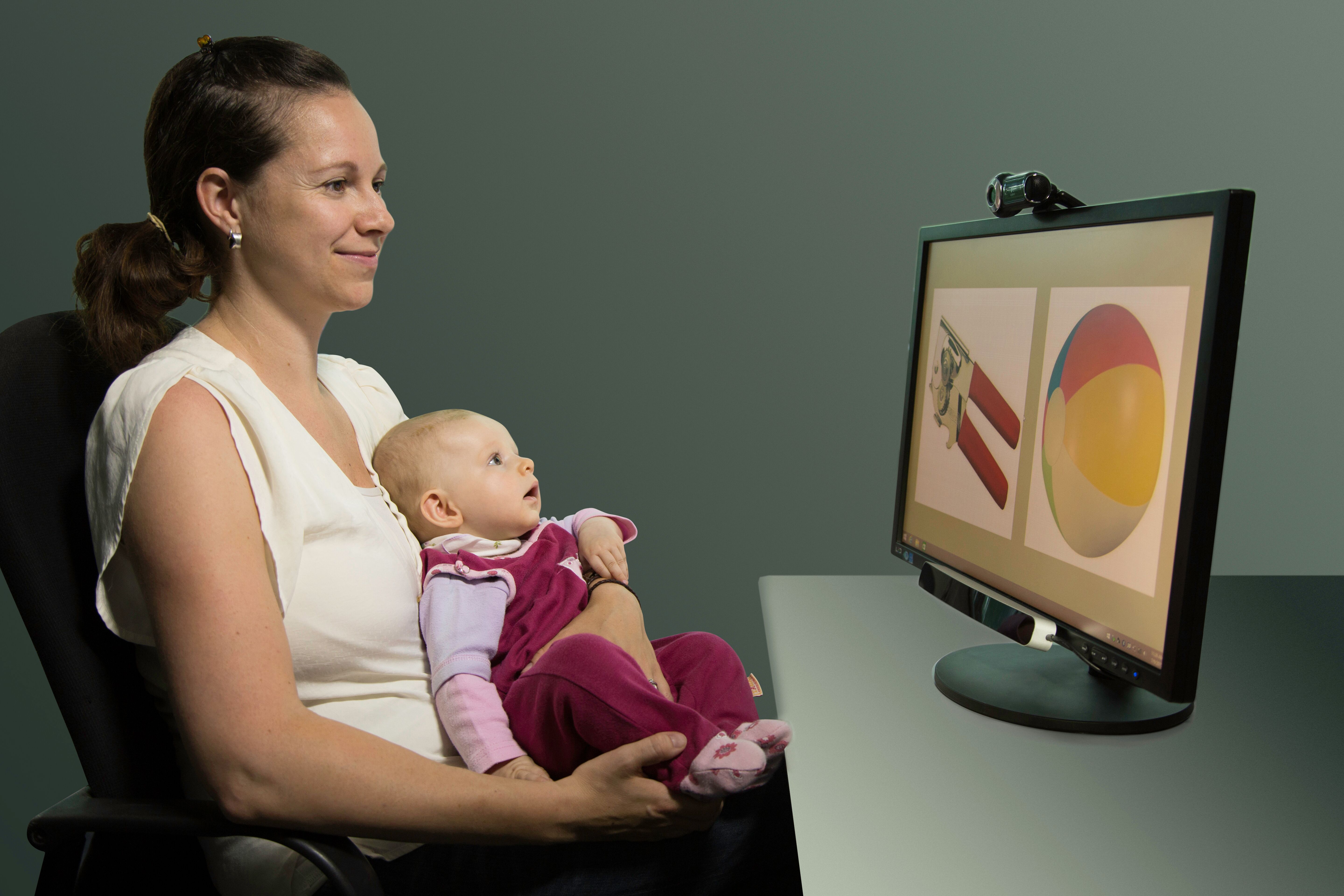
Dyadic ResearchSome researchers have found the best approach to tackling the challenges presented by these youngest participants is a combination of age-appropriate strategies and specialized equipment. A 2020 dyadic study published in the Journal of Cognitive Neuroscience examined the Effects of Maternal Singing Style on Mother-Infant Arousal and Behavior by using skin conductance (SC) levels as a measure of arousal in both the mother and infant participants. Both participants were fitted with pre-gelled electrodes attached to BioNomadix wireless EDA transmitters, which are less cumbersome than a fully wired solution. While the mother’s electrodes were attached at the neck, the infants were attached to the foot and secured in place with paper medical tape and covered with a sock. This ensured the electrodes would be held securely in place and reduce movement that could negatively impact signal quality.

We have only scratched the surface of the tools and strategies available for conducting lab research on very young participants. For additional information, watch our webinar on Research and Best Practices for Recording from Newborns, Infants, and Toddlers.


For additional information on research tools for young participants, contact your local BIOPAC sales representative.

Choosing the Right Signal: Easy as 1, 2, 3

In physiology and life sciences, researchers rely on a wide range of signals to gather data from humans and animals. These signals provide critical insight into how complex organisms function under a myriad of conditions. However, this wealth of potential data sources can also present a challenge for researchers: choosing the right signals on which to focus their data gathering. For those lacking a background in the disciplines and mechanisms behind these signals, sifting through the available methodology can be overwhelming. The best place to begin is by considering one or a combination of the “Big Three” physiological signals: ECG, EDA, and respiration.

In physiology and life sciences, researchers rely on a wide range of signals to gather data from humans and animals. These signals provide critical insight into how complex organisms function under a myriad of conditions. However, this wealth of potential data sources can also present a challenge for researchers: choosing the right signals on which to focus their data gathering. For those lacking a background in the disciplines and mechanisms behind these signals, sifting through the available methodology can be overwhelming. The best place to begin is by considering one or a combination of the “Big Three” physiological signals: ECG, EDA, and respiration.

One from the Heart

Changes in heart rate can provide information on both the physiological and psychological state of a research participant. Electrocardiography (ECG or EKG) is a biopotential measurement that provides direct metrics on electrical voltage changes in the heart. With the help of simple hardware and software, it is possible to record these voltage variations as waveforms to track how the heart responds to stimuli. Additional data can be provided by creating reoccurring reference points in the waveform, measuring the intervals between beats, and calculating heart rate variability to track changes over time or in response to stimuli.

Skin Deep

Electrodermal Activity (EDA) provides one of the best measurements of how the body responds to psychological stress and anxiety. It can be simply understood as a measure of changes in the electrical conductance of the skin in relation to the production of sweat. The physiological process of EDA comprises two components, skin conductance level (SCL) and skin conductance response (SCR). SCL is a tonic level that slowly changes over time, measuring the period for response to a stimulus to occur, from its onset to its peak. SCR reflects the phasic response to an arousing stimulus that is shown in faster changes of the skin conductance level. As an example of the application of EDA and ECG measurements in psycho-physiological research, a recent study applied both signals in measuring the cognitive states of gamers during human-computer interaction (HCI).

Breathe Easy

ArachnophobiaAs with changes in heart rate, variations in breathing cycle reveal how subjects respond to a wide range of stimuli and conditions. The signal produced by the respiratory cycle is a transduced physiological signal. As such we can apply mechanisms to create a voltage potential to measure phases of inhalation and exhalation in this cycle. This data can be represented in waveforms, providing data on respiratory rate, amplitude, and respiratory sinus arrhythmia (RSA). The latter combines respiratory data with ECG signal data to show changes in heart rate in response phases in the respiratory cycle. A 2020 machine learning study for the development of virtual reality applications combined respiration, EDA, and ECG signals to measure participant’s fear response to spiders, an example of how all three signals can be simultaneously utilized in psychophysiology research.

Choosing the right signal shouldn’t be the biggest hurdle to designing your next research project. Applying one or more of these “Big Three” can cover a wide range of data gathering goals.


For additional assistance, consult BIOPACS online resources on Life Science and Physiological Signals or contact your local BIOPAC representative.

Curriculum Roadmap: Guiding Students with HAPS Learning Goals

When planning any journey, choosing the right route can mean the difference between reaching one’s destination or becoming lost in the wilderness. It helps to know the purpose of the trip; is it merely to get from point A to point B, or is the journey itself as important as reaching the destination? What stops need to be made along the way?

These questions can also resonate for instructors as they guide students along their academic journeys, as the goal of instruction is not limited to just reaching a specific destination. It is often about what occurs along the way: experiences, obstacles, and challenges encountered that shape the student and their understanding of the subject matter.

To help students and instructors on the path to better life science education, BIOPAC created a roadmap with its curriculum for the Biopac Student Lab systems (BSL) for life science instruction. The BSL curriculum comprises more than 65 lessons that serve as carefully plotted points on a path to an in-depth and invigorating understanding of general life science disciplines.

This curriculum follows a route established by the Human Anatomy and Physiology Society (HAPS). HAPS has created a set of learning goals, guidelines, and outcomes that bring together a range of topics covered in Anatomy and Physiology I & II courses at the university level for both clinical and academic degree programs.

The advantages of following such a path are fourfold. First, students are provided with an outline of course expectations. Just as every journey should begin with a clear understanding of where one is going, knowing learning goals allows students to focus their efforts on what matters most. Furthermore, instructors are encouraged to relate learning goals to real-life applications to provide meaning in a relatable context.

Secondly, with straightforward goals in mind, the instructor will find it easier to manage course design by focusing on key elements of their curriculum. These can include the following:

  • Order of topics to cover
  • Pedagogical strategies
  • Lab experiments
  • Periodic assessments that help students stay on track

Taking this a step further, by following HAPS’s goals, guidelines, and outcomes, instructors will be better equipped to plan a comprehensive program of study. Well-defined goals allow the instructor to build a solid foundation for future courses and guide learning specific to the program’s discipline. Additionally, it can reveal redundancies, overlaps, and gaps in the degree track.Goal Planner

Lastly, just as a good cartographer’s map provides evidence of where the traveler has journeyed, carefully plotted goals, guidelines, and outcomes trace the students’ track within the academic program. This provides valuable information for accreditation while ensuring students graduate with the necessary skills for their chosen field in the sciences.


To learn more about how the BSL system and curriculum can help students and instructors get the most out of anatomy and physiology courses, contact BIOPAC today.

Projection VR: Virtual Rooms Turn Science Fiction into Reality

Fans of Star Trek have long marveled at the technological innovations spawned from the imaginations of its creators—from warp drives that bend space to transporters beaming individuals from point A to B. Few of these speculative innovations inspired more wonder than the “holodeck,” a room that creates a simulated sensory experience nearly indistinguishable from reality, where crew members can train, study, or engage in recreation within the space’s artificial environments.

The border between science fiction and fact can be fluid, as proven by the proliferation of virtual reality (VR) technology in recent years, providing an immersive experience like that promised by the fictional holodeck. When thinking about VR in the modern sense, one usually envisions a person fitted with a head-mounted display (HMD) that delivers visual stimuli directly to the eyes, with movement and views controlled by directional sensors in the HMD and hand controllers.

While this can certainly deliver an immersive experience, HMD systems have their limitations. HMDs and controllers are often tethered via cables to the high-end computers needed to generate realistic imagery. This can limit the user’s ability to move and interact naturally in a virtual environment. A variation is something much closer to the holodeck of science fiction—a room in which a virtual environment is projected onto walls rather than displayed on a headset screen. This is the world of projection VR.

Virtual reality innovators like WorldViz are taking projection VR to the next level by creating highly-customizable virtual environments that can be scaled and adapted to a variety of tasks. One application in which VR projection rooms are being used is to train emergency medical technicians (EMT) and other first responders in realistic environments, such as on a virtual roadside. Trainees can move about performing first aid as cars seem to rush by, safe from real traffic hazards.

Outside of training and educational applications, projection VR and VR rooms have proven to be affective in a variety of research studies. These technologies have been used in machine learning and behavioral studies focusing on children with autism spectrum disorders (ASD). In such applications, HMD systems may cause motion sickness or discomfort for the participants with ASD. Projection rooms, however result in no significant negative impacts on the participants’ comfort while still providing an immersive experience.

VR Driver StudyAs mentioned, projection VR experiences allow for additional technologies to be introduced seamlessly into the virtual experience. A 2020 study of driver gaze habits integrated driving environments created in Vizard 5.0 and projected on a screen with eye tracking to determine how drivers process visual information behind the wheel. Participants were able to interact with real driving controls while their “vehicle” moved through the virtual environment as they focused on visual cues.

VR projection rooms are no longer restricted to fictional flights of fancy. Their real world use is limited only by the imaginations of those applying these potent tools in the classroom and research lab.


For more information on how projection VR can benefit research, education, and training, join a webinar hosted by BIOPAC as we explore this virtual frontier where science fiction becomes science reality.

‘Tis the Season for Stress: Research Tools for Anxious Times

Holiday-related stress is as much of a seasonal tradition as family gatherings, frantic shopping forays, and over-indulgence in food and drink. Add to this potent mixture nearly two years of pandemic-induced tension and we have a volatile cocktail of emotions and anxiety awaiting us in November and December. What better time to study the causes and effects of stress?

Research has established strong links between stress and our moods and sense of well-being, not to mention overall mental and physical health. While certain types of stress during development can teach individuals to adapt to life’s many challenges, excessive and prolonged stress is shown to contribute to chronic disease and a range of psychological disorders. According to the American Psychological Association, which conducts an annual survey monitoring stress, the COVID pandemic has had an unprecedented impact on stress levels across all demographics, particularly among young people.

The pandemic is just one of many factors driving demand for new approaches to stress research. Thankfully, researchers now have access to a myriad of technologies to support any experiment design. In a recent webinar on techniques for measuring physiological changes related to stress, BIOPAC head of European Sales, Alex Dimov, outlined a range of stress-connected physiological responses that can be measured in the lab with available technologies. These include heart rate, heart rate variability (HRV), trans-radial-ulnar electrical bioimpedance velocimetry (TREV), cardiac output, blood pressure, EDA, pulse, respiration, EMG, EEG, and functional near infrared spectroscopy (fNIRS).

One of the challenges of measuring stress in a natural environment is finding the right data gathering tools to support the mobility of test participants. A 2019 study tackled this by examining a variety of techniques for measuring stress in day-to-day life using mobile phones and wearable sensors. Settings included the lab, automobile, office, school, and other daily life locations. Along with consumer products like smartphones, smartwatches, and fitness bands, researchers also employed wearable sensors, including the BioNomadix wireless PPG transmitter to measure blood volume pulse (BVP) using a photo plethysmogram (PPG) signal. Researchers found that the BIOPAC device provided a high level of accuracy (80%) when employed outside of the traditional lab environment.

Smelling RosesStress research technologies are not limited to data gathering. Stimulus presentation tools offer researchers options for both inducing and alleviating stress in the lab. A 2019 University of Wisconsin study investigating the role scents can play in reducing stress utilized EDA, heart rate, and respiratory rate to measure how rose scent reduced anxiety among students taking a difficult test. Respiratory rate was measured using a BIOPAC respiratory transducer while EDA data was collected with an EDA finger transducer. Data was collected and analyzed via the Biopac Student Lab System. While rose scent was delivered using scented lotion, it should be noted that BIOPAC offers a new modular scent delivery system that is capable of delivering up to eight scents per experiment, with more than 120 scent options.

With so many data gathering tools available, planning your next experiment should not leave you feeling on edge. Let your BIOPAC sales rep help you take the stress out of stress research.


BIOPAC offers a wide array of wired and wireless equipment that can be used in your research. To find more information on solutions for recording and analyzing signals such as ECG, heart rate, respiration, and more using any platforms mentioned in this blog post, you can visit the individual application pages on the BIOPAC website.

error: Content is protected !!