The Year in Review: Top Physiology and Neuroscience Research Stories 2022

Another calendar year has come and gone. COVID-19 remains a reality, but we continue to adapt to the ongoing pandemic and find new ways to overcome the challenges it poses at work, at school, at home, and in the lab. And speaking of research, 2022 proved to be a banner year for innovation and breakthroughs in the sciences, particularly in the physiological and neurological fields. Here is a quick round-up of some of the research stories that caught our attention.

Brain Implant Allows Locked-in Patient to Communicate

A man in his late 30s has been given the ability to communicate for the first time since being left in a completely locked-in state by amyotrophic lateral sclerosis (ALS). The patient, who was unable to move or even open his eyes, can communicate again thanks to a brain-computer interface (BCI) comprising two microelectrode arrays implanted in his motor cortex. The procedure was carried out by a team of researchers at the University of Tübingen in Germany. “This study answers a long-standing question about whether people with complete locked-in syndrome (CLIS) – who have lost all voluntary muscle control, including movement of the eyes or mouth – also lose the ability of their brain to generate commands for communication,” wrote Dr. Jonas Zimmermann, one of the co-authors of the study resulting from the procedure.

First Scan of Dying Human Brain Provides Insight into Near-death Experience

near-death experienceAn EEG recording from a dying 87-year-old patient has provided researchers with the first glimpse of the neurophysiological phenomena that occur shortly after cardiac arrest. While survivors of so-called near-death experiences have anecdotally reported intense and surreal experiences that include panoramic memory review, out-of-body experiences, and other hallucinatory phenomena, little is understood about what causes such occurrences. While experiments have been carried out on animal subjects showing a hypoactive state of brain activity under such conditions, this new data provided researchers with the first evidence “that the human brain may possess the capability to generate coordinated activity during the near-death period.”

Biomembrane Research May Advance Understanding of Memory in Computers and Humans

Drawing inspiration from the living realm, scientists at the U.S. Department of Energy’s Oak Ridge National Lab are applying biological concepts to next-generation computers with implications for both human health and cutting-edge computing. Researchers were able to demonstrate that an artificial cell membrane could imitate one of the hallmarks of biological memory and learning—long-term potentiation (LTP). The study, published in the Proceedings of the National Academy of Sciences, shows for the first time that a cell membrane by itself, without the benefit of proteins or other embedded biomolecules, can carry out LPT over several hours. The pure lipid membrane, which was formed using lipid-covered water droplets, represents the first nanoscale structure in which memory can be encoded. The discovery opens the door to neuromorphic computing, which imitates the way the human brain stores and processes information and is more efficient than traditional computing.

This is just a sampling of the exciting developments catching our attention in the sciences this year. For more explorations of the intersection of cutting-edge technology, physiology, and neuroscience, join BIOPAC at the T4 Human Physiology Conference in summer 2023. And have a happy New Year!


To learn more about BIOPAC’s role in exciting breakthroughs in neuroscience and physiology, and how we can assist you in achieving your research and teaching goals, contact your local sales representative today.


Evoked Potentials: Low-amplitude Signals for High-value Data

Researchers studying the nervous system have a host of physiological signals at their disposal to provide critical data on neurological function. Various signals present advantages and disadvantages, depending on the demands of the research being conducted. Evoked potentials offer a particularly effective means of measuring the nervous system’s response to certain stimuli, notably visual and auditory cues.

Evoked potentials are employed in both research and clinical settings to measure electrical activity in the central nervous system. Stimulation of specific sensory neural pathways produces this electrical activity, allowing researchers to see the speed and thoroughness with which these signals reach the brain. Evoked potentials provide researchers with heightened precision when isolating and localizing a participant’s response to stimuli.

Because evoked potential amplitudes tend to be low, from less than one to just a few microvolts, these signals can be difficult to differentiate from other higher amplitude signals and background noise. This is resolved by using signal averaging to separate the low-amplitude signals from the noise. Multiple readings are taken with evoked potential signals time-locked to the stimulus. Averaging these measurements allows the evoked potential to be filtered from the random signal noise.

Along with providing useful data for research, evoked potential tests are commonly used in clinical settings to diagnose and explore treatments for a range of disorders such as multiple sclerosis (MS), epilepsy, glaucoma, neuropathies, Alzheimer’s, and others. In both research and clinical applications, evoked potential tests provide several advantages over using other approaches since they are noninvasive, painless, and provide results more quickly than other neurological scans, such as MRI. Types of evoked potentials can be categorized by the stimuli with which they are associated. Visual evoked potentials (VEP) are triggered by visual stimuli such as light flashes or by participants viewing certain patterns. Auditory evoked potentials (AEP) measure responses to sound stimuli such as clicks. Somatosensory evoked potentials (SEP) are triggered by electrical stimulation of peripheral nerves.

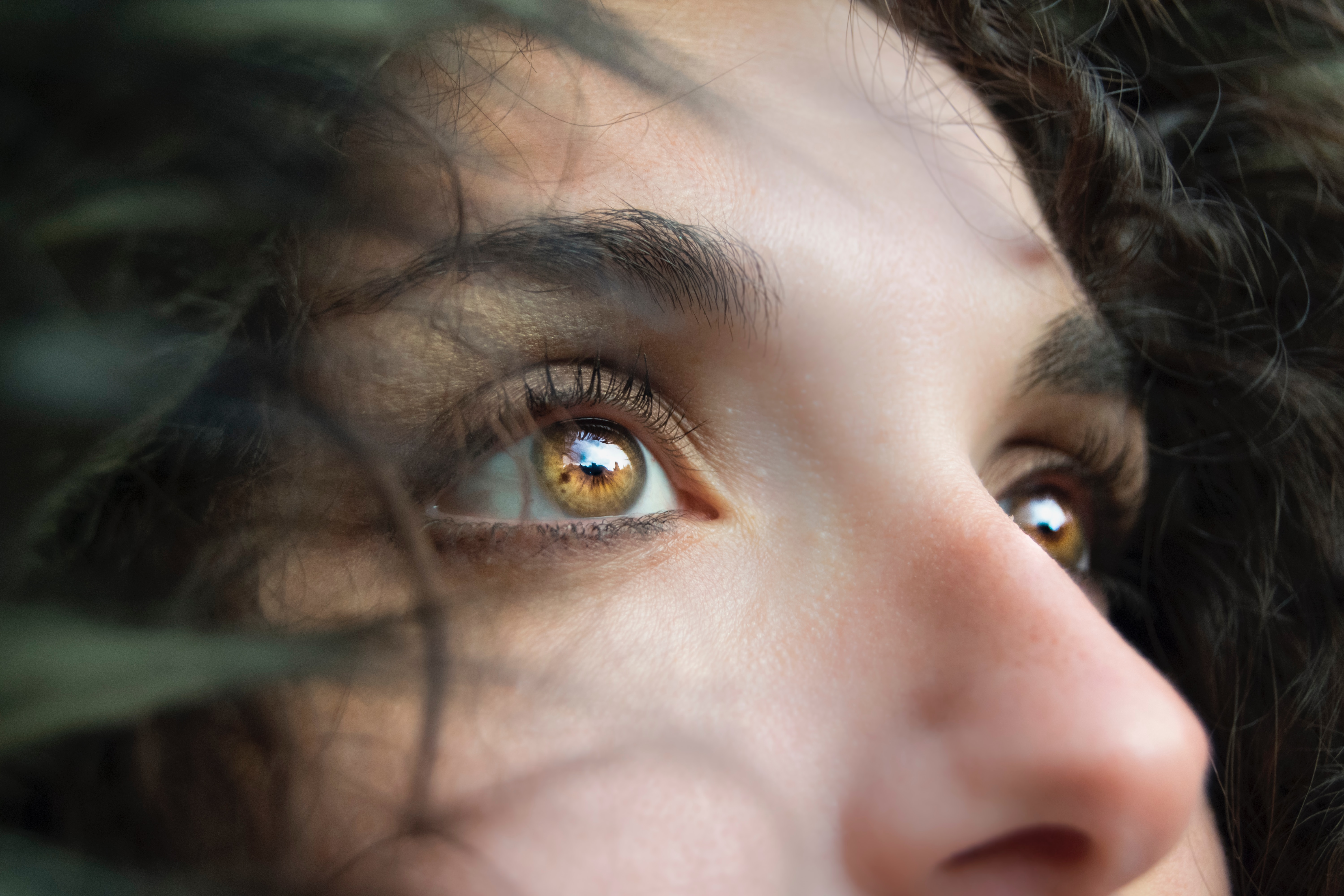
A 2022 study by researchers at the Autonomous University of Aguascalientes in Mexico evaluated the effectiveness of using VEP waveforms embedded in electroencephalographic (EEG) signals to determine the health of the optic nerve. The team employed an MP36 data acquisition system connected to an SS2LB lead set to record EEG signals from study participants. eyeThe signal data were used to create a database of VEPs that could be processed with analysis software for the early detection of glaucoma and other optic nerve-related illnesses.

A similar study combined the use of EEG signals with VEP to examine how participants recognize emotion in others. Researchers from two universities in China relied on a variation of VEP known as steady-state visual evoked potential (SSVEP), which exhibits distinctive frequency and phase properties. EEG signals were recorded via a BIOPAC data acquisition unit while participants were shown flickering images representing three emotional states—delight, sadness, and anger. The study found a positive correlation between SSVEP amplitude, alpha-frequency entrainment, and accurate identification of emotion.

These are just a few examples of how evoked potentials provide researchers with valuable insight into the central nervous system’s response to stimuli and its relationship to a wide range of physiological states. For additional information on how to use BIOPAC hardware and AcqKnowledge to stimulate a range of senses using electrical, auditory, visual, haptic, scent, and thermal stimuli and record evoked potentials, check out our webinar on Exogenous and Stimulus-related Evoked Potentials.


To learn more about how your study can benefit from using BIOPAC hardware and software to record and analyze evoked potential data, contact your local sales representative.

Fielding a Media Psychophysiology Team

Author: Ashley Churchill, M.A., BIOPAC Midwest Sales

Media psychophysiology research is being embraced by a variety of communication departments around the world. Media psychophysiology generally refers to the measurement of biological responses to messages in order to understand viewers’ cognitive and emotional processing of video, audio, or print messages. Media researchers often focus on measuring processes that occur as one interacts with media content over time and they look for insights into how and why certain media effects emerge (see Potter & Bolls, 2012 for more background).

Social science labs and communication researchers might feel daunted when starting up a lab if they are inexperienced in collecting physiology data. Here are three common strategies that will support the development of a media lab team and contribute to its success.

Ensure the research team has access to and is comfortable seeking out assistance from a variety of resources. Psychophysiology is a fundamentally interdisciplinary science. Incorporating it into your research requires that you draw on both technical and conceptual knowledge from disciplines outside of social science. To complicate things, formal psychophysiology resources can be difficult to find within the typical social science department. Effectively using external resources like subject librarians, university IT, faculty across departments, and BIOPAC’s support team, can help overcome this challenge.

Explain the conceptual aspects of media psychophysiology to your research assistants, whether they are volunteering for the semester or plan to stick around for a few years. Providing RAs with a conceptual background regarding relationships between media, psychology, and physiology can mean the difference between success and failure as far as effectively identifying and resolving problems during an experiment. It takes more than simple knowledge of electrode placement to make someone effective in a data collection role. Guiding RAs in a way that reflects this supports stronger research and researcher retention.

Preserve the practical knowledge generated by your team, do not let it leave with them. Learning how to conduct media psychophysiology research takes time and experience. As different team members rotate in and out of the lab over time, continue to support lab practices that preserve their knowledge and understanding of this research approach. Whether this takes the form of storing informative documentation or encouraging them to lead aspects of new member onboarding, these practices are important to improving the effectiveness of your team over time.

To find more information regarding media psychophysiology, check out our recent webinar featuring media psychophysiology expert Dr. Paul Bolls. To learn about signals that are common in media psychophysiology research— like fEMG, ECG, and EDA—visit our library of on-demand life science webinars.


BIOPAC offers a wide array of wired and wireless equipment that can be used in your research. To find more information on solutions for recording and analyzing signals such as ECG, heart rate, respiration, and more using any platforms mentioned in this blog post, you can visit the individual application pages on the BIOPAC website.

Photo by Vlad Hilitanu on Unsplash

Eye Tracking Insights

Try Not To Blink—This Won’t Hurt

What exactly, is eye tracking? Do I have to put on a helmet or stick something unpleasant into my eye? No, nothing so invasive, and not the least bit uncomfortable. Eye tracking is the process by which eye movements are recorded for the purpose of tracking where a subject is looking, what he/she is looking at, and the duration of the subject’s gaze on a given area. Eye tracking differs from electrooculogram (EOG) in that it records where and at what the subject is actually looking, whereas EOG primarily records the movements of the eye muscles. Rather than merely recording eye movement and position, eye tracking forges an invisible yet accurate connection between the subject and the objects within the subject’s field of vision. These “visual connections” of where the subject was looking during the study are clearly visible in the resulting data, as dramatic as tire tracks on an impressionable surface.

So how does eye tracking work? Using invisible near-infrared light combined with high-definition cameras, the eye tracking system projects invisible light onto the eye and records the direction from which it is reflected off the cornea. Eye tracking software is used to calculate the exact position and focal point of the eye. The sophisticated combination of eye tracking hardware and software allows for extremely accurate measuring and analyzing of visual behavior and fine eye movements. This makes it possible to create a detailed map of a subject’s field of vision and the specific visual elements honed in on by the subject. Eye tracking data is commonly acquired in tandem with other body signals for the purpose of determining the effects of what we are looking at has on our physiology. Common signals acquired during eye tracking studies are:

  • EEG (cognitive processes)
  • EDA (alertness)
  • ECG (anxiety)
  • EMG (muscle activity, conditioning)
  • EGG (pulse, respiration, temperature)

What are researchers looking for when conducting eye tracking studies? Eye tracking data is useful in a variety of different research fields including interface usability, reading analysis, psychological and emotional response, marketing research, training, clinical vision diagnosis, and more.

Eye tracking hardware mainly comes in two flavors. Screen based eye tracking involves having a subject sit in front of a computer monitor while being presented with various types of visual stimuli. A “bar type” eye tracker records the subject’s responses and interfaces with eye tracking software to present the stimulus and store the resulting data for analysis. BIOPAC’s AcqKnowledge software version 5.0.6 and higher, along with the BIOPAC MP160, MP36R, or Wireless Smart Center system, supports fully-integrated eye tracking functionality*. Rather than having to learn a second software program, the familiar AcqKnowledge interface has been extended to incorporate the EYE-TRACK-BAR and EYE-TRACK-BAR-XL remote screen eye tracking systems.

Mobile or “wearable” eye tracking systems utilize a pair of glasses with two types of built-in cameras. One camera faces the subject and tracks eye movements, gaze path, and other parameters, and a second camera (called a scene camera) records what the subject is looking at during the study. Mobile eye tracking allows the subject to engage in independent activities outside the lab, such as driving an automobile. Additional sensors record and log data from other body signals in real time, which can be loaded into the software for subsequent analysis.

For more information about eye tracking, we invite you to sign up for our free “Eye Tracking Fundamentals” webinar to be broadcast live on November 21, 2019 at 8:00 AM PST. Did you miss it or can’t attend? No problem. Follow the same link to watch it on demand.

*Eye Tracking Integration License for AcqKnowledge required.

Please also visit the following links for information about BIOPAC Eye Tracking products.

Eye Tracking Systems

Eye Tracking License


BIOPAC offers a wide array of wired and wireless equipment that can be used in your research. To find more information on solutions for recording and analyzing signals such as ECG, heart rate, respiration and more using any platforms mentioned in this blog post, you can visit the individual application pages on the BIOPAC website.

Photo by Marina Vitale on Unsplash

Neuroscience – A Brief History

What is neuroscience? In a nutshell, the study of neuroscience encompasses the physical and functional workings of the human brain and nervous system, drawing from a mix of cellular and molecular biology, anatomy, physiology, human behavior, and cognition. This field remains in a state of near-constant evolution, as the brain does not easily give up its secrets. Although the brain is home to a hundred billion neurons (brain cells), with nearly a quadrillion connections linking them, we have yet to fully and deeply understand the full breadth of a single cell.

But when and how did neuroscience originate? We know that as far back as 7,000 years ago, healers were boring holes into the skulls of their companions, a process known as trepanation. One can imagine the grim determination these ancients exhibited in their efforts to understand and relieve suffering, even if it was only with the intent of exorcising evil spirits. And it’s harder still to imagine how boring into someone’s living skull without anesthesia could possibly offer any measurable relief from suffering. However misguided these ministrations were, it’s clear that even millennia ago, the brain was recognized as an organ of great physiological significance.

For centuries, conventional wisdom recognized the heart as the seat of human consciousness. The Greek physician Hippocrates challenged this hypothesis, believing the brain to be the center of intelligence, yet the heart continued to hold sway with physicians and healers until long beyond Hippocrates’ time. This view persisted until the Roman physician Galen consistently observed that brain injuries sustained by gladiators often resulted in a marked decrease in cognition and mental acuity. (These injuries possibly foreshadowed today’s phenomenon of NFL CTE injuries.) Gradually, the brain began to take its rightful place in the pantheon of human physiology.

During medieval and Renaissance times, continuing important discoveries about the brain helped lay the foundation of what would become modern neuroscience, much of this new knowledge gleaned through illicit dissections, or by examining the brains of executed criminals. It’s safe to say that much scientific discovery—including that of human physiology—was hobbled by the religious constraints of the day. It wasn’t until the late 18th century, when Luigi Galvani observed the electrical activity of muscles and neurons, that key indicators about brain function became more apparent. Observations during this period often raised more questions than answers, however.

The 19th century brought further advances in the study of the brain and nervous system, notably by Emil du Bois-Reymond, who successfully demonstrated the electrical properties of nerve signals, and Paul Broca, who discovered through his pioneering work with brain-damaged patients that different regions of the brain were responsible for specific functions. This was later borne out by the German anatomist Korbinian Brodmann, who mapped the human cortex into 52 distinct areas, along with their corresponding functions.

In the 20th century, and particularly since the 1950s, neuroscience has evolved into an increasingly wide-ranging discipline, incorporating the brain and nervous system on a molecular level. This gave rise to many different types of neuroscience, which include affective, behavioral, molecular, cellular, clinical, computational, and a great many more. As the study of neuroscience has progressed, this field has produced no fewer than 19 Nobel Prize Laureates between 1904 and 2017. Neuroscience encompasses the most complex and sophisticated organ system in the body, a system that may possibly never be fully understood and certainly will continue to present wonders and challenges for centuries to come.

If you study neuroscience and want to learn more about how to get great data & meaningful life science measurements for human, animal, virtual, or MRI protocols, be sure to check out [button link=”https://www.biopac.com/webinars/” type=”small” newwindow=”yes”] BIOPAC Webinars[/button].

Photo by Jack Antal on Unsplash


T4 Conference Wrap-up

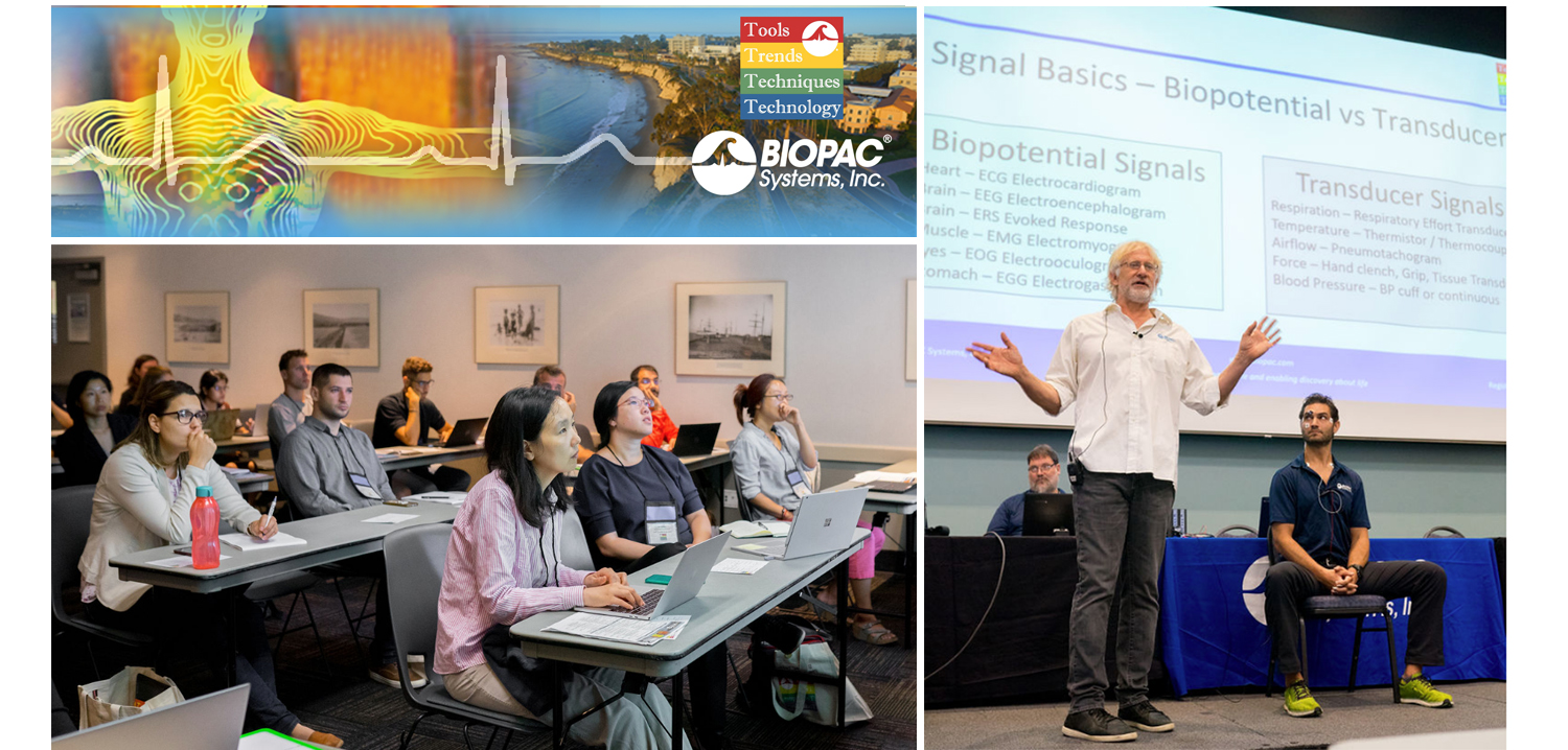
August is a month of barbecues, baseball, and last-minute vacations against the backdrop of a fading summer, and the eighth month of 2019 proved to be an equally exciting time at BIOPAC, with something of a pleasant vacation thrown in. On August 12-14, BIOPAC hosted its 2nd Biennial “Tools, Trends, Techniques, and Technology” Human Physiology Conference, known as “T4” to our friends and partners around the world. This three-day event, first held in 2017, featured lively, informative sessions hosted by BIOPAC staff, partners, noted academics, and industry experts, and covered a wide range of physiological techniques and disciplines. Over 150 attendees joined BIOPAC at the 2019 T4 Conference staged once again at the beautiful University of California at Santa Barbara campus. Participants and instructors winged in from China, Australia, Germany, Sweden, Italy, Spain, U.K., Ireland, and Egypt, as well as from wide regions of the U.S and Canada to soak up the idyllic surroundings of this ocean-side campus.

The focus of BIOPAC’s T4 Conference was to provide a fun, relaxed environment while hosting nearly 40 hands-on, human physiology training workshops chaired by over 20 distinguished instructors from around the globe. One of the many conference highlights was the “Mindfulness, Meditation, and Measurement: Mindfulness Exercises in Physiological Monitoring” session, hosted by Dr. Allan Weiss of the University of Southern California and  BIOPAC Co-founder and Director of R & D Alan Macy. These sessions, conducted in two parts over a two day period were booked beyond capacity, even though the early morning fog hadn’t yet lifted from the campus skyline.

The varied additional sessions covered a range from the basics, “Setting Up Your Lab, Getting Great Data and Troubleshooting,” (hosted by Dr. Terry Blumenthal of Wake Forest University, and BIOPAC’s Alan Macy and Tim Cook), and “Creating Your Own Lessons Using BSL PRO,” (hosted by Dr. Taffeta Elliot of the New Mexico Institute of Mining and Technology), to exciting new technologies such as “Integrating Screen-based Eye Trackers with AcqKnowledge,” with Keith Jackson of BIOPAC partner company EyeTech, BIOPAC CEO Frazer Findlay, and BIOPAC Engineering Manager Ed Peterlin. Additional wide-ranging physiological disciplines were also covered, including MRI techniques, stimulation, and exercise physiology. All sessions were conducted using BIOPAC hardware and software systems, as well as platforms provided by BIOPAC partners such as Cedrus, fNIR, WorldViz, Psychology Software Tools, and CNSystems. At the end of each day, it was time to relax and unwind with a beverage of choice, a a walk along the beach, and some good conversation. Each evening featured excellent meals and camaraderie in the relaxed yet elegant environment of some of Santa Barbara and Goleta’s most stunning surroundings. After three great days of immersive sessions, it came time for everyone to part ways, having forged new collaborative partnerships and an enhanced knowledge about the full potential of BIOPAC and BIOPAC partner products.

You don’t have to wait for T4 2021 to learn more about life science data—watch Fundamentals of Physiological Data Recording now!

For a closer look at T4, and a preview of what is sure to be an exciting 2021 event, we invite you to visit our T4 Human Physiology Conference page.

Looking for additional resources, tips, tricks, and expert advice on BIOPAC and BIOPAC Partner products? Please also visit our Webinars and Video Tutorial pages.


BIOPAC offers a wide array of wired and wireless equipment that can be used in your research. To find more information on solutions for recording and analyzing signals such as ECG, heart rate, respiration and more using any platforms mentioned in this blog post, you can visit the individual application pages on the BIOPAC website.

fNIRS Systems—a NIR miracle

At the dawn of the 19th century, pioneering astronomer Sir William Herschel discovered the existence of infrared radiation, which is electromagnetic radiation invisible to the human eye but identifiable via its ability to heat objects. Infrared radiation is commonly divided into three categories, near, mid, or far infrared waves. In ensuing years, infrared radiation was found to be useful for industrial, scientific, military, and law enforcement applications, such as heat signature tracking and night vision goggles. Over two decades ago, a new technology called near infrared spectroscopy (NIRS) was developed for non-invasively measuring specific aspects of human physiology using near infra-red waves. While far infrared waves lie closest to the microwave region, near infrared waves are closest in length to the visible light part of the electromagnetic spectrum, and are microscopic in size. Unlike far infrared waves, near infrared waves do not produce any heat. In fact, we can’t feel them at all. They are the small waves used by common devices such as a
remote control.

Today, near infrared waves are commonly used  for studying many application areas of human physiology, notably by the fNIRS Imaging Systems to gather precise, high-density brain function and muscle oxygenation data.

How does fNIRS work with regard to human physiology? It’s an optical technology which sends light into the tissue and records the light that scatters back via a photo-diode and sensor attached to the subject. The fNIRS continuous wave technology uses an optical window in the near infrared spectrum that enables light to pass through. For example, when you hold your hand in front of a strong white light, the tissue can appear red and almost translucent. fNIRS shines light into the skull or muscle tissue, and using special detectors, records the resulting light scatter that returns to the surface.  The light penetrates skin, bone, and alights on the brain before its return trip to the surface detectors, and accomplishes this painlessly without heat. In fact, the subject does not even feel the near infrared energy and the light is harmless. The result is startling images and functional data of the brain at work. The fNIRS Systems (ranging from Educational to High-Density versions) include fNIRsoft software for processing, analyzing and visualizing functional near infrared (fNIR) spectroscopy signals through a graphical user interface. Additionally, the fNIRS Systems work with the BIOPAC MP160 Research System and AcqKnowledge software in conjunction with other physiological signals such as ECG, respiration, cardiac output, blood pressure, electrodermal activity, and stimulus response markers. AcqKnowledge software also provides automated analysis tools for event related potentials and ensemble averaging.

fNIRS brain function application areas include workload assignment, VR,  stress, attention, geriatrics, economics, ergonomics, learning and performance, sports science, HCI, pain perception, and cognitive workload – low or high.

fNIRS Systems can also be used to measure muscle oxygenation data, including oxidative capacity, muscle load, lateral imbalances (left vs. right muscle group) oxygenated hemoglobin and de-oxygenated hemoglobin in muscle activity.

We invite you to enjoy a comprehensive free fNIRS webinar and to visit our fNIRS products page.

BIOPAC offers a wide array of wired and wireless equipment that can be used in your research. To find more information on solutions for recording and analyzing signals such as ECG, heart rate, respiration and more using any platforms mentioned in this blog post, you can visit the individual application pages on the BIOPAC website.

Synchronize Participant Video with Physiological Data

AcqKnowledge software includes a Media feature that allows you to record and link participant video with physiological data. A common issue that crops up when analyzing physiological data with linked participant video is a lack of synchronization between the resulting video playback and onscreen data. This most often occurs when a USB webcam-type video camera is used. These types of cameras typically support a 30 frame-per-second rate whereas AcqKnowledge and the MP System can sample at rates up to 200,000 Hz. These factors can introduce an unwanted delay or misalignment between the video playback and data in the resulting AcqKnowledge graph.

So how do we synchronize these disparate signals to dovetail perfectly before recording our data and video? Fortunately, AcqKnowledge provides adjustment tools for addressing this common issue. This simple procedure requires the use of the AcqKnowledge software’s powerful Stimulator feature and a BIOPAC OUT103 LED Video Sync Cable.

Here is the sync method in a nutshell: With the OUT103 LED connected to the Analog Output of the MP160 unit’s HLT100C module, the AcqKnowledge software’s Stimulator is used to output a 5-volt square wave pulse into an AcqKnowledge graph, Using this setup, the LED illuminates when the 5-volt pulse is applied. The trick now is to use the AcqKnowledge Media feature and webcam to record a short video of the LED illuminating while the 5-volt pulse is being applied.

In the resulting graph, it’s easy to see in the Media playback screen when the light comes on by slowly advancing the AcqKnowledge software’s I-beam cursor through the graph data. Does the LED in the recorded video illuminate before the cursor reaches the 5-volt pulse? After? Or at the same time? Chances are, the recorded light will pop on slightly before or after the cursor reaches the pulse.

To compensate for this, the AcqKnowledge Delta-T measurement is used to determine the time interval between the pulse and when the recorded LED illuminates. For example, if the recorded LED lights up an indicated 185 ms before the onset of the pulse, then this discrepancy can be removed by setting a delay of 185 ms in the Media setup tools. If the LED lights up after the pulse onset, a negative delay must be set (ex. -185 ms). Once the playback delay is eradicated, the experiment can continue with confidence that all video and physiological data will be properly aligned.

It’s features such as this that enhance the usability AcqKnowledge Media feature, making it a powerful tool for recording and synchronizing participant video and audio during experiments. After a bit of practice, dialing in the playback video for linked Media acquisitions becomes second nature.

To see AcqKnowledge Media Synchronization in action, we invite you to view a free on-demand webinar covering this procedure in detail.

BIOPAC offers a wide array of wired and wireless equipment that can be used in your research. To find more information on solutions for recording and analyzing signals such as ECG, heart rate, respiration and more using any platforms mentioned in this blog post, you can visit the individual application pages on the BIOPAC website.

Tips for implanting rodent pups with the Epoch wireless recording system

Implanting pups can be tricky, yet their value as neonatal models of human disease is key.

Tips for implanting rodent pups with the Epoch wireless recording system

The Epoch system enables recording 2 or 4 channels of EEG from neonatal rodents as young as postnatal day 10 (P10) in mice and P6 in rats using the 2-week transmitter. Implanting pups can be tricky—their skulls are very thin and the small size of the animals is difficult to work with. Yet, their value as neonatal models of human disease is key to developing new therapies or understanding neural dysfunction at such an early age.

Our experience has led to the following tips for maximizing the success of implanted pups:

  1. Allow pups time to recover. Although recordings can commence immediately, we recommend waiting 24 hours before commencing recordings to allow a fuller recovery.
  1. Limit recording time. Recording from rodent pups should be limited to about 2 h at a time, preferably in a temperature- and humidity-controlled incubator.
  1. Keep pre-weened pups in their dam’s presence as often as possible. Pups rely on the dam for complete care.
  1. Cull the litter to 4-6 pups. The dam may have issues with pups that are overly cold, not feeding often enough, or simply due to the presence of the transmitter.
  1. Implant all pups. It may be helpful to implant all pups in a litter either with Epoch transmitters or sham transmitters (sham transmitters are available, contact us).
  1. Use chew toys to prevent damage from the dam. Chew toys, such as nylabones, may help prevent the dam from chewing on the transmitters.
  1. Mind the state of the dam. Ultimately, the health and disposition of the dam may affect how she tolerates pups implanted with the Epoch transmitters.

Epoch, the smallest wireless EEG transmitter system available, is provided by BIOPAC to make quality EEG recordings from an early age possible. The Epoch transmitter is simply implanted to the skull of the animal with medical cyanoacrylate, requiring no bone screws. Surgery is quick and simple, requiring a small incision on the scalp, removal of the periosteum with 1% hydrogen peroxide, drilling small holes for the electrodes using a burr-type drill, implanting the transmitter, and re-absorbable sutures.

For more information on BIOPAC’s wide range of tools for recording, displaying, and analyzing EEG signals from human and animal subjects, including automated EEG analysis, seizure detection, evoked responses and many others, visit BIOPAC’s EEG Analysis page or view BIOPAC’s full line of electrodes, amplifiers, and wearable, wireless transmitters and loggers.

BIOPAC Systems, Inc. provides life science researchers and educators with data acquisition and analysis systems that inspire people and enable greater discovery about life. Visit us at www.biopac.com.

Stressing over the effects of stress

Several studies show chronic stress is typically detrimental to health, while acute stress may be linked to positive effects in addition to the negative counterparts

With April comes National Stress Awareness Month, and it bears noting that different types of stress have varying effects. Where chronic stress is typically detrimental to health, acute stress may be linked to positive effects in addition to the negative counterparts. But is this trade-off worth it?

In a biological psychology experiment testing the effect of oxytocin on acute social stress, researchers found different results between men and women. Subjects underwent several stress tasks while their electrocardiography and impedance cardiography were recorded with a BIOPAC MP Research System and amplifiers. The researchers found that after being given oxytocin during the stressful event, men had less negative affect and women had greater math performance (compared to the control group).

A neurobiology study examined the effect stress had on an extinction memory process when a competing association was paired with a previously aversive cue. To test this, participants were presented with two images, one that was paired with a shock from a BIOPAC bar electrode, and one that was not. Extinction memory training immediately followed this stimulus, with extinction retrieval testing following a day later. Data gathered by a BIOPAC data acquisition system suggested significantly lower retrieval in the group that experienced the acute stress stimulus.

Another biopsychology experiment measured the effect of acute stress on pro-inflammatory cytokine levels in the blood. BIOPAC equipment was used to measure ECG while participants were given the Trier Social Stress Test. After reviewing the data and blood samples, the researchers determined that levels of the cytokine cell, interleukin-1β, increased significantly immediately after the stress test.

The physiological and psychological impacts of stress are clearly multifaceted with many potential results. In some situations, a heightened positive affect or greater analytical thinking may be beneficial. But is this boost in mental performance worth the increase of inflammatory cells and lower memory retention? As with many aspects of life, moderation is the safest answer, ensuring the negative effects do not become chronic problems.

For more information on BIOPAC’s wide range of tools for recording, displaying, and analyzing ECG measurements from human and animal subjects including respiration, heart rate variability, and RSA, visit BIOPAC’s electrocardiography pages and view BIOPAC’s full line of electrodes, amplifiers, and wearable, wireless transmitters and loggers.

BIOPAC Systems, Inc. provides life science researchers and educators with data acquisition and analysis systems that inspire people and enable greater discovery about life. Visit us at www.biopac.com.

error: Content is protected !!