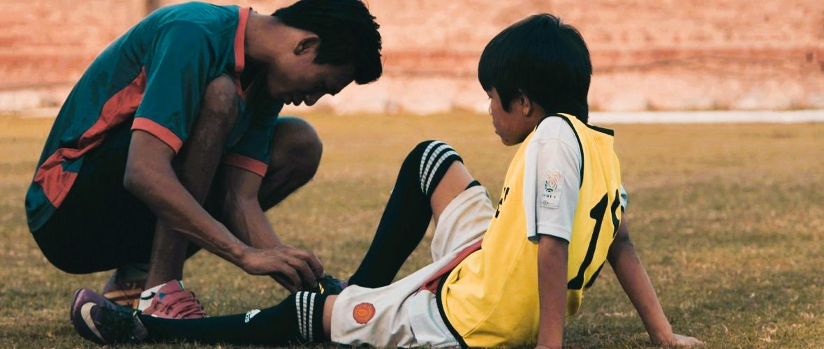
It can happen anywhere, anytime: on the pickleball court, during a pristine nature hike, or even while stepping off the front porch on the way to work. An ankle turns, a knee buckles, and suddenly you’re on the ground wondering what just happened. While such injuries often seem random, researchers who study human movement know they are rarely without warning.
Kinesiologists, scientists who study the mechanics and physiology of movement, have found that subtle changes in muscle activation, fatigue, coordination, balance, and neuromuscular control often emerge long before a strain, sprain, or overuse injury becomes apparent. To better understand these hidden danger signs, researchers increasingly rely on physiological measurements that reveal how the body’s muscular, nervous, cardiovascular, and biomechanical systems respond to activity. Such insights help identify injury risk factors and guide strategies designed to keep people moving safely and effectively.
Among the many tools used in kinesiology research, electromyography (EMG) is one of the most common because it reveals when and how muscles activate during movement. However, EMG is only one part of the story. Researchers also study force production, joint motion, movement acceleration, cardiovascular activity, respiration, muscle oxygenation, and neural activity to better understand how the body adapts to physical demands. Together, these measures provide a more complete picture of movement and help uncover patterns that observation alone may miss.
The applications of this work extend far beyond sports. Physiological monitoring helps clinicians evaluate rehabilitation progress, guide return-to-activity decisions, support fall-prevention efforts among older adults, and help design safer workplaces and training programs. Increasingly, researchers are shifting from simply treating injuries to identifying and addressing the factors that contribute to them before they occur.
 Researchers in India conducted a cross-sectional study comparing elite and recreational university badminton athletes to explore whether physiological and neuromuscular characteristics could serve as injury-risk markers. The investigators collected a range of performance and neuromuscular measurements, including surface EMG recordings, to examine differences between competitive levels and establish preliminary benchmarks for injury-risk stratification. Surface muscle activity was recorded using a BIOPAC two-channel data acquisition system as part of the neuromuscular assessment protocol. The study found that elite athletes exhibited distinct physiological and neuromuscular profiles compared to recreational players, suggesting that objective physiological markers may help identify movement patterns and performance characteristics associated with increased injury vulnerability. These conclusions support the growing use of physiological profiling to identify athletes who may benefit from targeted injury-prevention strategies before injuries occur.
A team of researchers in Tunisia investigated how fatigue of the ankle evertor muscles affects force perception and neuromuscular function in healthy adults. Ankle injuries are among the most common musculoskeletal injuries in both athletic and recreational populations, making ankle stability an important area of study. Using surface EMG to measure muscle activity before and after a fatigue protocol, the researchers examined how localized fatigue affected participants’ ability to accurately reproduce force levels. Researchers used a BIOPAC EMG amplifier and data acquisition system to record electrical activity from muscles involved in ankle stabilization. The researchers found that fatigue impaired force perception and altered neuromuscular responses, suggesting that fatigue may reduce the body’s ability to control ankle movements accurately. The study results add to growing evidence that fatigue-related physiological changes can increase injury risk, particularly during prolonged exercise or competition when neuromuscular control begins to decline.
A multi-institutional team of researchers from the United States examined neuromuscular function in individuals who had undergone anterior cruciate ligament (ACL) reconstruction, comparing them with healthy controls. The researchers wanted to better understand whether the relationships between muscle performance, muscle activation, and nervous system function change after ACL surgery. Participants completed a series of strength and neuromuscular assessments, with quadriceps muscle activity recorded using a BIOPAC data acquisition system with AcqKnowledge software and an EMG amplifier. BIOPAC isolated stimulators were also used during testing to help determine how fully participants could activate their quadriceps muscles. The study found that several relationships observed in healthy knees were no longer present after ACL reconstruction, suggesting that changes in neuromuscular control can persist long after surgery and rehabilitation are complete. These findings highlight how physiological monitoring can uncover hidden deficits that may not be apparent during routine recovery assessments, helping clinicians make more informed return-to-sport decisions and potentially reducing the risk of future injury.
As these examples demonstrate, physiological measurements give researchers powerful tools to identify injury risk before injuries occur, understand how fatigue alters movement control, and evaluate recovery following musculoskeletal injury. By revealing the physiological mechanisms that influence human movement, signals such as EMG and related neuromuscular measures help bridge the gap between laboratory research and practical injury-prevention strategies. As kinesiology research continues to evolve, physiological monitoring will likely play an increasingly important role in supporting safer movement, more effective rehabilitation, and improved long-term physical performance.
Are you planning a kinesiology-focused study? BIOPAC local sales representatives have the information you need to design and equip your research project.
