
In 1929, a German physician named Hans Berger discovered that electrodes placed on the scalp could detect various patterns of electrical activity. After verifying that the recordings were indeed recording from the brain, and were not artifacts of muscle or scalp, scientists began to study these “brain waves.” Today, EEG is still a medically useful recording for brain function. In medical and basic research, the correlation of particular brain waves with sleep phases, emotional states, psychological profiles, and types of mental activities is ongoing.
EEG records the electrical activity of the brain. Surface EEG electrodes provide a noninvasive way to measure brain function and regional brain activity. The EEG signal is made up of different frequency components and the amplitude of the signal varies in the different frequency bands: alpha, beta, delta, theta, gamma.
| Rhythm | Typical Frequencies (Hz) |
| alpha beta delta theta gamma |
8-13 13-30 1-5 4-8 >32 |
EEG can be recorded by placing electrodes on the scalp of the subject. The placement of the electrodes is important to ensure that you are recording data from the correct location when comparing and reporting results.The electrodes are placed on specific locations on the head. Each electrode location is given an alphanumeric name. Alpha is related to actual zone for electrode placement: F-Frontal, C-Central, T-Temporal, P-Parietal, O-Occipital zones. Numeric is based on the two hemispheres, odd numbers representing left side and even numbers representing right side.
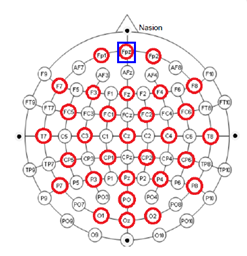 These locations are over certain areas of the brain that allow us to identify different types of activity:
- F7 near centers for rational activities
- Fz near intentional and motivational centers
- F8 close to sources of emotional impulses
- C3,C4,Cz deal with sensory and motor functions
- T3 and T4 emotional process
- T5 and T6 certain memory functions
- O1 and O2 for primary visual areas
An EEG cap can also be used to ensure proper electrode placement. The size of the EEG cap is critical in EEG recording. Having the correct cap size means that you can ensure that you are recording the correct brain location, otherwise you may think you’re recording F5 when you’re really recording F9.
Recording EEG is a bipolar recording (looking at difference between two electrodes). Electrodes are typically referenced to a physical electrode or site –
- vertex (Cz)
- linked-ears
- linked-mastoids
- ipsilateral-ear
- contralateral-ear
- C7 reference
- bipolar references
Recording can also be done using reference-free techniques when using 20 or more channels. The system will usually automatically take an average of all the electrodes rather than a physical location. The different averages include:
- Weighted average reference
- Common average reference
Once you have your EEG data, utilize AcqKnowledge software to run a number of fully automated EEG analysis functions. A range of fully automated routines provide quick, easy and reproducible results and include approximate entropy, delta power analysis, derive alpha RMS, derive EEG frequency bands, and EEG frequency analysis. The EEG frequency analysis provides a fully automated, epoch driven, analysis of the EEG signal. The system uses power spectral density to report Mean Power, Median Frequency, Mean Frequency, Spectral Edge, Peak Frequency values for each epoch.
For more information on BIOPAC’s wide range of tools for recording, displaying, and analyzing EEG measurements from human and animal subjects, visit BIOPAC’s electroencephalography pages and view BIOPAC’s full line of electrodes, amplifiers, and wearable, wireless transmitters and loggers.
BIOPAC Systems, Inc. provides life science researchers and educators with data acquisition and analysis systems that inspire people and enable greater discovery about life. Visit us at www.biopac.com.